Signalment:
Adult beaver,
Castor canadensisAdult male beaver found dead near a pond, March 2007.
Gross Description:
The animal presented in poor body condition. Throughout the plantar aspect of the hindlimbs and lateral margins of the tail, there are multiple cutaneous ulcers. On incision of the abdominal wall, there is approximately 5 ml of clear serosanguinous fluid. There was moderate enlargement of the spleen and liver and throughout the parenchyma, there are focally disseminated pale yellow white nodules.Â
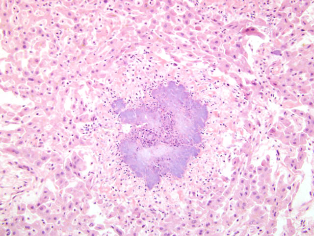
Histopathologic Description:
Immediately below the capsule and randomly throughout the parenchyma, there is multifocal to coalescing hepatocellular necrosis with lobulated colonies of coccobacilli frequently bound by variable neutrophilic infiltrates.Â
Morphologic Diagnosis:
Liver: Hepatitis, marked, necrotizing, multifocal to coalescing, acute, with florid lobulated colonies of extracellular coccobacilli
Lab Results:
PCR for
Francisella tularemia was negative. Aerobic culture yielded heavy growth of
Yersinia pseudotuberculosis from the spleen and liver.Â
Condition:
Yersinia pseudotuberculosis
Contributor Comment:
The cause of death of this animal is attributed to the cumulative effects of the necrotising hepatitis and splenitis and generalized emaciation. The heavy growth of
Yersinia pseudotuberculosis from the liver and spleen was considered significant. Although
Y pseudotuberculosis is endemic worldwide, infections are more commonly recognized in temperate zones, particularly in Europe, and have been reported in a wide variety of domestic and wild mammals, including rodents, rabbits, deer, cattle, goats and sheep and birds, such as turkeys, ducks, geese pigeons, pheasants and canaries.Â
Y pseudotuberculosis had previously been recovered in wildlife species between 1962 and 1973 (1 crow, 2 purple martens, 1 snowshoe hare, and 7 beavers) in Ontario, Canada and rat feces have been implicated as a source of human infection in Japan
4,5. Exposure is predominantly by consumption of fecal contaminated food or water, or alternatively, ingestion of infected prey
4. The epidemiology of infection is complex and not yet fully resolved; however, isolation of this bacterium from a beaver has important public health implications and the regional health authority has been notified and appropriate actions implemented. In humans, infection may result in mesenteric lymphadenitis, ulcerative ileitis, septicemia and erythema nodosum and most presenting patients are 4-15 year old males. From a wildlife population perspective, yersiniosis typically occurs in only select individuals in an area and, thus presumably has few implications for free ranging and marine mammals. In more densely populated communities, infection may be exacerbated by stress and reduced transmission distance.Â
JPC Diagnosis:
Liver: Hepatitis, necrotizing, acute, random, multifocal to coalescing, severe, with large colonies of coccobacilli, beaver (
Castor canadensis), rodent.
Conference Comment:
There are three species of
Yersinia that are pathogenic for rodents and humans:
Y. pestis (etiologic agent of bubonic and pneumonic plague),
Y. pseudotuberculosis, and
Y. enterocolitica.
6 All pathogenic
Yersinia spp. produce
Yersinia Outer Proteins (Yop), which enable extracellular survival and proliferation. A combination of various translocator, recognition, and effector Yops combine with a type III secretion system called Ysc to disarm macrophages and contribute to delaying the development of a cell-mediated immune response.
6
Type III secretion systems are found within a wide variety of Gram-negative bacteria, and are used to deliver bacterial proteins into host cells.
7 It consists of a base structure that spans the inner and outer bacterial membranes and a needle-like structure that provides a conduit for the transfer of bacterial proteins into the host cell.
7
There are 6 effector Yops, 5 of which are found in all three pathogenic
Yersinia species: YopH, YopM, YopE, YpkA/YopO and YopJ/YopP. YopT is exclusively found in
Y. enterocolitica.Â
3,7 YopB, YopD, and LcrV are required to translocate effector proteins into the host cell.Â
3,6 YopN and LcrG are part of a control and recognition system.Â
6
Yersinia Virulence Factors
| Type III secretion system | Composed of transmembrane base structure, and needle like conduit |
| Â | Â |
| PhoP | Plays a crucial role in ability to replicate within macrophages3 |
| Â | Â |
| Translocation Yops | Required for translocation of Yops into the host cell, but is not required for excretion of Yops into extracellular space3 |
| YopB | Â |
| YopD | Â |
| LcrV | Â |
| Â | Â |
| Control and Recognition Yops | Prevents Yop secretion prior to host cell attachement2 |
| YopN | Â |
| LcrG | Â |
| Â | Â |
| Effector Yops | Â |
| YopH | Disrupts actin structures including focal adhesions and prevents phagocytosis by macrophages and neutrophils3,7 , affects oxidative burst of macrophages and inhibits T- and B-cell signaling, and T-cell proliferation6 |
| YopM | Scaffolding protein, only Yersinia effector that lacks catalytic activity7 |
| YopE | Disrupts actin structures6, prevent phagocytosis by macrophages and neutrophils3 |
| YpkA/YopO | Disrupts actin skeleton, prevent phagocytosis by macrophages and neutrophils3,7 |
| YopJ/YopP | Induces programmed cell death in macrophages, and functions as an acetyltransferase7 resulting in inhibition of mitogen-activate protein kinase pathway3 |
| YopT (Y. enterocolitica) | Inactivates RhoA6, prevent phagocytosis by macrophages and neutrophils3 |
References:
1. Adkins I, K+�-�berle M, Gr+�-�bner S, Bohn, E, Autenrieth IB, Borgmann:
Yersinia outer proteins E, H, P, and T differentially target the cytoskeleton and inhibit phagocytic capacity of dendritic cells. Int J Med Microbiol 297:235-244, 2007
2. Day JB, Ferracci F, Plano GV: Translocation of YopE and YopN into eukaryotic cells by
Yersinia pestis YopN, tyeA, sycN, YscB and lcrG deletion mutants measured using a phosphorylatable peptide tag and phosphospecific antibodies. Mol Microbiol 47:807-823
3. Fisher ML, Castillo C, Mecsas J: Intranasal inoculation of mice with
Yersinia pseudotuberculosis causes a lethal lung infection that is dependent on Yersinia outer proteins and PhoP. Infect Immun 75:429-442, 2007
4. Fukushima H, Gomyoda M, Shiozawa K, Kaneko S, Tsubokura M:
Yersinia pseudotuberculosis infection contracted through water contaminated by a wild animal. J Clin Microbiol 26:584-585, 1988
5. Hacking MA, Sileo L:
Yersinia enterocolitica and
Yersinia pseudotuberculosis from wildlife in Ontario. J Wild Dis 10:452-453, 1974
6. Monnazzi LGS, Carlos IZ, de Medeiros BMM: Influence of
Yersinia pseudotuberculosis outer proteins (Yops) on interleukin-12, tumor necrosis factor alpha and nitric oxide production by peritoneal macrophages. Immunol Lett 94:91-98, 2004
7. Trosky JE, Liverman ADB, Orth K: Microreview:
Yersinia outer proteins: Yops. Cell Microbiol 10: 557-565, 2008