Signalment:
Gross Description:
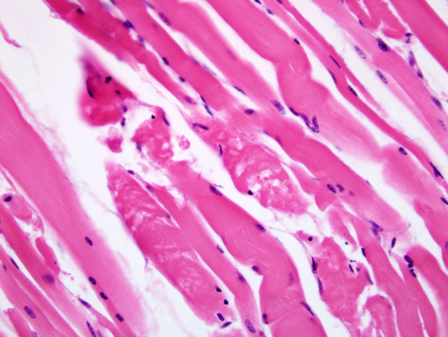
Histopathologic Description:
Morphologic Diagnosis:
Lab Results:
Condition:
Contributor Comment:
Microscopic lesions similar to those in skeletal muscle were also present in the heart of this donkey, but were more mild. Granular eosinophilic material compatible with hemoglobin or myoglobin was present in tubular lumens of the kidney. This material is most likely myoglobin released from the affected skeletal muscle fibers. Although gross lesions were not detected in the heart or skeletal muscle of this foal, pale areas in the heart and skeletal muscle can be observed in cases of nutritional myopathy of foals, however is not always observed.(7) Degenerate muscle may be very difficult to detect grossly when it is uncalcified, and is likely to escape detection.(3) Mineralization of muscle fibers is not always present in cases of nutritional myodegeneration in foals.(7)
There were approximately 50 female donkeys in this herd, with this being the only donkey diagnosed with nutritional myopathy, although foals had died in previous years with similar signs. The female donkeys were fed brome grass hay year-round, never being pastured on green grass, or supplemented with vitamins or minerals. Green, growing forages should provide adequate vitamin E as α-tocopherol, but the vitamin E content is greatly reduced in grass that is dried for hay. Mature plants contain less α -tocopherol than younger plants, and mature grass cut for hay can have loss of up to 80% of the tocopherols when dried in the sun for 4 days.(1) Plasma vitamin E status of horses is highest from May to August when fresh grass is being grazed and lowest when horses are fed harvested or stored feed during the same period.(2)
JPC Diagnosis:
Conference Comment:
Nutritional myopathy is common in calves, lambs, swine, and foals.(6) It can be caused by a lack of dietary intake of vitamin E or selenium or from competitive binding of selenium by copper, zinc, silver, or tellurium. Foods high in polyunsaturated fats such as fish require intake of more vitamin E to minimize oxidative damage from the metabolic processing of these foods.(6)
Vitamin E and selenium are important in preventing damage from free radicals from both within and from outside the cell. Free radicals are molecules with an odd number of electrons produced during normal cell functions or from tissue radiation, drug reactions, or inflammation. Free radicals are highly reactive molecules that can cause damage to mitochondria, endoplasmic reticulum, or the cytosol via damage to important cellular proteins or peroxidation and damage of cellular lipid membranes. When cellular membranes are damaged, ion gradients can not be properly maintained. Extracellular calcium moves into the cytosol and the cell responds by attempting to protect calcium-sensitive myofilaments by pushing calcium into mitochondria. Mitochondria quickly accumulate excess calcium and lose their ability to produce energy for the cell. Myofibrils exposed to leaking calcium hypercontract leading to degeneration and necrosis of myofibers.(6)
The attached table provides a non-comprehensive list of diseases considered to be associated with an imbalance or deficiency in either selenium or vitamin E.
| Cattle | Nutritional myopathy Retention of fetal membranes |
| Horse | Nutritional myopathy |
| Swine | Mulberry heart disease Hepatosis dietetica Exudative diathesis Iron hypersensitivity Nutritional myopathy Anemia |
| Sheep | Nutritional myopathy Infertility Poor growth potential |
| Dogs | Intestinal liposfuscinosis |
| Cats; mink; birds; pigs; rabbits; reptiles | Steatitis (yellow fat disease) |
| Chickens and Turkeys | Encephalomalacia (superficial cerebellar hemorrhage crazy chick disease) |
References:
2. Blakley BR, Bell RJ: The vitamin A and vitamin E status of horses raised in Alberta and Saskatchewan. Can Vet J 35: 297-300, 1994
3. Hulland TJ: In: Pathology of Domestic Animals, eds., Jubb JVF, Kennedy PC, Palmer N, pp. 228-234. Academic Press, Inc. San Diego, CA, 1993
4. Jones TC, Hunt RD, King NW: Nutritional deficiencies. In: Veterinary Pathology, 6th ed, pp. 789-794, Williams and Wilkins, Baltimore, MD, 1997
5. Radostits OM, Gay CC, Blood DC, Hinchcliff KW: Veterinary Medicine. In: A Textbook of the Diseases of Cattle, Sheep, Pigs, Goats and Horses, pp. 1515-1533. WB Saunders Company, London, 2000.
6. Van Vleet JF, Valentine BA: Muscle and tendon. In: Jubb, Kennedy, and Palmer's Pathology of Domestic Animals, vol 1 ed. Maxie MG, pp. 236-243. Elsevier Limited, Philadelphia, PA, 2007
7. Wilson TM, Morrison HA, Palmer NC, Finley GG, van Dreumel AA: Myodegeneration and suspected selenium/vitamin E deficiency in horses. J Am Vet Med Assoc 169: 213-217, 1976