Signalment:
Gross Description:
Histopathologic Description:
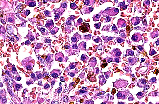
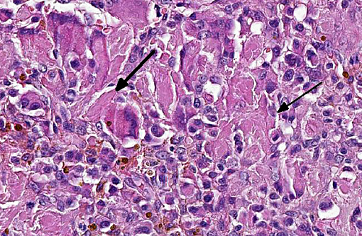
The cells are closely packed in sheets with a moderate amount of fibrovascular stroma. Single tumor cells are ovoid to polygonal, ranging in size from 20 to 30 μm in diameter. Cell borders are distinct. The moderate to abundant amount of eosinophilic cytoplasm is finely granular. Round to ovoid nuclei, ranging in size from 10 to 15 μm in diameter, are often located eccentrically within the cells. The chromatin is finely stippled, in some nuclei finely clumped with a distinct round centrally located eosinophilic nucleolus in each nucleus. Bi- and multinucleated tumor cells with up to three nuclei are frequent (> 5 / HPF). Anisocytosis and anisokaryosis are striking. The mitotic rate is low with up to 1 mitotic figure per high power field.
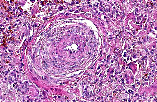
Preexisting vessels within the mass are thickened by increased numbers of spindle cells in the media (media hyperplasia) and deposition of extracellular, eosinophilic, fibrillar material (collagen).
Additionally, throughout the mass multifocal acute hemorrhages with extravasation of erythrocytes and fibrin, depositions of granular golden-brown pigment (hemosiderin) in macrophages and multiple areas of necrosis are present.
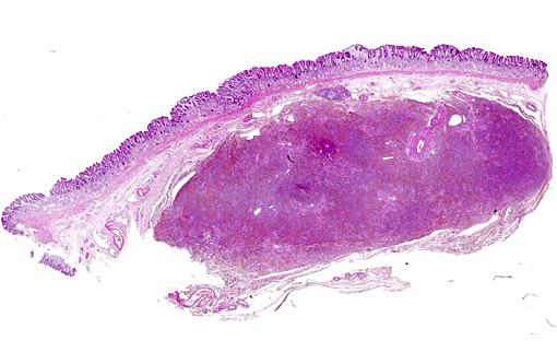
Submucosal tissue surrounding the neoplasm is expanded by edema, occasional hemorrhages, multiple hemosiderin-laden macrophages and moderate numbers of plasma cells.
Morphologic Diagnosis:
Condition:
Contributor Comment:
Clinically, gastric plasmacytomas appear nonspecific; occasionally epigastric pain, vomiting, anorexia weight loss and hemorrhages occur. Specific gross lesions are rare, as tumors cause more or less diffuse thickening of the stomach wall, like other round cell tumors. Histologically, gastric plasmacytomas are unencapsulated, but well demarcated, densely cellular masses, composed of variable mature, well- to poorly-differentiated round cells, which often contain typically eccentric located nuclei. Bi- and multinucleated cells with up to 5 nuclei per cell are a frequent finding.(5,8)
The deposition of AL-amyloid within tumor masses has been reported in few cases of EMP.(9,10,12) Congo red stain is useful to distinguish a gastric plasmacytoma from other round cell tumors, like malignant lymphoma or histiocytoma.(2,5) Congo red stain in this case failed to identify amyloid deposits.Â
The development of MM proceeding from EMP has not been determined in dogs, in contrast to humans.(8,13) In general, EMP located in the oral cavity tend to be benign, whereas EMP of the aboral digestive tract (esophagus, stomach, intestine, and rectum) tend to be more aggressive with potential to metastasize via the regional lymph nodes.(5)
JPC Diagnosis:
Conference Comment:
Conference participants agreed with the contributors assessment that the preexisting vessels in the submucosa are moderately thickened, though it was suspected amyloid is also responsible for this expansion in addition to spindle cells. There is abundant hemosiderin within this neoplasm, often within apparent neoplastic plasma cells. Iron accumulation in neoplastic plasma cells is a feature identified in previously reported cases, with one author speculating it may be due to a mutation of heme-containing enzymes.(11)
Plasmacytomas, in addition to multiple myeloma, lymphoma and ehrlichiosis, have all been associated with a monoclonal gammopathy.(1) Bence-Jones proteinuria is another clinical feature associated with plasma cell tumors; which are light chains of immunoglobulins small enough to permit their passage through the glomerulus.(1) A noted cytologic feature of plasma cell tumors is a rim of red globular material around the neoplastic cells, termed flame figures, which is another supportive diagnostic finding to these neoplasms.Â
References:
1. Allison RW. Laboratory evaluation of plasma and serum proteins. In: Thrall MA, Weiser G, Allison RW, Campbell TW, eds. Veterinary Hematology and Clinical Chemistry. 2nd ed. Oxford, UK: Wiley-Blackwell; 2012:469-470.
2. Brunnert SR, Altman NH: Identification of immunoglobulin light chains in canine extramedullary plasmacytomas by thioflavine T and immunohistochemistry. J Vet Diagn Invest: official publication of the American Association of Veterinary Laboratory Diagnosticians, Inc 3: 245-251, 1991.
3. Brunnert SR, Dee LA, Herron AJ, Altman NH: Gastric extramedullary plasmacytoma in a dog. Journal of the American Veterinary Medical Association 200: 1501-1502, 1992.
4. Fry MM, McGavin MD: Bone Marrow, Blood Cells and Lymphatic System. In: Pathologic Basis of Veterinary Disease, eds. McGavin MD, Zachary JF, 4th Edition ed., pp. 802-803. Penny Rudolph, St. Louis, Missouri, 2007.
5. Jacobs RM, Messick JB, Valli VE: Tumors of the Hemolymphatic System. In: Tumors in Domestic Animals, ed. Meuten DJ, Fourth ed., pp. 161-163. Blackwell Publishing Company, Ames, Iowa, 2002.
6. MacEwen EG, Patnaik AK, Johnson GF, Hurvitz AI, Erlandson RA, Lieberman PH: Extramedullary plasmacytoma of the gastrointestinal tract in two dogs. Journal of the American Veterinary Medical Association 184: 1396-1398, 1984.
7. Majzoub M, Breuer W, Platz SJ, Linke RP, Linke W, Hermanns W: Histopathologic and immunophenotypic characterization of extramedullary plasmacytomas in nine cats. Veterinary pathology 40: 249-253, 2003.
8. Nolan KD, Mone MC, Nelson EW: Plasma cell neoplasms. Review of disease progression and report of a new variant. Surgical oncology 14: 85-90, 2005.
9. Platz SJ, Breuer W, Geisel O, Linke RP, Hermanns W: Identification of lambda light chain amyloid in eight canine and two feline extramedullary plasmacytomas. Journal of comparative pathology 116: 45-54, 1997.
10. Ramos-Vara JA, Takahashi M, Ishihara T, Miller MA, Pace LW, Craft D, Common R, Watson GL: Intestinal extramedullary plasmacytoma associated with amyloid deposition in three dogs: an ultrastructural and immunoelectron microscopic study. Ultrastructural pathology 22: 393-400, 1998.
11. Rannou B, Heilie P, Bedard C. Rectal plasmacytoma with intracellular hemosiderin in a dog. Vet Pathol. 2009;46(6):1181-1184.
12. Rowland PH, Linke RP: Immunohistochemical characterization of lambda light-chain-derived amyloid in one feline and five canine plasma cell tumors. Veterinary pathology 31: 390-393, 1994.
13. Smithson CW, Smith MM, Tappe J, Beaudin A, Bradley M: Multicentric oral plasmacytoma in 3 dogs. Journal of veterinary dentistry 29: 96-110, 2012.
14. Vail D. Solidtary and extramedullary plasmacytic tumors. In: Withrow SJ, Vail ME, eds. Small Animal Clinical Oncology. Philadelphia, PA: WB Saunders; 2007:779-783.
15. Vasiliu T, Popa R: Forme gastrointestinale des tumeurs dites plasmacytomes. Comptes Rendus des S+�-�ances et M+�-�moires de la Soci+�-�t+�-� de Biologie 98: 738-740, 1928.