WSC 2023-24 Conference 11, Case 2
Signalment:
17-year-old, male, intact, Pacific pond turtle (Actinemys marmorata pallida)
History:
A wild-hatched turtle housed at the zoological institution had a history of severe chronic pitting and ulcerated lesions on the shell for over a year. Recently, it had a rapid clinical decline with anorexia and obtunded mentation. Given the poor prognosis, humane euthanasia was elected.
Laboratory Results:
Panfungal (ITS3-ITS-4) PCR assay on frozen shell yielded a 203 base pair fragment with 100% identity and 100% query coverage toMG780506, Emydomyces sp. isolate 13-1796.
Microscopic Description:
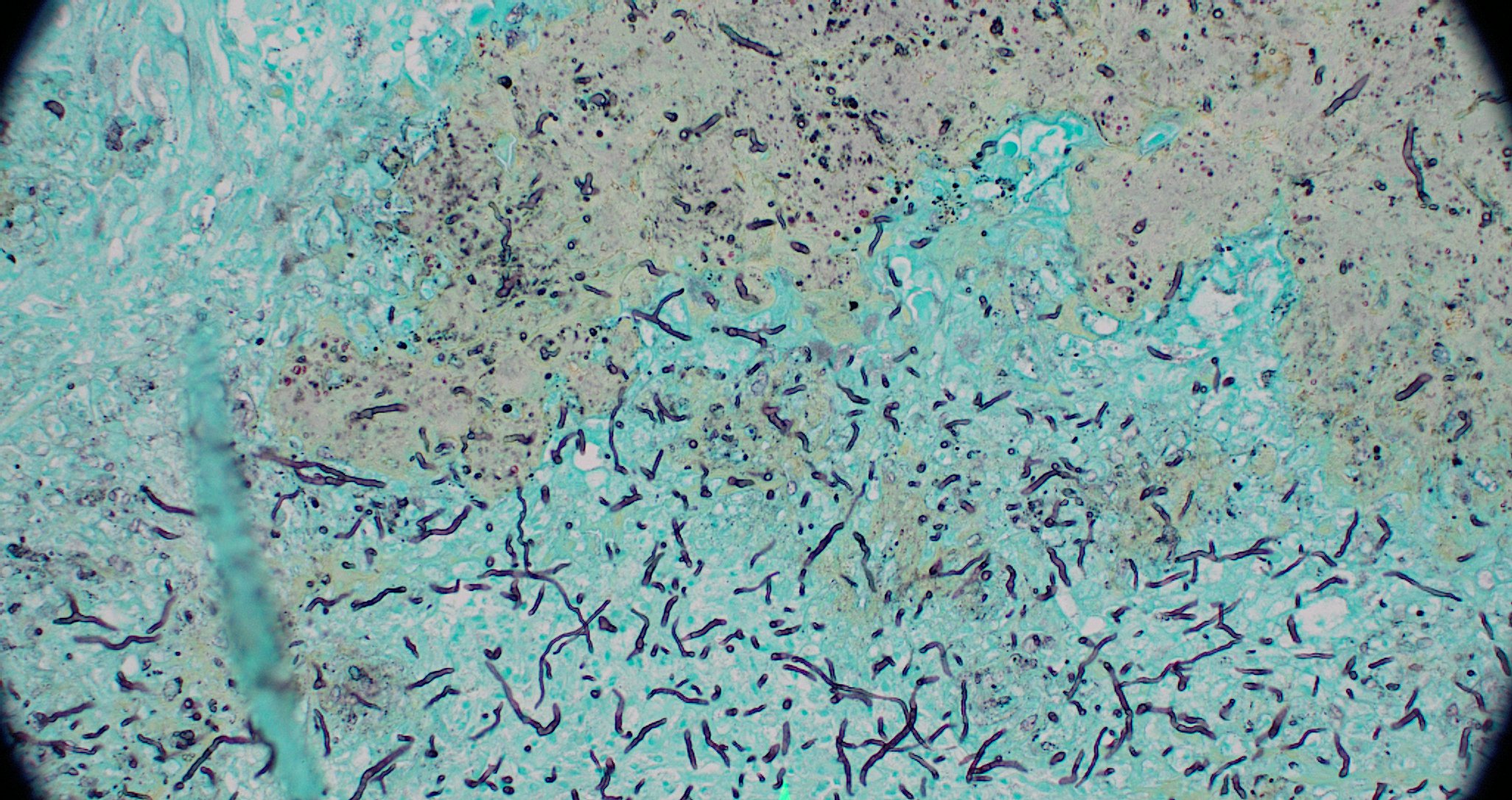
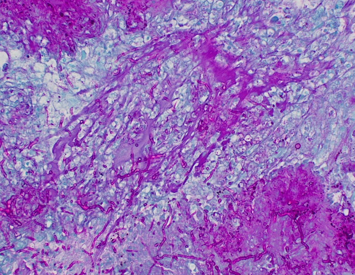
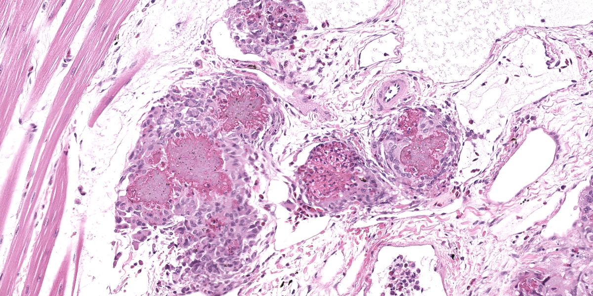
Head, dorsal: Extending from a focal ulcer on the skin surface and through the dermis to efface 60% of the dorsal calvarium, expand the meninges, and invade into the brain, as well as extending ventrally along the lateral aspect of the skull, are numerous coalescing epithelial inclusion cysts containing fungal hyphae. The epithelial inclusion cysts are lined by keratinized squamous epithelium and contain lamellated keratin, necrotic debris, entrapped necrotic bony fragments, fungal hyphae, and mixed bacteria. The fungal hyphae are 1 to 4 μm in diameter, regularly septated, and have acute to right-angle branching. The effaced bone has empty lacunae with loss of osteocytes (osteonecrosis), with moth-eaten margins and osteoclasts in Howship’s lacunae (osteolysis).
The affected meninges are also expanded by increased fibrous tissue. In this area, the epithelial inclusion cysts penetrate into the brain parenchyma and are associated with moderate heterophilic and histiocytic inflammation, necrotic debris, and rarefaction of the neuropil. In other sections, fungal invasion disrupts the ependymal lining of the ventricle. Variable numbers of heterophils, histiocytes, and lymphocytes are associated with the cysts and fungi throughout the section, but inflammation is most prominent in the skeletal muscle along the lateral aspect of the skull, where there are numerous, coalescing fungal heterophilic granulomas.
Contributor’s Morphologic Diagnosis:
Multiple tissues, dorsal head: Severe, regionally extensive, chronic-active, necro-ulcerative heterophilic and granulomatous dermatitis, myositis, osteomyelitis, and meningoencephalitis with epithelial inclusion cysts, osteonecrosis and osteolysis, and intralesional fungal hyphae (consistent with Emydomyces sp., presumably E. testavorans).
Contributor’s Comment:
Emydomyces testavorans, a newly described fungus, is strongly associated with ‘ulcerative shell disease’ or ‘pond turtle shell disease’ in free-ranging and captive aquatic turtles in North America.2,4,5 The classical shell lesions associated with emydomycosis are ulcerations in the carapace and/or plastron with increased pliability. In some cases, the turtles have expansile nodular masses within the shell, displacing the coelomic membrane, distorting the shell contour, and compressing internal viscera.4 The present case has an unusual presentation, in which the fungal infection affected the tissues of the head, in addition to the shell, and invades deeply into the brain causing meningoencephalitis with antemortem neurological signs.
Histologic features associated with E. testavorans infection include multilocular intradermal and intraosseous epithelial inclusion cysts, which are lined by keratinized stratified squamous epithelium and contain keratin debris and necrotic bone.2,4 The cysts are also associated with squamous metaplasia, hyperkeratosis, osteonecrosis, and inflammation, all of which are consistent with the present case of the Pacific pond turtle in our collection.4 Although E. testavorans is strongly associated with ulcerative shell disease in freshwater aquatic chelonians, causation has not been proven through experimental infections.
Very little is known about the pathogenesis and lesion progression of emydomycosis, and it is unclear if it is a primary pathogen or if a combination of factors is required to elicit disease. Reported histologic features do not appear to be consistently pathognomonic for E. testavorans infection. Instead, they could be non-specific and indicative of a chronic healing process with cysts representing walling off of the damaged tissue or dyskeratosis.4 Other differential diagnoses for ulcerative shell lesions in aquatic turtles include septicemic cutaneous ulcerative disease (SCUD), which is caused by a combination of trauma, bacterial infection, and poor water quality. Epithelial inclusion cysts have also been reported in one case of mycobacteriosis, with no evidence of concurrent fungal infection.4
Emydomyces testavorans is in the order Onygenales, along with other emerging pathogenic reptile keratinophilic fungi, such as Ophidiomyces ophiodicola and Nannizziopsis guarroi.5 Presumably, like many fungi in this order, E. testavorans is also an environmental saprophyte.5 Emydomyces testavorans was first reported in free-living turtles in California in 2020, but has been isolated from turtles in Illinois and Washington in samples from as early as 2016, suggesting a potentially broad geographic distribution.2,5 As Pacific pond turtles are listed as a vulnerable species, there is some concern that this recently described and likely underdiagnosed disease could pose a significant threat to already at-risk populations.1
Contributing Institution:
Disease Investigations
Institute for Conservation Research
San Diego Zoo Wildlife Alliance
PO Box 120551
San Diego, CA 92112
http://institute.sandiegozoo.org/disease-investigations
JPC Diagnosis:
Head: Epithelial inclusion cysts with osteonecrosis, granulomatous dermatitis, cellulitis, myositis, and meningitis, and fungal hyphae.
JPC Comment:
As the contributor notes, the key histologic features of emydomycosis are ulcerative dermatitis, necrotizing osteomyelitis, and inclusion cysts lined by keratinized stratified squamous epithelium containing keratin debris. Fungal hyphae that are thin-walled, regularly septate, and occasionally branched can be demonstrated with GMS or PAS staining, making for a relatively straightforward histologic diagnosis.5 Grossly, however, the lesions of emydomycosis may be difficult to identify as they can be highly localized lesions and may lurk under the shell surface after the initial shell defect has healed over.2 The contributor describes typical gross lesions in the excellent summary above, but gross lesions may also be rather subtle, and consist only of carapace flaking or bleaching.2 In a recent study of affected pond turtles in Washington state, emydomycosis was diagnosed based on external exam in 25-50% of wild caught animals, but in greater than 80% of animals based on CT scans, which revealed the inclusion cysts and osteolytic lesions that characterize the disease.2
Emydomycosis presents similarly to its main differential diagnosis, septicemic cutaneous ulcerative disease (SCUD), which can cause ulcerated lesions on the plastron and carapace, petechial hemorrhage, emaciation, lethargy, and death.3 SCUD is currently considered a multifactorial disease caused by poor water conditions and environmental stressors which predispose the animal to bacterial infection. SCUD was originally associated with Citrobacter freundii infection, but it is now understood that a variety of gram-negative organisms can cause the syndrome.
Conference discussion focused on reptilian dermatomycoses generally and on specific dermatologic disease of chelonids. In addition to SCUD and mycotic shell disease caused by E. testavorans, participants also discussed fibropapillomatosis in sea turtles, caused by chelonid herpesvirus-5, which are characteristic tumors of the skin around the eyes, mouth, limbs, shell, and cornea. These tumors can vary in appearance from flat, plaque-like lesions to exophytic or verrucous masses. They may also form on internal organs and grow large enough to impair buoyancy, leading to death.
Discussion of the morphologic diagnosis centered on whether the rather exuberant epithelial inclusion cysts were invading or merely compressing the brain. Participants felt the brain was only being compressed and thus preferred a diagnosis of meningitis rather than meningoencephalitis.
References:
- Adamovicz L, Allender MC, Gibbons PM. Emerging infectious diseases of chelonians: an update. Vet Clin North Am Exot Anim Pract. 2020;23(2):263-283.
- Lambert MR, Hernandez-Gomez O, Krohn AR, et al. Turtle shell disease fungus (Emydomyces testavorans): first documented occurrence in California and prevaelence in free-living turtles. Ichthyology & Herpetology. 2021;109 (4):958-962.
- Mitchell MA, Tully TN. Current therapy in exotic pet practice. Elsevier;2016.
- Woodburn DB, Kinsel MJ, Poll CP, et al. Shell lesions associated with Emydomyces testavorans infections in freshwater aquatic turtles. Vet Pathol. 2021;58(3):578-586.
- Woodburn DB, Miller AN, Allender MC, Maddox CW, Terio KA. Emydomyces testavorans, a new genus and species of Onygenalean fungus isolated from shell lesions of freshwater aquatic turtles. J Clin Microbiol. 2019;57(2).