CASE I:
Signalment:
A 2.5 year old primiparous Holstein Friesian cow (bos taurus).
History:
This dairy cow was from a herd of 360 animals, and had been exhibiting clinical signs consistent with toxic mastitis within the previous three weeks. Initially, this cow was treated with broad spectrum antibiotics, non-steroidal anti-inflammatories, and oral fluids, but failed to improve and was euthanized on welfare grounds.
Gross Pathology:
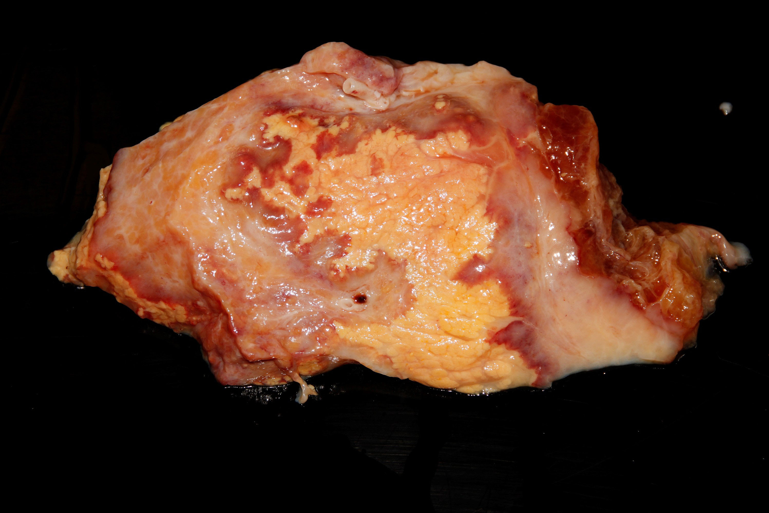
The right cranial mammary gland is enlarged and firm, and on the cut surface there is locally extensive fibrosis, with a distinct area of pallor (sequestrum) surrounded by a hyperemic halo. Within the teat cistern there is pale yellow, watery, fibrinosuppurative content. There is a focal, ~1-2cm diameter, capsulated abscess within the parenchyma as well. There is marked subcutaneous oedema surrounding the affected quarter.
Diagnostic Testing:
The aerobic bacteriology of the affected mammary gland identified Klebsiella pneumoniae, which was resistant to Ampicillin and sensitive to Neomycin, Streptomycin, Amoxicillin/Clavulanic acid, Cefpodoxime, Trimethoprim/Sulphamethoxazole, Enrofloxacin, Tetracycline. The culture and PCR-denaturing gradient gel electrophoresis (DGGE) testing for Mycoplasma was unrewarding.
Microscopic Description:
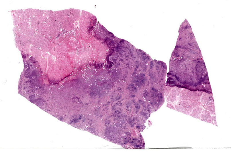
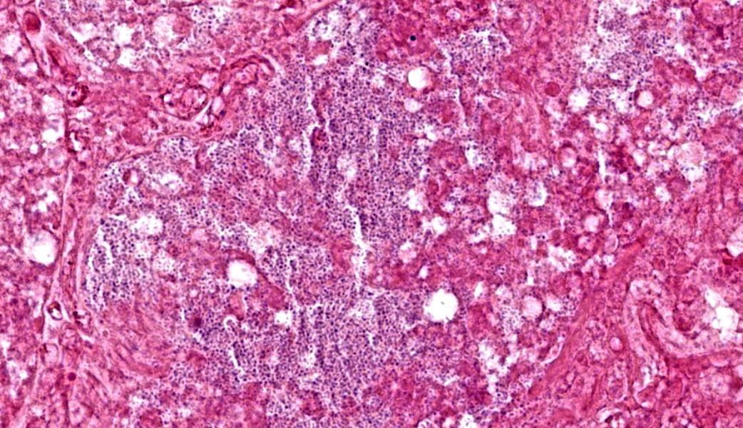
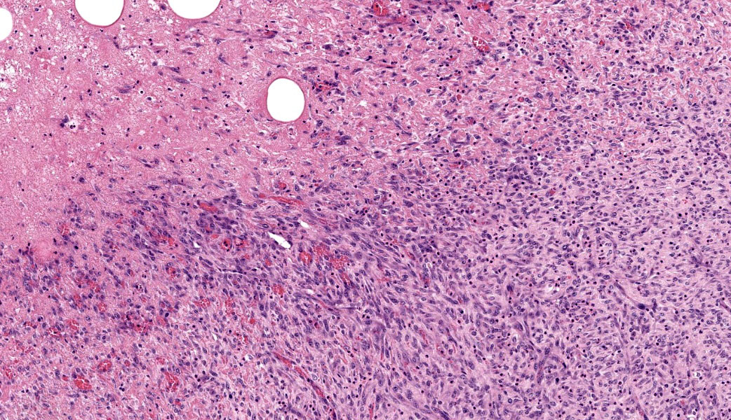
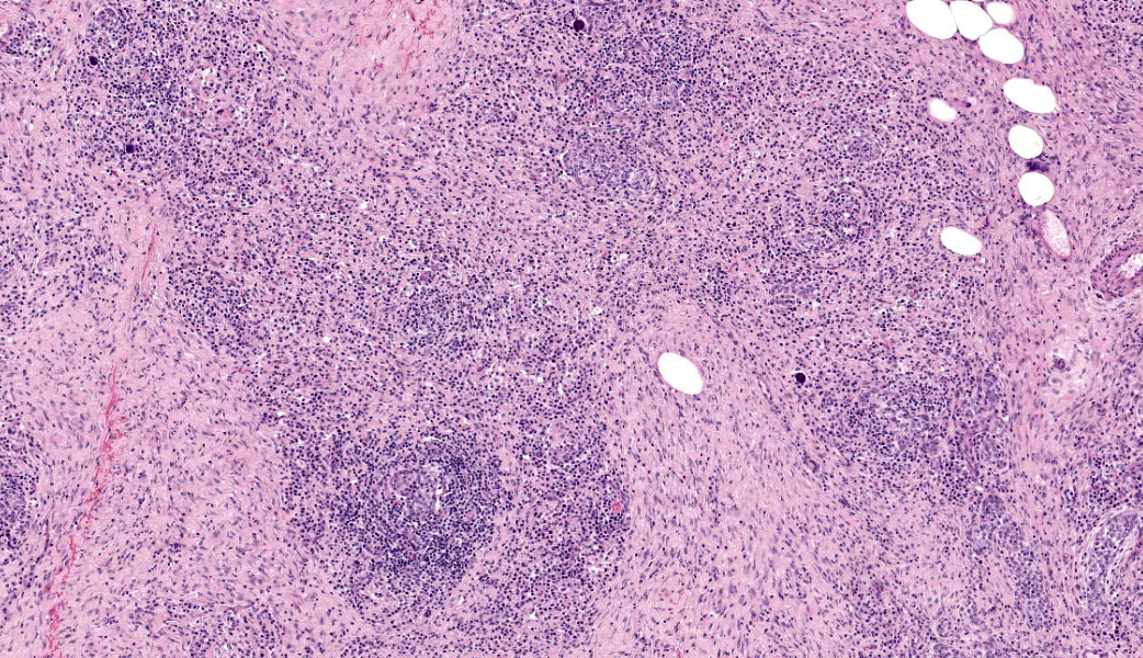
Mammary Gland - Right cranial quarter The mammary gland is effaced in a focally extensive manner by moderately well demarcated palely eosinophilic parenchyma with partial loss of differential staining and cellular detail whilst retaining preservation of the overall architecture (coagulative necrosis - sequestrum). Within the necrotic area, there are scattered pale basophilic, 2-3 micron in length, coccobacilli. The necrosed area is bordered by a thick rim of granulation tissue and nascent fibrosis. Throughout the remaining parenchyma, the mammary gland is multifocally effaced by extensive areas of abundant lytic and karyorrhectic leucocytes and debris (lytic necrosis) with abundant fibrin, alongside abundant granulation tissue, and a marked inflammatory infiltrate comprising of lymphocytes and plasma cells, often forming small clusters. Multifocally, there are small numbers of compressed and distorted interlobular ducts, remnants of compressed and atrophic mammary alveoli or basophilic mineral concretions of proteins and salts, that are sometimes accompanied by macrophages or very rare multinucleated giant cells (foreign body type). Within the inflamed granulation tissue, a small number of ducts are dysplastic, and scattered throughout the parenchyma are a moderate number of round optically empty spaces (lipid). Within some ducts, there are accumulations of lytic leucocytes and necrotic debris. A small number of mammary alveoli multifocally contain viable, degenerated and lytic neutrophils as well as necrotic debris that is bordered, dissected, and compressed by collars of fibrosis.
Morphological Diagnosis:
Mammary Gland: Severe, chronic-active, multifocal to coalescing neutrophilic, necrotizing and lymphoplasmacytic mastitis with intralesional bacteria, granulation tissue and sequestrum formation
Disease: Coliform mastitis
Aetiology: Klebsiella pneumoniae
Contributor's Comment:
The post-mortem findings are compatible with the history of mastitis. Based on the gross post-mortem appearance of the mammary glands, a coliform mastitis was considered most likely, with E. coli, Klebsiella spp., and Enterobacter spp. considered as the top differentials. Furthermore, the macroscopic and histopathological findings of necrosis and inflammation within the mammary parenchyma were compatible with coliform mastitis.
Bovine mastitis is one of the most prevalent and consequential diseases of dairy cows globally, carrying significant detrimental welfare and financial implications (1). Mastitis in dairy cows is caused by a wide variety of pathogens, including gram-positive cocci, gram-negative bacilli, Mycoplasma bovis, and the Prototheca species. Among these, gram-positive cocci and gram-negative bacilli are the main agents and the pathogenesis encompasses ascending infection. Streptococcus agalactiae and some types of Staphylococcus aureus are obligate parasites of the gland and inevitable pathogens, but the great majority of infections are opportunistic and environmental (see JPC 1210466).
Coliform bacteria such as Escherichia coli (the most common), and other Enterobacteriaceae (Enterobacter, Klebsiella, Citrobacter, Serratia, and Proteus). are ubiquitous in the environment and have the capacity to cause severe environmental mastitis via ascending infection from the teat.1 Coliforms produce endotoxins causing hemorrhage, oedema, thrombosis, and vascular damage, subsequently damaging tissues and potentially leading to death. Lactating cows are more likely to succumb to the effects of the endotoxins, as their response is determined by their reproductive cycle.5 The typical coliform mastitis in cows presents clinically with a sudden drop in milk production, watery or bloody milk, and a swollen, hot, and painful udder. Affected cows often show systemic signs such as fever, depression, dehydration, and, in severe cases, may become recumbent or die rapidly due to endotoxemia. As seen in this case, if the cow survives this acute and toxic stage, the affected mammary gland is then likely to sequester and slough off due to necrosis of the glandular tissue secondary to chronic coliform mastitis.12
Extensive investigations on the virulence profile of the E coli associated mastitis did not identify a clear set of virulence genes, and some authors prefer the use of mastitis associated E. coli (MAEC), with MAEC being more an "ecotype" rather than a "pathotype".6, 16
Klebsiella pneumoniae is one of the most common coliforms causing clinical bovine mastitis, but has been found to be one of the most damaging when considering milk production, treatment costs, and mortality rate. K. pneumoniae mastitis has a poor cure rate after antimicrobial treatment, and although it is commonly an environmental opportunist, lateral spread from diseased to healthy cattle is possible.11, 12
The pathogenicity of K. pneumoniae is related to its virulence factors, with the primary factors being capsular polysaccharide (CPs), lipopolysaccharides, siderophores, and fimbriae. Furthermore, Klebsiella species are a known reservoir for antibiotic resistance genes, which can spread to other Gram-negative bacteria.12, 16, 17 In vitro infection of bovine mammary epithelial cells with K. pneumoniae resulted in, showing rapid adherence to, and invasion of, mammary epithelial (milk-producing) cells by K. pneumoniae leading to cellular damage and apoptosis. K. pneumoniae was shown to promote the production of the following cytokines: IL8, IL-1β, and Tumor Necrosis Factor-α (TNF-α), as well as promoting the transcriptional expression of the pro-inflammatory genes for IL-6, IL-8, IL-1β, and TNF-α.10
Contributing Institution:
Veterinary Pathology Service University of Nottingham Sutton Bonington Campus College Road Sutton Bonington Leicestershire United Kingdom LE12 5RA Veterinary Pathology Service
JPC Morphologic Diagnosis:
Mammary gland: Mastitis, necrosuppurative, chronic-active, multifocal to coalescing, severe, with infarction and innumerable coccobacilli.
JPC Comment:
The one and only Dr. Corrie Brown, livestock infectious disease expert extraordinaire from the University of Georgia, moderated the JPC's 22nd conference this year before presenting the keynote lectures at the Northeastern Veterinary Pathology Conference in Washington, D.C. She put WSC participants through their paces with a quartet of excellent ruminant cases, starting off with this classic, but deceptively nuanced, case of coliform mastitis. This served as a springboard to review the anatomy, immunology, and epidemiology of mastitis in dairy cattle. One participant astutely reminded everyone that, in the lactating mammary gland, the term "acinus" should be substituted in favor of the term "alveolus" to help differentiate between the stages. This is a small but important distinction when describing the functional unit of milk production.
Participants initially considered Staphylococcus aureus as a potential cause in this case. However, the absence of Splendore-Hoeppli material and the overwhelming necrosuppurative characteristic of the lesion made this etiology less likely. Trueperella pyogenes was also suggested as a differential, but its hallmark abscess formation was not present. Instead, presentation of overwhelming necrosis pointed towards coliforms, which, as the contributor mentioned in their comment, include E. coli, Enterobacter, Klebsiella, Citrobacter, Serratia, and Proteus.1,2
Dr. Brown emphasized that mastitis is primarily a disease of dairy cattle, not because other animals don't get mastitis, but because dairy cows experience far more teat trauma through repeated hand- or machine-milking. The teat canal and cistern normally provide excellent immune protection, but chronic mechanical stress compromises this barrier. This is why pre- and post-milking teat dipping remains one of the most effective mastitis prevention strategies in modern dairy management.
A key teaching point was that effectively all mastitis pathogens enter the mammary gland through the teat with one important exception: highly pathogenic avian influenza.14,15 This virus is currently the only virus known to infect the bovine mammary gland. HPAI was recently seen in the WSC this year during Conference 17, Case 2.
Dr. Brown contrasted the clinical behavior of the major mastitis pathogens as important clues to assist the pathologist in narrowing down differentials. Staphylococcus aureus and Streptococcus spp. typically cause subclinical or mild clinical mastitis - the cow feels fine, but the milk looks and smells abnormal. Dry cow therapy has nearly eradicated Streptococcus mastitis in the United States, although Staphylococcus continues to be one of the most common mastitis pathogens affecting dairy cattle.7 Coliform mastitis, however, is entirely different. Cows become acutely, profoundly ill, often immediately postpartum, with systemic endotoxemia driving the severity of disease.2 Recovery rates are poor even with aggressive therapy.2,11,12
Participants differed in opinion regarding the use of the term "sequestrum." While the contributor provides a cogent explanation for why this term may be used in cases of mastitis - due to the body walling off the affected quarter and eventually sloughing it - some participants opined that the sharply demarcated, devitalized region of mammary tissue was better described as an infarct given the endotoxin-induced thrombosis that is characteristic of coliform mastitis. This distinction mattered, they argued, because the pathogenesis is vascular, not traumatic.
Discussion then turned to Klebsiella pneumoniae, the major pathogen in this case. Klebsiella thrives in wood products such as sawdust and shavings, making bedding a common source of exposure.8 While sometimes described as "contagious," Dr. Brown clarified that transmission is environmental, not cow to cow.11 Participants debated whether Klebsiella can enter mammary epithelial cells; while Salmonella and Shigella are well-established as intracellular pathogens, the evidence for Klebsiella is less clear.4 There are, however, some in-vitro studies that suggest possible invasion, although whether this translates in-vivo has yet to be fully elucidated.4
One participant noted that the bacterial morphology in this case did not match their mental image of Klebsiella, which is typically produces a capsule. The capsule of Klebsiella is one of its most important virulence factors.10,17 The organisms here appear to lack a capsule on H&E, which contributed to diagnostic hesitation. Others wondered whether a Staphylococcus aureus co-infection might be present, which is certainly possible given that there were several gram-positive bacteria present on a Gram stain. However, capsule production by Klebsiella may be downregulated secondary to certain host factors, such as increased extracellular iron as occurs with severe tissue damage, so reliance on capsule presence may be deceptive.10
References:
- Bianchi RM, Schwertz CI, de Cecco BS, Panziera W, De Lorenzo C, Heck LC, Snel GGM, Lopes BC, da Silva FS, Pavarini SP, Driemeier D. Pathological and microbiological characterization of mastitis in dairy cows. Trop Anim Health Prod. 2019 Sep;51(7):2057-2066.
- Burvenich C, Van Merris V, Mehrzad J, Diez-Fraile A, Duchateau L. Severity of E. coli mastitis is mainly determined by cow factors. Vet Res. 2003;34(5):521-564.
- Cheng J, Zhang J, Han B, Barkema HW, Cobo ER, Kastelic JP, Zhou M, Shi Y, Wang J, Yang R, Gao J. Klebsiella pneumoniae isolated from bovine mastitis is cytopathogenic for bovine mammary epithelial cells. J Dairy Sci. 2020 Apr;103(4):3493-3504.
- Clegg S, Murphy CN. Epidemiology and Virulence of Klebsiella pneumoniae. Microbiol Spectr. 2016;4(1):10.1128/microbiolspec.UTI-0005-2012.
- Foster, R.A., 2017. Female reproductive system and mammae. In: J.F. Zachary (ed), Pathologic basis of veterinary disease, 6th ed, (Elsevier, St. Louis, MO), p. 1147-1193.
- Germon P, Foucras G, Smith DGE, Rainard P. Invited review: Mastitis Escherichia coli strains-Mastitis-associated or mammo-pathogenic? J Dairy Sci. 2025 May;108(5):4485-4507.
- Keefe GP. Streptococcus agalactiae mastitis: a review. Can Vet J. 1997;38(7):429-437.
- Morales-Ubaldo AL, Rivero-Perez N, Valladares-Carranza B, Velazquez-Ordonez V, Delgadillo-Ruiz L, Zaragoza-Bastida A. Bovine mastitis, a worldwide impact disease: Prevalence, antimicrobial resistance, and viable alternative approaches. Vet Anim Sci. 2023 Jul 24;21:100306.
- Munoz MA, Welcome FL, Schukken YH, Zadoks RN. Molecular epidemiology of two Klebsiella pneumoniae mastitis outbreaks on a dairy farm in New York State. J Clin Microbiol. 2007;45(12):3964-3971.
- Paczosa MK, Mecsas J. Klebsiella pneumoniae: Going on the Offense with a Strong Defense. Microbiol Mol Biol Rev. 2016;80(3):629-661. Published 2016 Jun 15.
- Pollock J, Foster G, Henderson K, Bell J, Hutchings MR, Paterson GK. Antimicrobial resistance profiles and molecular epidemiology of Klebsiella pneumoniae isolates from Scottish bovine mastitis cases. Epidemiol Infect. 2025 Jan 17;153:e15
- Schlafer, D.H., Foster, R.A. 2016 Female genital system. In: M.G. Maxie (ed|), Jubb, Kennedy, and Palmer's Pathology of Domestic Animals, 6th ed, vol. 3, (Elsevier, St. Louis, MO), 358-464.
- Schukken Y, Chuff M, Moroni P, Gurjar A, Santisteban C, Welcome F, Zadoks R. The "other" gram-negative bacteria in mastitis: Klebsiella, serratia, and more. Vet Clin North Am Food Anim Pract. 2012 Jul;28(2):239-56.
- Shi J, Kong H, Cui P, et al. H5N1 virus invades the mammary glands of dairy cattle through 'mouth-to-teat' transmission. Natl Sci Rev. 2025;12(9):nwaf262.
- Sordillo LM, Streicher KL. Mammary gland immunity and mastitis susceptibility. J Mammary Gland Biol Neoplasia. 2002;7(2):135-146.
- Tong X, Barkema HW, Nobrega DB, Xu C, Han B, Zhang C, Yang J, Li X, Gao J. Virulence of Bacteria Causing Mastitis in Dairy Cows: A Literature Review. Microorganisms. 2025 Jan 15;13(1):167.
- Wusiman, M., Zuo, J., Yu, Y. et al. Molecular characterization of Klebsiella pneumoniae in clinical bovine mastitis in 14 provinces in China. Vet Res Commun. 2025;49(18).