Signalment:
12-year-old mixed breed female spayed cat.This cat had a lumpectomy in November of 2010. One month later, regrowth was found, and the mass had grown fast since. Mastectomy was recommended and performed on July 7, 2011. At operation, pyometra was detected and an OHE was also performed. The uterus was submitted for examination along with the ovaries.
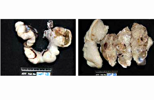
Gross Description:
The uterine horns and body were submitted in formalin. The uterus was bilaterally swollen and the uterine horns multifocally enlarged. Bilaterally, ovaries were shrunken and interpreted to be atrophic. Upon sectioning, the uterus was diffusely and bilaterally thickened, had a cystic nature, and the lumen was filled with inspissated chocolate-colored material.
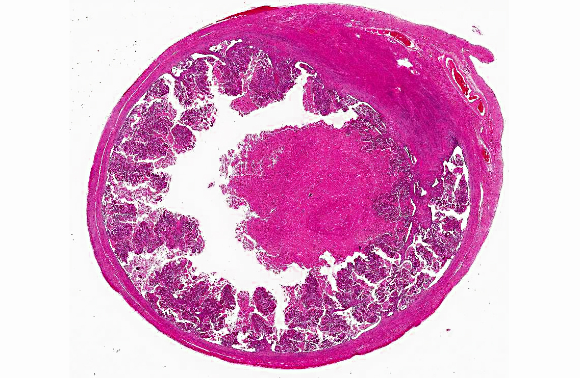
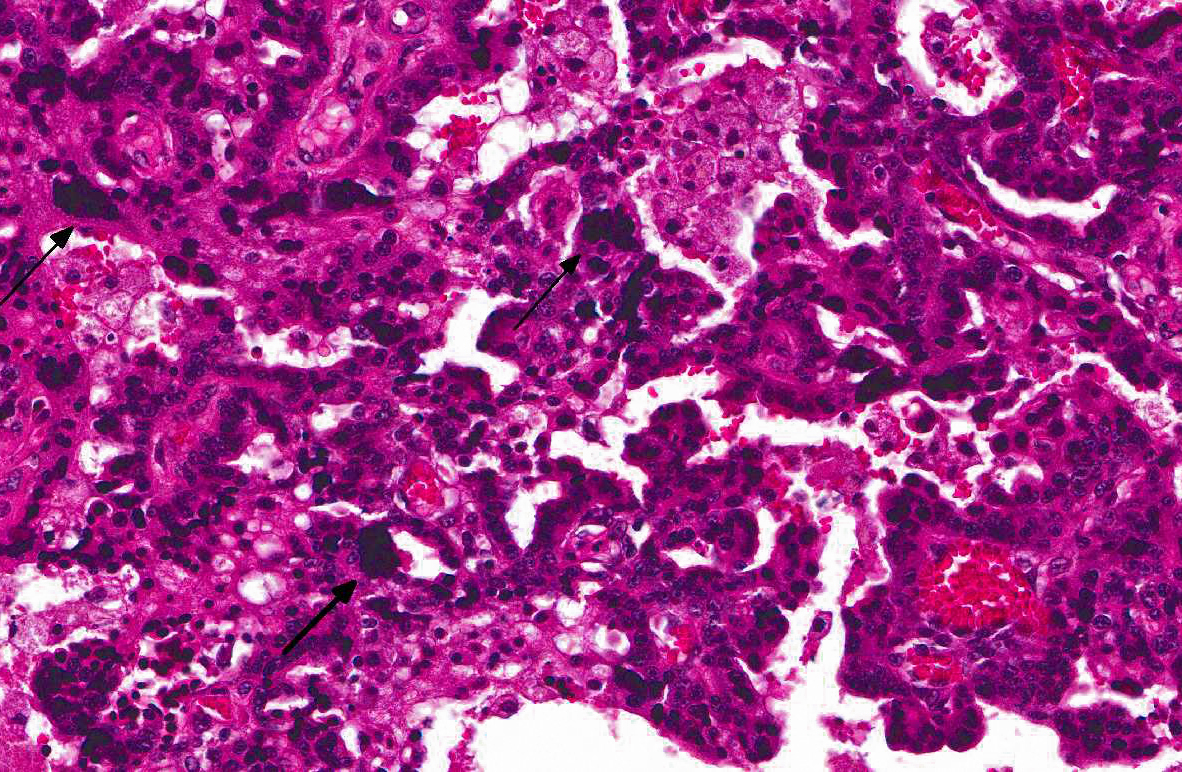
Histopathologic Description:
Uterus: Expanding the endometrium and encroaching upon the lumen is a well-demarcated, unencapsulated, poorly circumscribed, infiltrative, moderately cellular neoplasm. Neoplastic cells form florid papillary and micropapillary projections into the lumen; neoplastic cells are supported by a moderate fibrous stroma. Neoplastic cells are cuboidal to columnar with indistinct cell borders and a moderate amount of finely granular eosinophilic cytoplasm. Nuclei are round to oval with finely stippled chromatin and 1-2 basophilic nucleoli and there is mild anisocytosis. Mitotic figures are rare. Throughout the neoplasm, there are multinucleated neoplastic cells with dense, hyperchromatic nuclei. These giant cells range up to 250 μm in diameter. The uterine lumen and spaces between papillary projections are filled with brightly eosinophilic cellular and basophilic nuclear debris, admixed with small amounts of hemorrhage and mineral. Within the underlying endometrium, there are numerous mildly ectatic glands containing bright red secretory material.
Morphologic Diagnosis:
Uterus: Endometrial adenocarcinoma, giant cell variety.
Condition:
Endometrial adenocarcinoma, giant cell type
Contributor Comment:
Epithelial malignancies of the uterus are considered to be rare in domestic species. In domestic species, uterine adenocarcinoma is most commonly seen in the cow, but has also been reported in the mare, ewe, bitch, and queen.(2,8) In the bitch and queen, uterine malignancies constitute less than 1% of all neoplasms. In the ox, uterine carcinomas are solitary, often scirrhous neoplasms which invade the endometrium and myometrium, and often metastasize to the iliac and sublumbar lymph nodes, and ultimately to the lung and liver. Endometrial adenocarcinoma has been reported rarely in the queen.(3) In the largest review 8 of 13 uterine neoplasms arose in the endometrium; one of these was a mixed Mullerian neoplasm.(3) Myometrial invasion was variable in these cases, and carcinomatosis was noted in three, and pulmonary metastasis in one. Only two of the cats with endometrial adenocarcinoma had disease-free intervals longer than five months. Neoplastic cells are immunopositive for cytokeratin, vimentin, smooth muscle actin, COX-2, beta catenins, progesterone and estrogen receptors and caveolin-1.(3,7) In laboratory species uterine adenocarcinomas are most commonly reported in the rabbit and mouse. In the mouse, endometrial carcinomas are infiltrative neoplasms which invade the endometrium and myometrium, may occlude the uterine lumen, and may metastasize to distant sites. Squamous differentiation is seen with endometrial adenocarcinoma in the B6C3F1 mouse, usually in association with chemical administration. Uterine adenocarcinomas are considered to be the most common spontaneous neoplasm in the rabbit, usually seen in animals four years or older. In this species, multiple neoplasms may arise in both horns of the uterus and metastasize readily to the peritoneal cavity and ultimately to lungs and viscera. In humans, a variety of endometrial carcinomas have been diagnosed, based on the predominant cell-type (serous, clear cell, and poorly-differentiated endometrial carcinoma).(4) One of the many variants of poorly-differentiated endometrial carcinoma in the human is the endometrial giant cell carcinoma which morphologically resembles the neoplasm of this case. In a limited number of cases in one study, visceral metastasis was seen in one of five individuals. The presence of giant cells is of special interest in this case. In addition to the human cases listed above, giant cells have been rarely reported in endometrial carcinoma in domestic species, with one reported in the bitch.
JPC Diagnosis:
Uterus: Endometrial carcinoma.
Conference Comment:
The contributor provided a very good summary of endometrial carcinoma in various species. Interestingly, in women, post-menopausal hormone replacement (estrogen) is understood to be a risk factor for developing endometrial adenocarcinoma. Other species in which endometrial adenocarcinomas have been reported (i.e. rabbits, cats) are induced ovulators, which can result in extended periods of estrogen stimulation similar to post-menopausal women receiving estrogen replacement therapy . Rabbits have a high incidence of uterine adenocarcinoma, with 79% of females affected after 5 years of age.(3) The lower prevalence of uterine adenocarcinoma in cats may be attributable to the common practices of either neutering females, or breeding intact queens, thus reducing the number of individuals that experience the prolonged increases in estrogen. In women, there are estrogen-dependent and estrogen-independent adenocarcinomas, of low and high grade, respectively. In one study, feline tumors with marked nuclear atypia and metastasis usually did not express estrogen receptors, suggesting that estrogen independence may indicate a worse prognosis.(3)
References:
1. Gil da Costa RM, Santos M, Amorim L, et al. An Immunohistochemical Study of Feline Endometrial Adenocarcinoma.
J Com Path. 140:254-259, 254-259.
2. Maclachlan NJ, Kennedy PC. Tumors of the genital systems, teratoma. In: Meuten DJ, ed.
Tumors in Domestic Animals. 4th ed. Ames, IA: Iowa State University Press; 2002:554.
3. Miller MA, Ramos-Vara JA, Dickerson MF, et al. Uterine neoplasia in 13 cats.
J Vet Diagn Invest. 2003;15:51 5-522.
4. Mulligan AM, Plotkin A, Rouzhahman M, et al. Endometrial giant cell carcinoma: a case series and review of the spectrum of endometrial neoplasms containing giant cells.
Am J Surg Pathol. 2010;34:1132-1138.
5. Davis BJ, Dixon D, Herbert H. Ovary, oviduct, uterus, cervix and vagina. In: Maronpot RR, ed.
Pathology of the Mouse, Reference and Atlas. 1st ed. St. Louis, MO: Cache River Press, Inc.; 1999:433-434.
6. Pena FJ, Gines JA, Duque J, et al. Endometrial adenocarcinoma and mucometra in a 6-year-old Alaska malamute dog.
Reprod Dom Anim. 2006;41:189-190.
7. Pires MA, Seixas F, Palmeira C, et al. Histopathologic and immunohistochemical exam in one case of canine endometrial adenocarcinoma.
Reprod Dom Anim. 2010;45:545-549.
8. Schlafer DH, Miller RB. Female genital system. In: Maxie MG, ed.
Jubb, Kennedy, and Palmer's Pathology of Domestic Animals. 5th ed. Vol. 3. St. Louis, MO: Saunders Elsevier; 2007:453454.