1999-2000 Wednesday Slide Conference 27
Diagnoses
NOTE: Click on images for larger views. Use
browser's "Back" button to return to this page.
Return to WSC Case Menu
-
- Case 1 Contrib# 99-1240 AFIP# 2674720
Bovine
- Morphologic Diagnosis: Cerebral cortex: Neuronal
necrosis, laminar and segmental, with fibrinoid vascular necrosis
and edema.
- Etiology: Hypernatremia.
- Disease: Salt poisoning / water deprivation.
-
- Case 2 Contrib# TAMU99-1 AFIP# Bovine
- Morphologic Diagnosis: Haired skin: Dermatitis,
proliferative, chronic-active, focally extensive, severe, with
hyperkeratosis, epithelial ballooning degeneration, and eosinophilic
intracytoplasmic inclusion bodies.
- Etiology: Parapoxvirus (genus Parapoxvirus, family
Poxviridae).
- Disease: Parapox virus infection (previously undescribed).
-
- Case 3 Contrib# 989-4037 AFIP# 2694690
Deer
 Immunohistochem
Mab89/160.1.5
Immunohistochem
Mab89/160.1.5
- Case 27-3. This photomicrograph is of the dorsal motor
nucleus of the vagus nerve of this deer stained with a monoclonal
antibody (Mab 89/160.1.5). The positive red staining is interpreted
to be scrapie-associated prion protein or an antigenically similar
protein that has been found with chronic wasting disease of deer
and elk. (Legend and image from Colorado State
University Diagnostic Lab, Ft. Collins, CO, USA and reproduced
with permission.)
 20x
obj
20x
obj
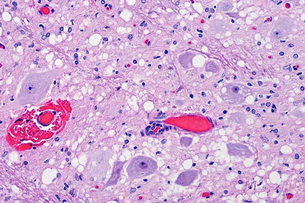
- Case 27-3. Brain stem. Multifocally, neurons and neuropil
contains round clear vacuoles (spongiform degeneration). There
is focal perivascular hemorrhage (probably an artifact) and mild
gliosis.
- Morphologic Diagnosis: Brain stem, medulla oblongata:
Neuronal degeneration, vacuolation, and loss, with spongiform
change of gray matter neuropil, and astrocytosis, consistent
with spongiform encephalopathy.
- Etiology: Prion protein (PrP-Sc, scrapie associated
prion protein)
- Disease: Chronic wasting disease of deer and elk.
-
- Case 4 Contrib# 17077-99 AFIP# 2689003
Porcine

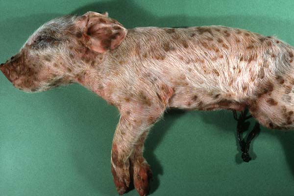
- Case 27-4. This newborn piglet (note umbilical cord)
has multiple, diffusely distributed, slightly raised, reddish-brown
epidermal papules.
- Morphologic Diagnosis: Haired skin: Dermatitis
and folliculitis, necrotizing, subacute, focally extensive, severe,
with vasculitis, acanthosis, epithelial ballooning degeneration
and eosinophilic intracytoplasmic inclusion bodies.
- Etiology: Swine pox virus, (genus Suipoxvirus, family
Poxviridae).
- Disease: Swine pox
Return to WSC Case Menu
 Immunohistochem
Mab89/160.1.5
Immunohistochem
Mab89/160.1.5
 20x
obj
20x
obj