Signalment: 4-year-old, male, Chinese Crested, canine.
History: This dog was presented to the neurology department at the Veterinary Hospital at the University of Pennsylvania with history of circling to the right, blindness and seizures.
Gross Pathology: At necropsy there was marked alopecia. The upper canine teeth had failed to erupt but were found within the gums and angled rostrally. The lower right canine tooth was missing. A necrotizing encephalitis affecting the right side of the brain was the cause of death.
Contributor's Diagnosis and Comments: Ectodermal (epidermal) dysplasia - Chinese Crested - canine.
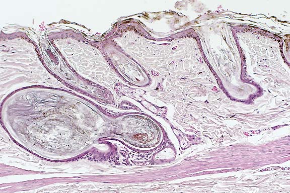
There is moderate to severe orthokeratotic hyperkeratosis, which extends into the follicular infundibulum with dilation of the follicular ostium. A few catagen and telogen follicles can still be found. The follicular papillum can be identified in abnormal locations, such as adjacent to the sebaceous ducts and at the base of follicles showing severe follicular dilation secondary to keratin accumulation. Some of the hairs are found at very unusual angles. Occasional melanophages are present in the dermis. The sebaceous glands and apocrine glands are within normal limits.
Conference Note: Additional histologic features of this
interesting case were discussed in conference. These include:
a. Arrector pili muscles are often oriented parallel to the skin
surface.
b. Apocrine ducts sometimes enter follicles at abnormal locations.
c. Mesenchymal cells of the dermal papillae occasionally surround
the epidermal bulb, rather than forming a bulb- or cone-shaped
projection surrounded by the hair bulb epidermis.
d. There is remarkable comedo formation.
Some participants considered a differential diagnosis of hyperadrenocorticism. While some features are present in this case, there are usually fewer sebaceous glands and less dermal collagen than is seen here.
The Chinese Crested dog is devoid of hair except for the crown of the head, the lower portions of the limbs, and the tail. A dominant gene for hypotrichosis (Hr) in combination with the gene for long hair (l) produces the breed. The homozygote HrHr is a prenatal lethal, hence the Chinese crested dog is an obligate heterozygote.2
Contributor: Laboratory of Pathology, University of Pennsylvania, School of Veterinary Medicine, 3800 Spruce Street, Philadelphia, PA 19104-6051.
Signalment: 18-year-old, castrated male, Domestic Shorthair, feline.
History: This cat was presented to the dermatology department
at the Veterinary Hospital at the University of Pennsylvania with
a 6 month history of a cutaneous mass and a crusting dermatosis
of 3 months' duration. The mass was present on the right dorsal
lumbar area of this cat's body and was a cutaneous horn. Four
small dry crusted lesions (approximately 2 mm in diameter) were
noted on the ventral neck (2 lesions), left scapular area (1 lesion)
and inguinal area (1 lesion). Thoracic ultrasound and radiographs
showed a large mediastinal mass
(10 x 4.5 cm) which displaced the cardiac silhouette caudally,
and a 1 cm soft tissue opacity between the sixth and seventh ribs.
Abdominal ultrasound revealed a 5 x 2.5 cm hepatic mass in the
left medial lobe and some irregularities in the splenic margins.
About 8 months after the initial visit, the cat died at home and
was presented for necropsy.
Gross Pathology: At necropsy, the crusted skin lesions
had increased in size. One of these is submitted as the unknown
slide. The anterior mediastinal mass was a thymoma and the hepatic
mass was a hepatoma. The cause of death was hemoperitoneum due
to rupture of the hepatoma.
Laboratory Results: The complete blood count (CBC), feline
leukemia virus (FeLV) and feline immunosuppressive virus (FIV)
serology by ELISA, skin scrapings, chlorphenolac preps of hairs,
bacterial culture, fungal cultures and Wood's light examination
showed no abnormal findings. Serum chemistry results showed elevations
in liver enzymes [ALT=129 U/L (N=13-57) and SAP=94 U/L (N=10-37
U/L)].
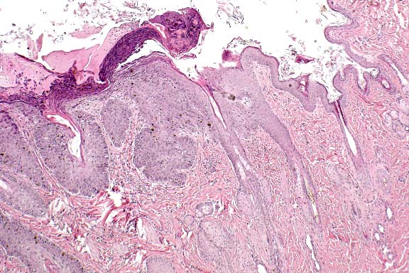
Contributor's Diagnosis and Comments: Squamous cell carcinoma in situ (Bowen's disease).
Squamous cell carcinoma (SCC) in situ is considered to be a premalignant condition. In humans, SCC in situ has been associated with an internal malignancy or arsenic exposure. This form of squamous cell carcinoma does not require ultraviolet light exposure and a sparsely haired/poorly pigmented skin for its development, as is seen in most cases of SCC in the cat which has a predilection for eyelids, ears and planum nasale. We do not know if there is any relationship between the presence of the mediastinal thymoma and hepatoma and the development of these lesions.
Conference Note: Immunohistochemical procedures to detect human papilloma viruses were applied to this tissue at the AFIP, but results were negative.
SCC in situ is considered to be rare in cats and very rare in dogs.2 Clinically, affected animals present with one or more erythematous, pigmented, nodular plaques which may be eroded or ulcerated. The plaques are well demarcated from the surrounding haired skin. Lesions may occasionally show local invasion or microinvasion, but metastasis has not been reported. Older cats are most often affected, and there is no known breed predilection.
SCC in situ is distinguished from actinic keratosis primarily on clinical grounds, i.e. actinic keratosis affects only sun-exposed, lightly pigmented skin. A histologic feature of actinic keratosis which is not seen in this case is fragmentation and replication of elastin fibers.
In contrast to feline cases, typical human cases of Bowen's disease involve singular lesions that often occur in sun-exposed skin. Approximately 5% of human patients develop an invasive squamous cell carcinoma.5
Signalment: 3-year-old, male, guinea pig.
History: A 3x5x2 cm cutaneous mass, located dorsolaterally to the base of the tail, was surgically removed from a guinea pig.
Contributor's Diagnosis and Comments: Haired skin: Complex cystic trichofolliculoma.
Trichofolliculoma is the most common benign skin tumor found in guinea pigs. They are commonly located in the dorsal lumbar region. Their characteristic cystic nature, nonencapsulation, extension from the epidermis to the subcutis, presence of sebaceous glands and arborizing pattern of secondary follicular structures along with abortive hair production are well represented in all submitted slide sections of this tumor.
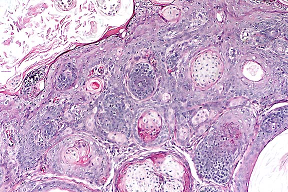
Conference Note: In contrast to guinea pigs, trichofolliculomas
are relatively rare in dogs and cats.3 Differentiation of this
lesion from other follicular neoplasms is simplified by the fact
that the arborizing pattern of the secondary follicular structures
is unique to trichofolliculoma. However, a trichofolliculoma with
predominantly mature secondary follicles may resemble a dermoid
cyst. Features helpful in differentiating trichofolliculoma from
a dermoid cyst include:
a. The central cyst of a dermoid cyst is entirely lined by squamous
epithelium, whereas the central cyst of a trichofolliculoma may
contain epithelial segments resembling isthmus or matrical portions
of a hair follicle.3
b. Follicles radiating from the central cyst of a trichofolliculoma
are more numerous, and also often show secondary branching.3
Contributor: Texas Veterinary Medical Diagnostic Lab, Post Office Drawer 3040, College Station, TX 77841
Signalment: 6-year-old, castrated male, Angora, goat.
History: This goat was presented for thick, dry, crusty, scaly skin which was moderately pruritic initially and continued to worsen. Skin was biopsied at the time of shearing, approximately two months after initial signs were noted.
Gross Pathology: The skin lesions extended from the dorsal neck, over the back to the tail, with irregular patches on the sides and rear quarters.
Laboratory Results: Skin scrapings were negative for mites and fungi.
Contributor's Diagnosis and Comments: Pemphigus foliaceus.
The microscopic lesions in the skin of this goat are common to those which characterize pemphigus foliaceus, that is, subcorneal pustules which contain acantholytic keratinocytes and neutrophils; lymphocytes, histiocytes, neutrophils and some eosinophils in the adjacent superficial dermis; some involvement of the external root sheath of hair follciles; and adherence of overlying crust on the surface. The goat was treated with systemic corticosteroids and had a favorable response. However, five weeks after administration of a long-acting corticosteroid, lesions began to recur.
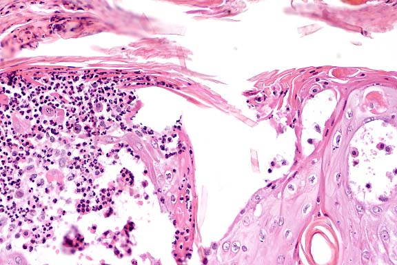
Conference Note: Conference participants considered a differential diagnosis of pemphigus foliaceus (PF) and cutaneous acariasis. Although many of the features of PF are present, there are very few acantholytic cells in many of the sections. Acantholysis is a consistent feature of PF; therefore, participants concluded that a definitive diagnosis could not be made based solely on the histopathology of the submitted section. Cutaneous acariasis can cause a similar histologic lesion, and although no mites were seen, this remains a plausible differential diagnosis.
Pruritus is seen in less than 50% of dogs with PF, and lesions are typically present on the dorsal muzzle, planum nasale, pinnae, periobital skin, and footpads.2 However, bilaterally symmetrical truncal lesions are occasionally seen. In cats, lesions are often restricted to the face, ears, interdigital webs, and nipples.2 In one report of a pygmy goat with PF, pruritic lesions with a similar distribution to the presented case were seen.4
In humans, PF is the result of production of autoantibody against a cadherin adhesion molecule, desmoglein I, which is located in desmosomes.1 The binding of pemphigus autoantibodies to keratinocyte antigens stimulates secretion of urokinase-type plasminogen activator, that in turn activates plasminogen and is associated with loss of cohesion between keratinocytes.
Contributor: The Upjohn Company, 301 Henrietta Street, Kalamazoo, MI 49002.
International Veterinary Pathology Slide Bank:
Laser disc frame #2318-2321, 2359, 2369, 2425, 2426, 3306, 3307,
4086, 4087, 9732, 10946, 11814, 11861, 13417, 13421, 14398, 18648,
18660, 18661, 19520, 21763, 21793, 21794, 22735.
Terrell W. Blanchard
Major, VC, USA
Registry of Veterinary Pathology*
Department of Veterinary Pathology
Armed Forces Institute of Pathology
(202)782-2615; DSN: 662-2615
Internet: blanchard@afip.osd.mil
* The American Veterinary Medical Association and the American College of Veterinary Pathologists are co-sponsors of the Registry of Veterinary Pathology. The C.L. Davis Foundation also provides substantial support for the Registry.