
Signalment: 4-5 week, Suffolk, both sexes, sheep.
History: Lambs exhibited sudden hind-limb paresis or fractures of long bones. Lambs were housed outdoors.
Gross Pathology: One lamb had a compression fracture of a lumbar vertebra. A second lamb had a fracture of a distal femoral metaphysis involving the physis. Both lambs had a few firm swellings of midshaft ribs, the ribs were soft and bent easily without snapping, and vertebrae were soft and easily sliced with a knife.
Laboratory Results: Milk replacer analysis (dry weight): Calcium: 0.34%; Phosphorus: 0.41%.
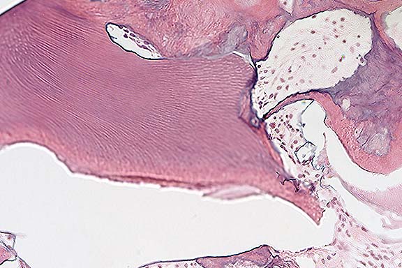
Contributor's Diagnoses and Comments: 1. Bone (vertebra, rib): Osteoporosis, severe. 2. Bone (vertebra): Compression fracture, acute (pathologic).
Etiologic diagnosis: Dietary calcium deficiency.
Conference Note: Osteopenia indicates reduced bone mass. Osteoporosis, or bone atrophy, signifies a pathologic loss of bone, usually with clinical significance. Osteoporosis results when bone resorption predominates over bone formation. It is a common lesion in farm animals, and in these animals its origin is usually nutritional.
Nutritional imbalances which can result in osteoporosis include starvation; deficiencies of calcium, phosphorus, or copper; and excess of molybdenum. Calcium deficiency uncomplicated by vitamin D and/or phosphorus deficiency rarely, if ever, occurs as a natural disease. In contrast, complicated calcium deficiency is common.2 In both young and adult animals, this results in a loss of cancellous bone. Bones with a high trabecular component, such as verebrae, are most severely affected. In mature animals, calcium deficiency may be asymptomatic, but in young animals pathologic trabecular bone fractures may occur. Often the parathyroid glands are enlarged due to secondary hyperplasia. This leads to increased osteoclastic resorption. Fibrous osteodystrophy may result, as in this case, but is usually less extensive than that seen in the osteodystrophy of calcium deficiency secondary to phosphorus excess.2 The vertebra from this lamb exhibits marked endocortical and mild marrow cavity fibrosis, both resulting from fibrous osteodystrophy. Although few osteoclasts are demonstrable, multiple scalloped edges on bone trabeculae are convincing evidence of increased resorption.
International Veterinary Pathology Slide Bank:
Laser disc frame #8117, 8118, 12834, 19138, 19139.
Signalment: 1-year-old, male, Sprague-Dawley rat.
History: This rat was a control animal in a 2-year carcinogenicity study. Rats were fed a commercial diet of ground rat chow. The animal was euthanatized.
Gross Pathology: The animal was moderately thin. A hard
mass, 4 cm in diameter, was present on the left side of the mandible.
Contributor's Diagnosis and Comments: Mandible: Odontoma,
compound, Sprague-Dawley rat.
Conference Note: This case was also studied by the Department of Oral and Maxillofacial Pathology of the AFIP; most of the staff members of that department favored a diagnosis of complex odontoma. They noted that clinicopathologic correlation with radiographs would be helpful in confirming the diagnosis. They stated that both types of odontomas are well differentiated, but complex odontomas are less "organized" than compound odontomas. According to one author, distinction of the two types may be arbitrary.4
Odontomas are usually located in the mandibular or maxillary arch, and are more common in cattle and horses than in other domestic species.
Contributor: The Procter & Gamble Company, Miami Valley Laboratories, P.O. Box 398707, Cincinnati, Ohio 45239-8707
International Veterinary Pathology Slide Bank:
Laser disc frame #20059
Signalment: 5-year-old, female, Chesapeake Bay Retriever, canine.
History: This dog had an eight month progressively worsening lameness of the right rear leg. Radiographic examination revealed lysis of the right tibial tarsal bone. Radiographic examination of the thorax revealed no lesions. The right rear limb was amputated.
Gross Pathology: The medullary cavity of the right tibial tarsal bone was mostly filled with a firm (not hard), brown-white mottled material. The contour of the bone appeared normal.
Contributor's Diagnosis and Comments: Tibial tarsal bone; chondrosarcoma, moderately differentiated.
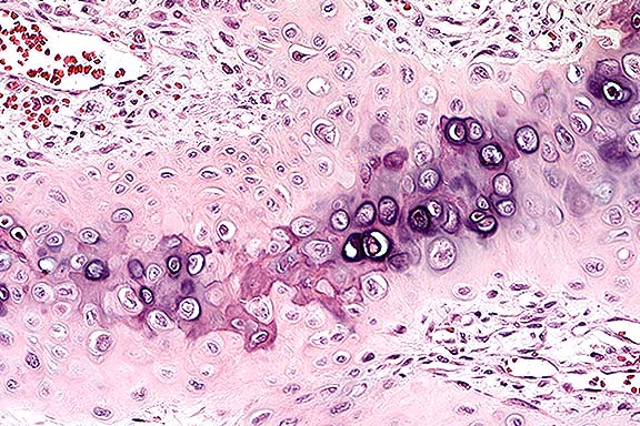
Conference Note: Chondrosarcoma (CS) is the second most common skeletal neoplasm in dogs and man.1 As with osteosarcoma (OS), CS most frequently affects large breed dogs. However, in contrast to OS, CS rarely affects the giant breeds such as the Great Dane or St. Bernard.
Primary chondrosarcomas arise from existing normal cartilage and from perichondrium. Secondary chondrosarcomas develop from abnormal cartilage, such as that which exists in osteochondromas. Chondrosarcomas are usually found in mature animals, and occur more often in the pelvis, nasal cavity, sternum, and ribs than in the axial skeleton.5 In contrast with OS, CS grow more slowly and metastasize later. They may be associated with recurrences many years after initial surgery.1 A recent report by Hahn et al. described bilateral renal metastases of a nasal chondrosarcoma in a dog, which occurred one year following nasal surgery and radiotherapy for the primary tumor.6
International Veterinary Pathology Slide Bank:
Laser disc frame #678, 758, 1030-31, 1392-93, 1790-91, 2258, 2863,
3690-91, 3769-70, 5601, 7920, 24656.
Signalment: 4-week-old, Quarterhorse, male, equine.
History: Progressive lameness and swelling of the carpus. Radiographs of the distal radius showed bilateral radiolucency and irregular profile of the distal radial physis.
Gross Pathology: Sections across the distal radius showed an irregular profile of the metaphyseal side of the distal physis. The physis showed irregular thickness and interrupted areas of fragmented and disjointed ossification.
Contributor's Diagnosis and Comments: Osteochondrosis of the distal physis of the radius, equine.
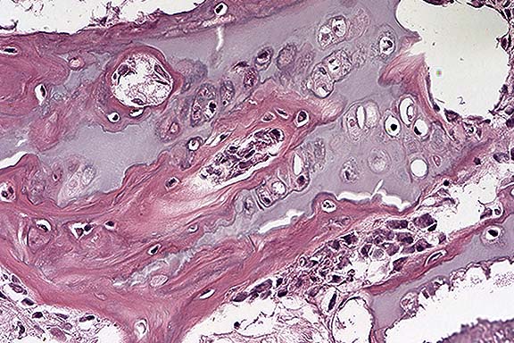
Conference Note: Some sections contain an incomplete metaphyseal fracture with associated callus formation. Most sections contained a roughly triangular locus of trabecular bone bounded by cartilage near one end of the physis. This lesion was interpreted to be a focus of retained cartilage that had undergone partial mineralization and ossification and subsequent fracture. Possible reasons for the cartilage retention include 1) primary idiopathic cartilage retention (the dysplasia form of osteochondrosis), or 2) an initial fracture, with interference of blood vessels and resultant inhibition of mineralization of the cartilage.
The causes of osteochondrosis are not well understood. Multiple factors are considered to be involved, including genetic predisposition, rapid growth, degree of physical activity, gender, and nutritional status.2
Contributor: School of Veterinary Medicine, University of Wisconsin, 2015 Linden Drive West, Madison, WI 53706
International Veterinary Pathology Slide Bank:
Laser disc frame #8014-15, 10760-64, 21033-34.
Terrell W. Blanchard
Major, VC, USA
Registry of Veterinary Pathology*
Department of Veterinary Pathology
Armed Forces Institute of Pathology
(202)782-2615; DSN: 662-2615
Internet: blanchard@email.afip.osd.mil
* The American Veterinary Medical Association and the American College of Veterinary Pathologists are co-sponsors of the Registry of Veterinary Pathology. The C.L. Davis Foundation also provides substantial support for the Registry.