Signalment: Approximately 10-month-old, Salers cross, female ox (Bos taurus).
History: The heifer was losing weight despite anthelmintic treatment and preferential feeding. Physical examination revealed a thin, weak, 152 kg animal with a temperature of 37.8o C and depressed ruminal motility.
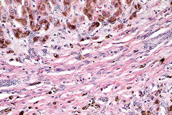
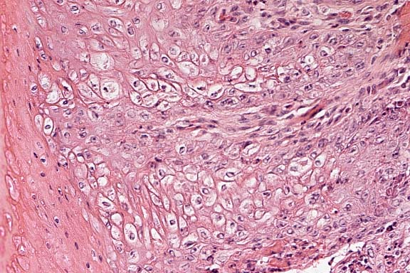
Gross Pathology: The entire liver was firm, with a thickened, opaque capsule and thin arborizing white bands that faintly delineated hepatic lobules. The gallbladder wall was thickened and had mild papillary mucosal projections. The cut surface of submandibular and hepatic lymph nodes was darkly tinted.
Laboratory Results:
PCV 27% WBC 11.0 X 10³ / ul
HGB 8.3 g/dl segs 5390 / ul
MCHC 30.7 g/dl bands 550 / ul
Retics not observed lymphs 4070 / ul
Plt 253 X 103 / ul eos 550 / ul
Alb 2.8 g/dl monos 440 / ul
TP (serum) 9.1 g/dl fibrinogen 400 mg / dl
Glob 6.3 g/dl Na 151 mmol
ALP 652 IU K 4.5 mmol
AST 118 IU Cl 104 mmol
TBil 2.17
Serum Iron 718 ug/dl
Total Iron Binding Capacity 718 ug/dl
Unbound Iron Binding Capacity 0%
Transferrin saturation 100%
Hepatic iron concentration: 6480 ppm, wet weight
Hepatic copper concentration: 1 ppm, wet weight
Hepatic molybdenum concentration 3.7 ppm, wet weight
Hepatic zinc concentration 22.3 ppm, wet weight
Aerobic bacterial culture of liver and bile: neg
Bovine virus diarrhea virus culture of blood and multiple organs:
neg
Contributor's Diagnoses and Comments:
1. liver: hepatic degeneration, chronic, diffuse, severe, characterized
by profound hepatocellular hemosiderosis, bridging portal and
centrilobular fibrosis, marked reduction of hepatocellular mass,
and biliary hyperplasia.
2. lymph node (hepatic and submandibular): lymphadenopathy, chronic,
diffuse, severe, characterized by profound intraphagocytic hemosiderosis,
effacement of nodal parenchyma by sheets of histiocytes, and mineralization
of arterioles and connective tissue.
3. lymph node (hepatic): lymphadenitis, granulomatous, multifocal,
mild, with associated, mineralized fungal hyphae.
Etiologic Diagnoses:
1. Hemochromatosis of Salers cattle (autosomal recessive trait).
2. Mycotic infection (secondary).
Hemochromatosis is a rare, inheritable disease caused by excessive
absorption of normal dietary iron. It has been reported in Salers
cattle and is thought to be an autosomal recessive trait. In this
case, the same bull (3/4ths Salers) was both sire and grandsire
of the affected heifer. Hemosiderosis affected the liver and lymph
nodes of this calf, and to a much lesser extent the lung and kidney
(brain and pancreas were not examined). The spleen contained unremarkable
quantities of intraphagocytic hemosiderin within periarteriolar
lymphoid sheaths, and hemosiderin deposits were not observed in
the red pulp. The iron content in the liver was extremely elevated,
especially considering the high proportion of connective tissue.
If expressed on a dry weight basis, the iron concentration would
exceed 20,000 ppm.
Similar conditions occur in people and birds. Secondary iron overload is caused by excessive parenteral iron or repeated blood transfusions. Dietary iron overload is important in Bantu people that drink a high iron beverage and are genetically predisposed to iron accumulation.
It is possible that the mycotic infection in this calf occurred secondary to immunosuppression. The fungal hyphae observed in the lymph node appear dead and are often mineralized. The rising iron concentration in the lymph node may have eventually become toxic to the fungi.
Conference Note: The brown granular pigment stained blue with Perls iron stain, confirming that it is hemosiderin.
Primary hemochromatosis in humans, though similar to the condition seen in Salers cattle, differs in several ways. In cattle, iron accumulates in pancreatic acini, whereas in humans both acini and islets are affected. In fact, 75-80% of affected humans develop diabetes mellitus.3 Other findings in cattle that differ from those in humans include mineralization of portal blood vessels, lack of pancreatic fibrosis, and lack of deposition of hemosiderin in myocardial, joint, or epidermal tissues.1
Definitive diagnosis of hemochromatosis requires evidence of liver damage, as well as confirmation of excessive iron stores through liver biopsy. In humans, symptomatic patients usually have levels in excess of 10,000 ppm dry weight, and in those with complicating cirrhosis, levels generally exceed 22,000 ppm.
Though the mechanisms for regulation of intestinal iron absorption are obscure, the fundamental disease mechanism in hemochromatosis is believed to be a direct iron toxicity to host tissues. The following mechanisms have been proposed: (1) lipid peroxidation via iron-catalyzed free radical reactions (the Fenton reaction); (2) iron stimulation of collagen formation; and (3) direct interactions of iron with DNA, leading to lethal alterations.3
Contributor: Wyoming State Veterinary Laboratory, 1174 Snowy Range Road, Laramie, Wyoming 82070
References:
International Veterinary Pathology Slide Bank:
Laser disc frame #19513-4.
Signalment: Adult zebra fish (Brachydanio rerio).
History: This is one of approximately 100 fish divided between 2 tanks used in a reproductive physiology research program. Affected fish stopped eating and had evidence of respiratory distress. Representative fish from the tanks were euthanized by immersion in MS 222 and complete necropsies were immediately performed.
Gross Pathology: In affected fish, the gill lamellae
were thickened and diffusely red-purple with pinpoint yellow foci.
There were no other gross abnormalities.
Laboratory Results: Wet mounts of gill clippings obtained
at necropsy demonstrated large numbers of round to pear-shaped,
50-100 :m diameter, brown organisms that were attached to the
gill lamellae by a thin stalk (rhizoid).
Contributor's Diagnosis and Comments: Gill: Branchitis, proliferative, subacute, with myriad protozoan trophonts consistent with Piscioodinium spp.
Histologic findings were similar in all fish examined. In addition
to the proliferative branchitis, affected fish had a multifocal,
acute dermatitis associated with organisms identical to those
observed in the gills. The organisms in histologic section were
up to 60 :m wide with abundant foamy cytoplasm and a prominent
eosinophilic nucleus. In some sections, the parasite is observed
attached to the gill lamellae by a thin, almost transparent stalk
or rhizoid. Fine, brown granules are observed within most of the
parasitic trophonts and are interpreted as photosynthetic pigment.
These findings are considered compatible with oodinosis or "velvet
disease" caused by the dinoflagellate parasites Piscinoodinium
spp in freshwater fish or Amyloodinium ocellatum in saltwater
fish. A wide variety of teleost species as well as elasmobranchs
can be affected. Infections with the freshwater forms have also
been described in aquatic amphibians. Oodinosis can be the cause
of extensive morbidity and mortality in aquarium systems and can
be difficult to eradicate.
The life cycle of the parasite is very similar to another common parasite of fish, Ichthyophthirius multifiliis, the cause of "Ich". Nonmotile, feeding trophonts are firmly attached to the gills or to the skin by attachments called rhizoids. After a period of a few days, the trophonts detach from the host and become tomonts, which undergo division to produce the biflagellated infective dinospores. The motile dinospores attach to the host and begin the life cycle anew.

Conference Note: At least three morphologic characteristics allow one to differentiate Piscinoodinium, a dinoflagellate, from Ichthyophthirius, a ciliate, in histologic section. Ichthyopthirius (Ich) trophonts are often encysted within the host epithelium of gills or skin; Noga3 considers this to be a pathognomonic finding. Ich has a horseshoe-shaped macronucleus and the trophonts are generally larger than those of Piscinoodinium, ranging up to 1 mm.3,5
Contributor: National Zoological Park, 3001 Connecticut Avenue NW, Washington, D.C. 20008
References:
International Veterinary Pathology Slide Bank:
Laser disc frame #19889-91, 23232, 24411
Signalment: 5-year-old, female, brown-eared pheasant (Crossoptilon mantchuricum)
History: This animal was from a zoo, and was found dead. Its general body condition was poor.
Gross Pathology: The cecal wall was diffusely thickened and nodular.
Contributor's Diagnosis and Comments: Cecum: Typhlitis, granulomatous, with mesenchymal proliferation around Heterakis isolonche.
Histology: The submucosa is widely expanded by the presence of well-circumscribed, discrete, irregularly-sized, and highly cellular nodules which impinge on the mucosa and tunica muscularis. Most of them are composed of small interlacing bundles of elongated fibroblast-like cells around one or a few sections of a nematode parasite. Onion-like arrangements around blood vessels are sometimes observed. The cells show an open-faced nucleus, a prominent nucleolus and an eosinophilic, sometimes vacuolated, cytoplasm.
The nematode is typical of Heterakis isolonche: polymyarian coelomyarian musculature, triradiate intestine, uninucleate cuboidal to columnar intestinal cells with a low microvillar border, large unstalked lateral chords and prominent cuticular alae extending almost through the entire length of the body. Numerous thick-shelled eggs are present within a few nodules.
Some nodules are composed of variable proportions of finely vacuolated, slightly pigmented macrophages (interpreted to be lipofuscin pigment) and vacuolated polygonal cells with hyperchromatic nucleus (interpreted to be degenerated fibroblast-like cells). Numerous small collections of pigmented macrophages are interspersed between the large nodules; they are considered to be the footprints of previous parasitic nodules which have degenerated.
Although inflammatory cells are generally scant around the parasites, areas of necrosis and foreign-body macrophagic reaction (notably around degenerated eggs) are sometimes observed. Atrophy of the mucosa, mild inflammatory cells infiltrating in the lamina propria, and hypertrophy of the muscularis mucosa and tunica muscularis are associated histological changes.

Conference Note: Conference participants discussed the controversial histogenesis of the spindled to polygonal cells comprising many of the nodules containing adult worms. A Masson's trichrome-stained section viewed in conference demonstrated very little collagen within these nodules. An immunohistochemical stain for lysozyme, an histiocytic marker, showed multifocal positive staining within some spindle and vacuolated polygonal cells.
In addition to pheasants, Heterakis isolonche infection has been reported in three species of quail in the United States, and in chickens in Tanzania. Another Heterakis species which may be found in cecal nodules is H. beramporia, which infects chickens in Asia and the Pacific area.
Contributor: PFIZER, B.P. 109, 37401 AMBOISE (France)
References:
Signalment: Three dead fetuses and one live (few hours old) mixed breed piglet (porcine) from the same litter.
History: Three hundred sows on the farm. Increased numbers of stillborn piglets over the last two weeks. Three stillborn or mummified fetuses in this litter of fifteen.
Gross Pathology: Four porcine animals were received. One animal was a 20 cm crown-to-rump length, moderately macerated fetus. A second animal was a 22 cm crown-to-rump length, mildly macerated fetus. The third animal was a 27 cm crown-to-rump length, fully haired, term, moderately autolyzed fetus. The remaining fourth animal was a live, fully developed, newborn piglet. The placentas of the three fetuses were also submitted and were severely autolyzed. All of the placentas contained abundant, creamy, opaque, white to yellow material. All four animals had numerous, 3 to 6 mm diameter, focal lesions, disseminated over the skin. The lesions had an elevated margin and depressed center. In the three fetuses, these foci were also covered with the same creamy, pale material found in the placentas. In the live piglet, the skin lesions were dry and scaly. The piglet also had elevated, red, circular lesions on the planum nasale and the palmar surface of the front claw.
Laboratory Results:
Bacteriology Results: Placenta #1: 4+Escherichia coli, 3+Streptococcus equisimilis, 2+Citrobacter freundii
Placenta #2: 2+Escherichia coli, 1+Streptococcus equisimilis,
1+Citrobacter freundii
Stomach fluid: Negative culture
Lung #1: ONE COLONY Escherichia coli
Lung #2: ONE COLONY Escherichia coli
Virology Results: Lung x 3 : FA Test for porcine parvovirus: NEGATIVE
Contributor's Diagnoses and Comments:
Conference Note: The development of swine pox lesions progresses through the classical stages of macule, papule, vesicle, and pustule. Lesions usually resolve within 3-4 weeks after the appearance of the macule, unless they are complicated by secondary bacterial infection which may prolong the clinical disease. Young animals are more severely affected than adults, and lesions are generally more widespread. Neonatal pigs may have a generalized disease and widespread lesions, whereas adults usually develop a milder form of the disease with lesions on the udder, ears, snout, and vulva. Swinepox may have high morbidity (up to 100%) in young animals raised in unhygienic conditions. The mortality rate is usually less than 5%. In addition to direct contact and transplacental transmission, mechanical transmission by the pig louse (Hematopinus suis), as well as flies and mosquitoes, has been documented.5
Contributor: Department of Veterinary Pathology, Western College of Veterinary Medicine, University of Saskatchewan, 52 Campus Drive, Saskatoon, SK Canada S7N 5B4
References:
International Veterinary Pathology Slide Bank:
Laser disc frame #1501, 1736-38, 2782-83, 4481-83, 4573-74, 4603,
5180-81, 6989-90, 7608-09, 9601, 9674, 13556, 20769, 21049-50.
Terrell W. Blanchard
Major, VC, USA
Registry of Veterinary Pathology*
Department of Veterinary Pathology
Armed Forces Institute of Pathology
(202)782-2615; DSN: 662-2615
Internet: blanchard@email.afip.osd.mil
* The American Veterinary Medical Association and the American College of Veterinary Pathologists are co-sponsors of the Registry of Veterinary Pathology. The C.L. Davis Foundation also provides substantial support for the Registry.