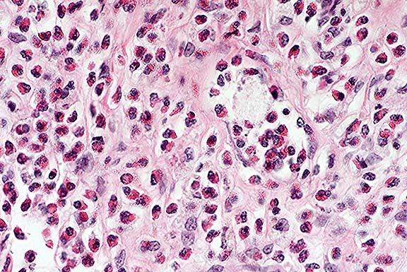
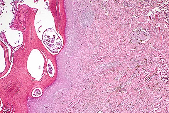
AFIP Diagnosis: Haired skin, dermis and subcutis: Eosinophil infiltrate, severe, with fibrosis and fewer lymphocytes, plasma cells, mast cells and neutrophils, New Zealand white rabbit, lagomorph.
Signalment: 16-month-old, female, New Zealand white rabbit (Oryctolagus cuniculus)
History: The rabbit developed an ulcerated mass under the tail.
Gross Pathology: There was a firm lobulated ulcerated mass approximately 7-8 cm in diameter under the tail that partially surrounded the rectum dorsally and laterally. On section the tumor was tan, lobulated, and partially encapsulated.
Contributor's Diagnosis and Comments: Eosinophil granulocytic sarcoma.
Conference Note: Although the contributor's diagnosis
was carefully considered, and it is agreed that the submitted
case resembles that reported by Perkins et al., it was concluded
that the nature of the process represented in the skin sections
is uncertain. Since the infiltrate does not contain atypical cells,
blasts or immature myeloid cells, a reactive lesion cannot be
excluded. Possible causes include hypersensitivity, parasites,
drugs and non-eosinophil neoplasia.
In humans, eosinophilic proliferations are uncommon. Most have
been described as "chronic eosinophilic leukemia" and
are characterized by widespread organ involvement, especially
in skin, heart, and lymph nodes accompanied by marked peripheral
blood eosinophilia. Cytogenetic studies are often needed to demonstrate
that the eosinophilia is neoplastic and not reactive.
Granulocytic sarcoma, or chloroma, is the extramedullary growth of focal granulocytic neoplasms, and may be of neutrophil or eosinophil type. These have been reported in dogs and cattle.2 In dogs, the most commonly involved organs are lung, gut, and skin. In cattle, skeletal muscle is characteristically affected. Affected animals are initially aleukemic, and the neoplastic proliferation exhibits variable differentiation. If there is minimal differentiation, this lesion is usually misclassified as lymphoma. If 1-2 µm sections are examined, cytoplasmic granulation is more readily apparent.
Contributor: Department of Comparative Medicine, M.S. Hershey Medical Center, PennState University, 500 University Drive, Box 850, Hershey, PA 17033
International Veterinary Pathology Slide Bank:
Laser disc frame #: none
Signalment: 17-year-old, neutered, female, Domestic Shorthair cat.
History: Euthanasia was performed on this aged cat after she becameseverely depressed and began bleeding from her mouth and nose.
Gross Pathology: The liver and spleen were enlarged. The liver was pale yellow with an accentuated lobular pattern and multiple, variably-sized, areas of hemorrhage disseminated throughout all lobes.
Laboratory Results: Prior to euthanasia: anemia (Hct = 14%) with polychromasia; leukocytosis with lymphopenia; thrombocytopenia (63,000/microliter); hypoproteinemia (5.5 g/deciliter); azotemia; hypokalemia.
Postmortem cytology of bone marrow: Mast cells were observed among other hematopoietic cells.
Contributor's Diagnosis and Comments: Liver: Systemic mastocytosis ("mast cell leukemia") with periacinar hepatocellular necrosis and bilirubin accumulation (cholestasis).
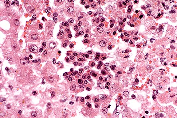
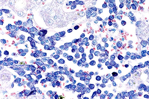
Conference Note: Although this feline lesion is often called mastocytosis, the diagnosis of malignant mast cell tumor was preferred because the term mastocytosis does not clearly indicate that the condition is neoplastic.
In cats with this form of mast cell neoplasia, splenomegaly due to massive infiltration of mast cells is consistently found. Other organs often involved include liver, lymph nodes, and bone marrow. Approximately 50% of affected cats have mastocytemia.
The origin of mast cells is controversial. Although they are functionally similar to basophils, there is no evidence that the basophil is a precursor of the tissue mast cell; neither is there proof of a common basophil-mast cell progenitor.4
Mast cells are essential in the development of type 1 hypersensitivity reactions. Mast cells possess high-affinity receptors for the Fc portion of IgE molecules, which are formed by B lymphocytes upon first exposure to an allergen. On subsequent exposure to the specific allergen, multivalent antigens bind to more than one IgE molecule and cause cross-linkage of adjacent IgE antibodies. This bridging of IgE molecules bound to the mast cell leads to perturbation of the Fc receptor and initiates two parallel and interdependent processes, one leading to mast cell degranulation with discharge of preformed (primary) mediators, and the other leading to de novo synthesis and release of secondary mediators. Primary mediators comprise four categories: biogenic amines, such as histamine (and serotonin in rodents); chemotactic mediators, primarily eosinophil chemotactic factor (ECF); enzymes, both proteases (chymase, tryptase) and acid hydrolases; and proteoglycans, including the well-known anticoagulant heparin. Secondary mediators synthesized by mast cells include leukotrienes B4, C4, and D4; prostaglandin D2; platelet activating factor; and various cytokines.
Contributor: Animal Disease Diagnostic Laboratory - 1175, Purdue University, West Lafayette, IN 47907-1175.
International Veterinary Pathology Slide Bank:
Laser disc frame #5962, 5963, 6010, 6011
Signalment: 1.5-year-old, female, European ferret (Mustela putorius furo)
History: This ferret came from a household of 8 ferrets. The owner said the animal had stopped eating and had begun to show respiratory distress. Upon presentation to a veterinarian, the ferret was euthanatized.
Gross Pathology: The lungs were diffusely firm and mottled tan and red. The spleen was enlarged.
Laboratory Results: No bacterial growth was obtained in cultures of lung. Fluorescent antibody tests for canine distemper virus were negative on lung.
Contributor's Diagnosis and Comments: Severe diffuse lymphohistiocytic interstitial pneumonia due to Aleutian disease virus.
These sections of lung contain large numbers of plasma cells located in perivascular, peribronchial, and peribronchiolar spaces as well as in alveolar septa. There is multifocal type II pneumocyte hyperplasia. Large numbers of macrophages are present in alveoli which obscure normal alveolar architecture. In some areas, there are acicular clefts in alveoli which are surrounded by macrophages. Airways are filled with a mixture of macrophages, lymphocytes, desquamated epithelial cells, and proteinic material. Gomori's methenamine silver stain did not reveal any fungal or protozoal organisms. Lesions in other tissues consisted of moderate to marked periportal lymphoplasmacytic infiltration, and the splenic red pulp was replaced by large numbers of lymphocytes and plasma cells.
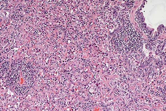
Conference Note: Aleutian Disease Virus (ADV) causes two disease syndromes in mink and ferrets. In classic Aleutian Disease (AD), adults develop a chronic persistent infection characterized by hypergammaglobulinemia, plasmacytosis, and immune-complex glomerulonephritis and arteritis. The lung lesion in the present case is characteristic of classic AD. The prominent perivascular and peribronchiolar lymphoplasmacytic infiltrates are significant features of the interstitial pneumonia seen in classic AD. ADV has also been shown to cause an acute interstitial pneumonia in mink kits, in which immune complexes apparently are not involved. In this syndrome, naive kits infected as neonates develop severe lung lesions consisting of diffuse hypertrophy and hyperplasia of type II pneumocytes, interstitial edema, and hyaline membrane formation. Intranuclear inclusions are often found in type II pneumocytes. These inclusions are round or irregular, vary in size, and can completely fill the nucleus as a basophilic or amphophilic material, or can be surrounded by a clear halo and marginated chromatin. A prominent histologic difference in the lung lesions between the two syndromes is that there is a notable absence of interstitial mononuclear cell infiltrates in the acute kit pneumonia. Also in the latter syndrome, gross and microscopic lesions are confined to the lung.
The differential diagnosis of interstitial pneumonia in mustelids should include canine distemper virus infection. In this disease, there is diffuse thickening of the pulmonary interstitium due to an infiltrate of mixed inflammatory cells, proteinaceous material, and congested alveolar capillaries. Alveolar spaces are frequently attenuated and contain increased numbers of macrophages, sloughed pneumocytes, fibrin, and edema. Usually, numerous intracytoplasmic and fewer intranuclear eosinophilic inclusions are present within bronchiolar epithelial cells, pneumocytes, and macrophages. Occasional syncytial cells are present within alveoli.
Contributor: Veterinary Diagnostic Center, Fair St. and East Campus Loop, University of Nebraska-Lincoln, Lincoln, NE 68583-0907
Signalment: 1- to 2-year-old Chinese silky chickens (Gallus domesticus)
History: The birds were submitted for evaluation of the skin lesions present on the shanks and feet.
Gross Pathology: The birds had locally extensive exfoliative dermatitis affecting the unfeathered skin of the legs and feet.
Laboratory Results: Parasitology: Skin samples were processed using KOH digestion and mites identified as Knemidocoptes mutans were seen.
Contributor's Diagnosis and Comments: Marked hyperplastic superficial perivascular dermatitis with intra-corneal arthropod parasites (Knemidocoptes mutans).
Conference Note: Birds are host to diverse mites. Philips1 classifies many of these by their preferred habitats, which include feathers, quills, skin, subcutaneous tissue, and the respiratory tract. Food sources available to mites include blood, tissue fluid, skin and feather lipids, keratin, fungi, algae, and other mites. Many avian mites are not pathogenic. Others are often secondary problems associated with stress or dietary deficiency.
Knemidocoptes spp., members of the Class Arachnida, Order Acarina,
Suborder Sarcoptiformes, and Family Sarcoptidae, are important
mange mites of birds. Knemidocoptid mites invade feather follicles
and the stratum corneum, primarily affecting the face, feet, and
cere. The disease that results, often referred to by the lay terms
‘scaly leg ' or ‘scaly face' is characterized by hyperkeratosis,
pruritus, and feather loss. Knemidocoptes pilae parasitizes the
cere and legs of psittacines. Knemidocoptes jamaicensis infects
the feet of many passerines. K. mutans, and K. gallinae (also
called the depluming mite), infect chickens; in addition to the
signs described above, these infections can cause crippling, weight
loss, and a decrease in egg production.1
Contributor: Animal Health Laboratory, Laboratory Services
Division, University of Guelph, Box 3612, Guelph, Ontario, Canada
N1H 6R8
Terrell W. Blanchard
Major, VC, USA
Registry of Veterinary Pathology*
Department of Veterinary Pathology
Armed Forces Institute of Pathology
(202)782-2615; DSN: 662-2615
Internet: blanchard@email.afip.osd.mil
* The American Veterinary Medical Association and the American College of Veterinary Pathologists are co-sponsors of the Registry of Veterinary Pathology. The C.L. Davis Foundation also provides substantial support for the Registry.