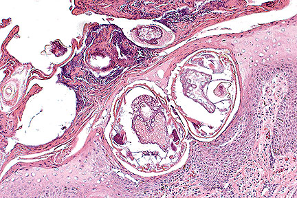
 Cross
sections of two intraepidermal acarids from the pinna of a fox.
(HE, 200X, 122K)
Cross
sections of two intraepidermal acarids from the pinna of a fox.
(HE, 200X, 122K)Signalment: Adult Red Tail Fox, wild.
Cross
sections of two intraepidermal acarids from the pinna of a fox.
(HE, 200X, 122K)
History: This fox was killed by farm dogs in South Dakota.
Gross Pathology: Only the head was submitted for a rabies exam. There was severe encrustation which involved the ears and face.
Laboratory Results: None submitted.
Contributor's Comments:
Morphologic Diagnosis: Auricular dermatitis, with marked purulent and hyperkeratotic epidermitis and associated mange parasites, chronic, severe.
Etiology: Sarcoptes scabiei
In the spring of 1994, 3 foxes were submitted with similar severe sarcoptic mange.
AFIP Diagnosis: Ear: Dermatitis, plasmacytic, chronic, diffuse, moderate, with severe orthokeratotic and parakeratotic hyperkeratosis, intracorneal abscesses, epidermal hyperplasia, and numerous intracorneal acarid parasites, red fox (Vulpes vulpes), canid, etiology consistent with Sarcoptes scabiei.
Conference Note: This case was reviewed by Dr. C.H. Gardiner, parasitology consultant for the Department of Veterinary Pathology, AFIP. Sarcoptes scabiei is the cause of scabies in humans and sarcoptic mange in most domestic animals; each variety of mite is named for its host, for example Sarcoptes scabiei var. canis. The species adapted mites may infect other host species; however, in most cross-infections, the mites do not complete their life-cycle. In the normal host, the mites burrow into the stratum corneum and feed on cells of the stratum granulosum and stratum spinosum. Epidermal damage induces epithelial hyperplasia and the development of parakeratotic crusts. The females lay 40 to 50 eggs which develop into larva and then nymphs in the intracorneal burrows. Neither adults nor eggs survive well in the environment.
Transmission is predominantly by direct contact, although indirect contact with fomites is another possible mechanism. The initial lesions of sarcoptic mange are localized and nonpruritic. After a period of 7 to 11 weeks, there is a generalized urticarial eruption and extreme pruritus; these lesions coincide with the development of immediate and delayed hypersensitivity reactions. Immunosuppressed animals do not mount adequate hypersensitivity reactions and develop severe lesions with large numbers of mites. Common causes of immunosuppression in animals include malnutrition and concurrent disease; in Maryland, canine distemper is diagnosed in approximately 50% of the wild canids presenting with sarcoptic mange.
Animals infected with Sarcoptes mites develop erythematous macules and crusts on the skin. Immunosuppressed animals may develop massive infections with alopecia, lichenification, and accumulation of thick scales and fissuring of the skin. Histologically, the mites can be seen within the parakeratotic stratum corneum. The adult mites have striations on their cuticles and large dorsal cuticular spines; Sarcoptes is the only mite with these prominent dorsal spines. There is epidermal hyperplasia and an overlying thick crust composed of orthokeratotic and parakeratotic debris, serum, and viable and degenerate neutrophils. Dermal vessels are dilated and lined by hypertrophic endothelium. There is often a perivascular infiltrate of lymphocytes and eosinophils. Chronic lesions have dermal fibrosis and a predominantly mononuclear inflammatory response.
Contributor: South Dakota State University, Department of Vet. Science, Brookings, SD 57007.
References:
1. Yager JA, Scott DW: The skin and appendages in Pathology of
domestic animals. Jubb KVF, Kennedy PC, and Palmer N eds., Academic
press, San Diego, pp. 681-682, 1993.
International Veterinary Pathology Slide Bank: Laser disc frame #4357-8, 4571-2, 4818, 9941, 11805, 13608-10, and 18667-8.
Signalment: Wild-caught red-spotted newt (Notophthalmus viridescens) from Vermont; adult male of breeding age; weight: 2.2 g; snout-vent length: 45 mm.
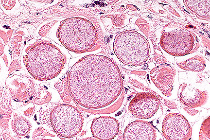 Spores of Icthyophonus-like
within the skeletal muscle of a red spotted newt. (HE, 400X, 150K)
Spores of Icthyophonus-like
within the skeletal muscle of a red spotted newt. (HE, 400X, 150K)
History: At one pond, breeding newts were noted to be lethargic in June. Many had subtle, raised, non-ulcerated, asymmetrical, plaque-like swellings on the dorsum of the rump (pelvic region), proximal half of tail, and distal half of body. Although no dead newts were found, the population seemed to be declining.
Gross Pathology: 1. Liver: Pigmentosis (melanosis), diffuse, severe. 2. Muscles of pelvic region: Myositis, pigmented and proliferative, disseminated, moderate.
Laboratory Results: 1. Cytology (blood smear): Trypanosomiasis, mild. Etiology: Trypanosoma diemyctyli. 2. Fungal culture results (rump skin and muscle): Aureobasidium pullulans.
Contributor's Diagnosis and Comments:
Morphological Diagnosis:
Skeletal muscle: Myositis, granulomatous and mycotic, chronic, diffuse, severe.
Etiology: Ichthophonus-like fungus.
This fungal infection may be more familiar to fish pathologists than veterinary pathologists. Attempted fungal cultures of 3 affected newts were non-productive; a contaminant fungus, Aureobasidium pullulans, was isolated from several affected newts. In the absence of cultural isolation and identification of the organism, it is designated Ichthyophonus-like.
Only the skeletal muscles of newts harbored this fungal infection. The fungus was most abundant in muscles of the pelvic region, with gradually decreasing numbers of fungi in muscle groups from the proximal tail, distal half of body, proximal half of body, hindlimbs, neck, head, and forelimbs. In some newts, there was little or no inflammatory cell response to large numbers of intra-muscular organisms, while other newts showed marked degeneration of myocytes and an intense, nodular and coalescing, granulomatous inflammatory response. In some newts, there was a mixture of inflammatory cell responses: some organisms elicited intense inflammation, while adjacent organism showed no inflammatory cell response. Inflammation consisted mostly of epithelioid macrophages, macrophages, lymphocytes, and a few neutrophils and eosinophils. The organisms were PAS-positive, GMS-positive, acid fast-negative, and pale blue in the Giemsa stains.
All provided sections showed the fungal organism only in cross section. The shape of the fungus actually is strikingly cylindrical (with rounded ends). The fungus lacks internal structures such as spores, daughter spherules, cleavages, septa, vacuoles, buds and hyphae. The large cross-sectional diameter of the fungus (35-150 microns) makes it unlikely to be confused with any other organisms, except, possibly, Chrysosporium sp.(formerly Emmonsia sp.), the etiologic agent of adiaspiromycosis. However, Chrysosporium is distinctly spherical and has not been reported from ectothermic vertebrates; spontaneous infections are mostly restricted to the lungs (of mammals). Ichthyophonus has been described as a cylindrical organism (Reimshuessel, 1993). Other fungi, protozoa, and mircosporidial cysts which could be confused with Ichthyphonus, have internal structures, such as spores, sporonts, cleavages, daughter spherules, etc (see Green et al., 1995).
An error in a publication by Elkan (1976) is worth noting. Elkan illustrated this infection in an unidentified "newt", and called it "Ichthyosporidium." The genus name, Ichthyosporidium, is now restricted to a taxon of microsporidians which, so far, infect only fish (see Canning &Lom, 1986).
With diligent search, rare trypanosomes may be found within blood vessels. About 80% of newts from the pond in Vermont were infected by Trypanosoma diemyctyli, one of the largest of all trypanosomes. No specific gross or histologic lesion of visceral organs could be attributed to this protozoan.
AFIP Diagnosis: Transverse section through the base of the tail, skeletal muscle: Numerous fungal organisms, with mild multifocal granulomatous myositis, red-spotted newt (Notophthalmus viridescens), amphibian.
Conference Note: Ichthyophonus hoferi is the most common marine fungal pathogen of fish. Infected fish swim abnormally, are lethargic, and listless. The scaled skin may become granular and roughened over affected areas. Some fish develop a dark coloration on the lateral line which may progress to discoloration of the entire fish. Histologically, fungal spores are bounded by a granulomatous inflammatory reaction. The spores are spherical in cross-section, varying from 10 to 250 æm in diameter and frequently occur in the liver, spleen, kidneys, muscle, and skin. The spore wall is PAS-positive and ranges from 2 to 11 æm in thickness.
Contributor: Maryland Dept of Agriculture, Animal Health/Diagnostic Lab, 8077 Greenmead Drive, College Park, MD 20740.
References:
1. Green DE, J Andrews, and JM Abell. 1995. Preliminary investigation
on mycotic myositis in red-spotted newts (Notophthalus viridescens)
from Vermont. Herpetopathologia (Proceedings of 5th International
Colloquium on Pathology of Reptiles & Amphibians) 1995:49-62.
2. Elkan E. 1976. Pathology in the Amphibia, Chapter 6. IN: Lofts,
B (Ed) Physiology of the Amphilbia, Volume 3. Academic Press,
New York. Pp. 273-312.
3. Herman R.L. 1984. Ichthyophonus-like infection in newts (Notophthalmus
viridescens Rafinesque). Journal of Wildlife Disease 20:55-56.
4. Canning EU, and J Lom.1986. The Microsporidia of Vertebrates.
Academic Press, Harcourt Brace Jovanovich, New York. Pp. 146-151.
5. Reimschuessel, R. 1993. Fungal diseases of marine tropical
fishes (chapter 79). IN: Stoskopf, M.K. (Ed) Fish medicine, W.B.
Saunders, Philadelphia. 639.-642.
6. Larone DH. 1987. Medically Important Fungi, A guide to Identification,
2nd Ed. Elsevier Science Publishing, New York. P. 157.
Signalment: Fourteen-week-old Broad Breasted White turkey.
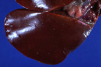 Multifocal hepatic necrosis in a
turkey. (18K)
Multifocal hepatic necrosis in a
turkey. (18K)
History: One-day-old Male Broad Breasted White turkeys were obtained from a commercial hatchery and raised in isolation rooms at the National Animal Disease Center. At 14 weeks of age, the turkeys were inoculated intramuscularly with 0.1 ml of tryptose broth containing 105 colony-forming units of Pasteurella multocida strain P-1059. Clinical signs were seen at 18 hours post-inoculation and consisted of ruffled feathers, anorexia, and watery to mucoid diarrhea. Signs progressed to rapid respiratory rate, mucus discharge from the mouth, and marked cyanosis. Turkeys became moribund and died within 24 hours of experimental infection.
Gross Pathology: The livers had multiple, randomly distributed, circumscribed, <1 mm to 3mm foci of necrosis on capsular and cut surfaces. In addition, the livers bulged on cut surface and bled freely. There were scattered petechial and ecchymotic subserosal hemorrhages of abdominal viscera and subepicardium. Hemorrhages were also seen in the lungs and intestinal lumina.
Laboratory Results: Pasteurella multocida was isolated from whole blood and identified by biochemical tests.
Contributor's Diagnosis and Comments:
Morphologic Diagnosis and Etiology: Acute, multifocal, mild to moderate heterophilic hepatitis with necrosis and large numbers of intravascular and sinusoidal short rod-shaped bacteria. Fowl cholera, Pasteurella multocida.
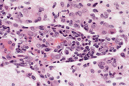
There are multifocal areas of hepatocellular necrosis. Most sections contain multiple 50 m to 500 m foci of coagulative necrosis with infiltrates of low to moderate numbers of heterophils. In addition, there is a moderate degree of single-cell necrosis. Sinusoids are dilated and contain massive numbers of short rod-shaped bacteria. Most, if not all, bacteria are extracellular. There is disruption of hepatic cords with individualization of hepatocytes. There is moderate diffuse congestion.
AFIP Diagnosis: Liver: Hepatitis, necrotizing, heterophilic, multifocal, moderate, with myriad intrasinusoidal and intrahistiocytic bacilli, Broad Breasted White Turkey, avian.
Conference Note: The etiology of fowl cholera is Pasteurella multocida, a gram-negative, nonmotile bacillus. Fowl cholera affects a wide range of avian species, including turkeys, chickens, ducks, geese, game birds, and companion birds. The wide range of avian hosts suggests that all birds are susceptible. Chronically infected birds are considered to be a major source of infection; however, pathogenic strains of P. multocida have been isolated from swine, raccoons, and free-flying birds that have access to poultry. Dissemination of P. multocida within flocks is primarily by secretions from the mouth, nares, and conjunctiva of infected birds.
Although birds infected with P. multocida may die before any lesions can be detected, large numbers of bacilli may be present in vascular spaces. Acutely infected birds develop generalized congestion of abdominal viscera; multifocal serosal petechial and ecchymotic hemorrhages are usually present. Subepicardial and subpleural hemorrhage is common. Pneumonia and hepatic necrosis may be seen. Disseminated intravascular coagulation may develop and result in formation of fibrin thrombi and generalized hemorrhage. The intestine is frequently expanded by large amounts of mucus. If the bird survives the acute disease, foci of chronic localized infection often develop. These local infections are characterized by caseous exudate and commonly occur in the respiratory tract, sinuses, conjunctiva, joints, foot pads, peritoneal cavity, and oviduct. Additionally, the middle ear is often infected, sometimes resulting in torticollis.
Contributor: USDA/ARS National Animal Disease Center, P.O. Box 70, 2300 Dayton Road, Ames, Iowa 50010-0070.
References:
1. Rimler RB: Cross-protection factor (s) of Pasteurella multocida:
Passive Immunization of turkeys against fowl cholera caused by
different serotypes. Avian Dis 31:884-887, 1987.
2. Rimler RB, Rebers PA, Rhoades KR: Fowl cholera: Cross-protection
induced by Pasteurella multocida separated from infected turkey
blood. Avian Dis 23:730-741, 1979.
3. Rhoades KR: The microscopic lesions of acute fowl cholera in
mature chickens. Avian Dis 8:658-665, 1964.
International Veterinary Pathology Slide Bank: Laser disc frame
#1726-7, 3135, 3381, 3389, 7795-8, 20536-7, 23381-2, and 23473.
Signalment: Eight-week-old female Toggenburg goat.
History: This kid exhibited posterior paresis and was presented for euthanasia. Her doe had chronic mastitis.
Gross Pathology: The lungs were irregularly mottled red, pink, and gray (mild interstitial pneumonia). The mucosa of the jejunum was thickened by white plaques (coccidiosis).
Laboratory Results: Serology was positive for caprine arthritis- encephalitis (CAE) virus by agar gel immunodiffusion.
Contributor's Diagnosis and Comments:
Morphologic Diagnosis and Etiology: Spinal cord (transverse section): Leukomyelitis, nonsuppurative, multifocal, with demyelination and lymphocytic perivasculitis. Caprine arthritis-encephalitis virus.
The nature of the lesion in the spinal cord was compatible with CAE. This diagnosis was supported by positive serology. Interstitial pneumonia and mastitis (in the doe) have also been associated with CAE.
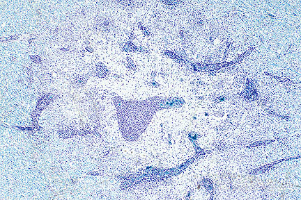
AFIP Diagnosis: Spinal cord: Leukomyelitis, lymphohistiocytic, multifocal, moderate, with demyelination and mild lymphocytic meningitis, Toggenburg, caprine.
Conference Note: Caprine arthritis-encephalitis (CAE) is caused by caprine lentivirus; a retrovirus closely related to the ovine lentivirus responsible for ovine progressive pneumonia (OPP) and visna. The encephalitic form of CAE affects kids between 2 and 4 months of age is often fatal.The virus is transmitted from an infected doe to its offspring by virus infected cells in milk; in utero transmission may be another means of spread. Kids that survive the nervous syndrom or have early inapparent infections usually develop synovitis and periarthritis in adulthood. Like the ovine lentiviruses, CAE is also associated with an interstitial pneumonia, arthritis, and mastitis in adult animals.
Onset of CAE is indicated by rear-limb paresis and ataxia, which progresses to paralysis. There are no indications of cerebral dysfunction. The white matter of the brain and spinal cord is most severely affected, developing perivascular and meningeal nonsuppurative inflammatory infiltrates and gliosis. In addition, there is patchy, discontinuous demyelination within the spinal cord. The lesions are widely distributed but tend to be more severe in the subependyma and beneath the pia of the spinal cord; however, the lesions of CAE do not have the proclivity for the periventricular lesions seen in sheep with visna.
Caprine arthritis-encephalitis virus also induces arthritis, interstitial pneumonia, and mastitis. Arthritis produced by CAE is characterized by vascular injury to synovium-lined structures with exudation of protein into synovial spaces. There is synovial villous hypertrophy, edema, and infiltration by plasma cells, lymphocytes, and macrophages. Hyalinized masses of fibrin form within the joint. Formation of carpal hygromas is common. Tendons and tendon sheaths can be similarly affected.
There have been no detailed studies of the respiratory lesions associated with CAE. The respiratory component of CAE occurs only in adult goats and is characterized by lymphoid interstitial pneumonia and hyperplasia of bronchiolar smooth muscle, similar to OPP. Affected goats also have extensive alveolar filling by dense, acidophilic, proteinaceous material, and widespread lining of alveolar septa by alveolar type II pneumocytes; these two lesions are absent in OPP.
Mammary lesions consist of interstitial inflammatory infiltrates of lymphocytes, plasma cells, and macrophages, with degeneration and loss of acinar and ductular epithelium. In contrast to OPP in which mastitis plays an important role in the disease process, mastitis is not a prominent component of CAE infection.
Contributor: Animal Disease Diagnostic Laboratory, Purdue University, 1176 ADDL, West Lafayette IN, 47907-1175.
References:
1. Jubb KVF, Huxtable CR. The nervous system. In Jubb KVF, Kennedy
PC, Palmer N, eds. Pathology of Domestic Animals, fourth ed, vol
1. San Diego: Academic Press Inc. 1993, p416.
International Veterinary Pathology Slide Bank: Laser disc frame #3303, 7475-8, 20445-7, 20834, and 22912.
* The American Veterinary Medical Association and the American College of Veterinary Pathologists are co-sponsors of the Registry of Veterinary Pathology. The C.L. Davis Foundation also provides substantial support for the Registry.
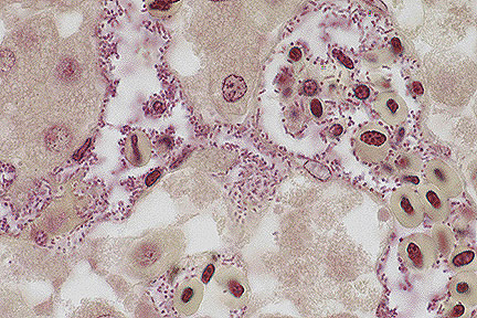 Myriad gram-negative
bacilli within the an area of heaptic necrosis in a turkey, consistent
with Pasteurella multocida. (BH, 1000X, 105K)
Myriad gram-negative
bacilli within the an area of heaptic necrosis in a turkey, consistent
with Pasteurella multocida. (BH, 1000X, 105K)