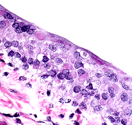
 Numerous intracytoplasmic morbilliviral
inclusions in the renal pelvic epithelium of a mink. (40X, HE,
64K)
Numerous intracytoplasmic morbilliviral
inclusions in the renal pelvic epithelium of a mink. (40X, HE,
64K)Signalment: 2-year-old female standard mink, Mustela vison
History: This mink came from a herd of 1800 breeding females in which 5 had crusty eyes, mouths and feet and exhibited anorexia.
Gross Pathology: Besides crusty eyes and nose seen clinically, the lungs were dark red and firm.
Laboratory Results: None
Contributor's Diagnoses and Comments: Renal tubular medullary mineralization, moderate and diffuse. Renal pelvic epithelial apoptosis with intracytoplasmic inclusions. Etiology: canine distemper virus.
Renal tubular mineralization in the medulla is often seen in anorexic mink, probably due to dehydration. Cytoplasmic inclusions were also present in bronchiolar epithelium and in the urinary bladder mucosa. Severe bronchopneumonia was also present. The host range of canine distemper virus includes the dog, coyote, fox, wolf, dingo, jackal, raccoon, panda, weasel, mink, ferret, badger, skunk and otter. It was felt that raccoons may have transmitted the infection to mink in this case. A similar, if not identical virus, has been reported to cause distemper-like illness in captive and wild leopards, tigers, lions, jaguars and civets. Similar viruses infect harbor seals, porpoises and javelinas.
AFIP Diagnoses:
Conference Note: Canine distemper virus (CDV) is a morbillivirus in the Paramyxoviridae family. Infection with CDV occurs by inhalation with the virus localizing in tonsils and bronchial lymph nodes. After 2-5 days there is a cell associated viremia, with the virus infecting lymph nodes, spleen, thymus, bone marrow, and macrophages in the lamina propria of the stomach and intestine. At this stage of infection, severe depletion of lymphocytes may develop with concomitant immunosuppression. At 8-10 days post-infection, the virus again disseminates, with continued infection of mononuclear cells and epithelial cells, causing hyperkeratotic dermatitis, diarrhea, pneumonia, and keratitis. The brain is sometimes affected, usually after the visceral infection has ended. The virus first infects macrophages in the meninges and later spreads to ependymal cells, glial cells, and neurons. Neuronal involvement leads to behavioral changes and varying degrees of muscular spasm or paresis. Forty to sixty days after apparent recovery, some dogs develop demyelinating lesions within the brain. This complication of CDV infection is usually fatal.
Morbilliviruses possess two proteins which facilitate binding to host membranes, hemagglutinin and F protein. The F factor mediates fusion of the viral envelope with the cellular membrane and assists in viral attachment. It also causes host cell fusion and is responsible for the formation of syncytial cells. The ability to fuse host cells allows the virus to spread without being exposed to antibody. To be biologically active the F protein must be cleaved by a host protease into two disulfide-linked polypeptides, F1 and F2. If a host cell lacks the necessary proteases, the virus formed is not infectious, since the F factor is required for viral attachment.
Conference participants also noted numerous lipid vacuoles within the renal tubular epithelium. This is a common finding in anorectic or fasted mustelids and is related to the high metabolic rate of these animals and their ability to rapidly mobilize fat stores.
Fatty change of renal tubular epithelium also occurs in "nursing disease" of mink. Nursing disease occurs around 40 days after whelping and is characterized by dehydration, renal insufficiency and death. There may be a genetic predisposition for this disease in light color mutations. Affected females also frequently have a concurrent subclinical mastitis.
Contributor: Utah Veterinary Diagnostic Laboratory, 950 East 1400 North Logan, Utah 84322-5700
References:
International Veterinary Pathology Slide Bank:
Laser disc frame #13013, 13012, 13011, 6898, 6897, and 4810.
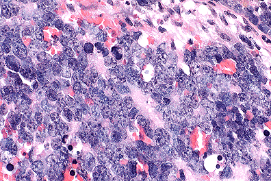 Retinoblastoma cells in a mouse.
(40X, HE, 103K)
Retinoblastoma cells in a mouse.
(40X, HE, 103K)
Signalment: 4-month-old male FVB line 19 (àAHPV16E6/E7 Line 19) transgenic mouse (Mus musculus), one of a group maintained at a commercial breeding establishment for use in human cervical cancer research.
History: Several mice of the above strain presented with unilateral proptosis. All mice have bilateral cataracts.
Gross Pathology: The globe was enlarged (0.5cm diameter) and protruded from the palpebral fissure. The eye was red-brown and solid. No extraocular structures appeared to be involved grossly. In one case (not the submitted case) a metastatic mass was detected in the subcutis of the neck.
Morphologic Diagnosis: Eye: Retinoblastoma, FVB line 19, Mus musculus, rodent.
Laboratory Results: None
Contributor's Comments: The ocular mass, which in most sections is confined to the globe, is composed of pleomorphic, poorly differentiated, neoplastic retinoblasts arranged in cords and sheets. The cells are polygonal with scant cytoplasm and large "open-faced" basophilic nuclei with prominent nucleoli. There are numerous normal and bizarre mitotic figures and large foci of necrosis within the body of the mass.
Multinucleate cells are frequent and randomly distributed throughout the mass; some of these are degenerate. In some sections, extension of neoplastic cells into the optic nerve can be seen and in other sections, the degenerate lens is seen.
The one cell-embryo of the FVB/N mouse has a large pronucleus which facilitates transgenic manipulation of DNA constructs into the gene pool. The FVB line 19 was a transgenic mouse developed to aid in research into human cervical cancer. The transgenes this particular strain carries are human papillomavirus type 16 E6 and E7 oncogenes.
The E6 and E7 oncoproteins exert their affects, at least in part, by association with and inactivation of cellular tumor suppressor gene products including the retinoblastoma susceptibility gene product, Rb.
The FVB line 19 has the expression of E6 and E7 targeted to the lens which results in all mice having bilateral cataracts and microphthalmia. Some authors report a 40% incidence of lens tumors in adult mice with the highest level of E6 and E7 expression but as yet there have been no cases of lens tumors in the strain held at this breeding facility.
AFIP Diagnosis: Eye: Primitive neuroectodermal tumor, FVB line 19 transgenic mouse, rodent.
Conference Note: The retina is formed from neuroectodermal cells which line the floor of the primitive forebrain. Retinoblastomas arise from the neuroectoderm of the optic cup. In this case, the tumor appears to originate at the optic cup and expands into the vitreous chamber, elevating and detaching the retina. The tumor also invades the optic nerve in some sections, a common finding in retinoblastomas. This case was reviewed by the staff of the Department of Ophthalmic Pathology, AFIP. They believe that this tumor is neuroectodermal, but lacks sufficient differentiation to be definitively classified as a retinoblastoma. Thus, a diagnosis of primitive neuroectodermal tumor is favored.
The Rb gene is a tumor suppressor gene. Tumor suppressor gene protein down regulates cell proliferation. In humans, 60% of retinoblastomas occur sporadically, and the other 40% are familial. This ratio of occurrence has been explained by the "two hit" hypothesis. In familial cases of retinoblastoma, an individual is born with a mutant Rb gene on a single allele, the first hit. The second hit is a mutation of the second allele of a retinal cell that carries the original mutant allele. In sporadic cases, mutation of both alleles must occur in a single retinal cell. In either case, the retinal cell loses both copies of the Rb gene and unregulated proliferation ensues. As the contributor notes, FVB 16 mice carry the human papilloma virus 19 E6 and E7 genes. The gene products of E6 and E7 interact with the Rb gene product, interfering with the regulatory mechanism of the Rb gene product. Although the mechanism of inactivation is different between naturally occurring cases in humans and the transgenic mouse model, the end result is the same; loss of down regulation by Rb gene product and tumor formation.
Several other cancer suppressor genes act in a manner similar to Rb gene and have been associated with cancer development. Suppressor genes associated with cancer include p53 gene, which is associated with most human cancers; adenomatous polyposis coli (APC) gene, which is associated with carcinomas of the colon, stomach, and pancreas; and WT-1 gene, which is associated with Wilm's tumor.
Contributor: Murdoch University, School of Veterinary Studies, Murdoch Drive, Murdoch Beth, West Australia, AUSTRALIA 6015
References:
Signalment: 19-year-old Arabian male/castrate equine
History: This horse had a history of epistaxis. Rhinoscopy revealed a mass in the left nostril. A large mass that appeared to compress the nasopharyngeal area was seen in the right nostril.
Gross Pathology: A soft, reddish-brown mass obliterated the sphenopalatine sinus and ethmoid bone on the left side. A larger mass with similar features occupied the sphenopalatine sinus and ethmoid space on the right side. In addition, the turbinate part of the right frontal sinus and right maxillary sinus were excavated and filled with the reddish-brown tissue.
Laboratory Results: None.
Morphologic Diagnosis: Ethmoid hematoma (progressive hematoma of the ethmoid region, hemorrhagic nasal polyp) in the horse.
Contributor's Diagnosis and Comments: Histologically, there is evidence of recent and remote areas of hemorrhage and a spectacular foreign body granulomatous inflammatory reaction. Macrophages and giant cells contain phagocytosed RBC and digested blood pigments as well as yellow to yellow-brown isotropic rectilinear, squiggly and fragmented foreign body fibers. Extensive iron-positive deposits are seen in the collagenous stroma and in walls of small muscular arteries. Iron encrusted connective tissue fibers appear to provide the nidus for eliciting giant cell formation. Some engulfed and free connective tissue fibers also stained positive for calcium deposits. Spherical concretions found free or within giant cells are believed to consist of bilirubin. Organizing fibrous connective tissue may be seen beneath the respiratory epithelium at the lesion periphery.
Ethmoid hematoma may cause pressure necrosis of surrounding bone. Cellular pleomorphism encountered on examination of tissue impression smears may simulate features of neoplasia.
AFIP Diagnosis: Nasal mucosa: Polyp, with hemorrhage, hemosiderosis, granulomatous inflammation and mineralization (ethmoid hematoma), Arabian horse, equine.
Conference Note: Progressive ethmoid hematomas are nonneoplastic, locally aggressive masses that usually progressively enlarge. Grossly, the lesion presents as a smooth polyp arising from the ethmoid turbinates; it may be uni- or bilateral and expand into the frontal, maxillary or sphenopalatine sinuses. Commonly, there is necrosis of underlying bone.
The pathogenesis of ethmoid hematoma is poorly understood. Possible predisposing factors include chronic infection, repeated episodes of hemorrhage, and congenital or neoplastic conditions. Although ethmoid hematomas are relatively uncommon, there are documented cases in Thoroughbreds, Arabians, quarter horses and "warm-blooded" horses. Epistaxis is the most consistent clinical sign and results from ulceration or discontinuity of the epithelium covering the lesion. Diagnosis is best confirmed by biopsy. Treatment is difficult and the hematomas often recur after surgical excision, warranting a poor prognosis for a cure. However, it appears that Nd:YAG laser ablation of ethmoid hematomas may reduce post-treatment recurrence.
Grossly, fungal granulomas, nasal amyloidosis, nasal polyps and neoplasia must be differentiated from ethmoid hematoma.
Contributor:
University of Minnesota
College of Veterinary Medicine
Department of Veterinary Diagnostic Medicine
1333 Gortner Avenue
St. Paul, Minnesota 55108
References:
International Veterinary Pathology Slide Bank:
Laser disc frame #0391, 15377-80, 15386-88, and 15418.
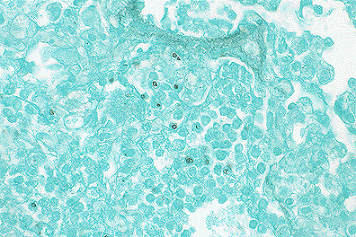 P. carinii cyst walls
stain well with GMS. (40X, GMS, 103K)
P. carinii cyst walls
stain well with GMS. (40X, GMS, 103K)
Signalment: Adult female ferret (Mustela putorius furo).
History: This pet ferret was observed by the owner to be constantly scratching. Physical examination revealed the animal to be infected with fleas and to have a dermatitis over the posterior half of the body, compatible with allergic flea dermatitis. Eight weeks later, the animal started to have difficulty breathing and died before she could be re-examined by the practitioner.
Gross Pathology: The lungs were mottled with gray compressed area and were of a rubbery consistency.
Laboratory Results: Culture: several coliform bacteria were isolated from the lung.
Morphologic Diagnosis & Etiology:
Contributor's Comments: It is felt the corticosteroid treatment rendered the animal susceptible to developing pneumonia. The acute inflammatory reaction is attributed to a bacterial infection, probably coliform and the interstitial pneumonia is attributed to P. carinii. P. carinii cysts were seen with GMS stain.
AFIP Diagnosis:
Conference Note: Pneumocystis carinii has a worldwide distribution and inhabits the respiratory tracts of man and many animals. Pneumocystis has caused clinical pneumonia in man, horses, primates, dogs, pigs, goats and other mammals; affected animals and humans are immunosuppressed. Whether P. carinii should be classified as a protozoan or as a fungus has been controversial. However, recent studies of ribosomal RNA, mitochondrial proteins, and genes which encode for thymidylate synthetase suggest that this organism is a fungus.
Pneumocystis carinii possesses two prominent antigens, major surface glycoprotein (MSG), and another 45-55kd molecule of unknown function. Either of these antigens is readily recognized by an immunocompetent host and stimulates both humoral and cell mediated immune responses. In the absence of an immune response however, P. carinii adheres to type I pneumocytes; this attachment may be facilitated by bridging between MSG and fibronectin. Once attached, P. carinii maintains an extracellular, intra-alveolar existence. P. carinii binds surfactant, which is thought to"hide" surface antigens and prevent recognition by the host's immune system. P. carinii pneumonia is suggested by the presence of large amounts of intra-alveolar surfactant, which appears as a foamy, eosinophilic material when stained with hematoxylin and eosin. The organisms stain with methenamine silver, periodic acid- Schiff, and Gram's stain.
Concurrent infections with bacteria, other fungi, or viruses are common in immunocompromised hosts. In this case, coliform bacteria were cultured from the lung and very likely were the cause of the neutrophilic inflammation.
Contributor:
St. Jude Children's Research Hospital
Comparative Medicine Division
332 North Lauderdale
Memphis, Tennessee 38105
References:
International Veterinary Pathology Slide Bank:
Laser disc frame #5290-1, 8448, 8450, 8493, 9295, 12374, 19657,
and 24541-2.
Dana P. Scott
Captain, VC, USA
Registry of Veterinary Pathology*
Department of Veterinary Pathology
Armed Forces Institute of Pathology
(202)782-2615; DSN: 662-2615
Internet: Scott@email.afip.osd.mil
* The American Veterinary Medical Association and the American College of Veterinary Pathologists are co-sponsors of the Registry of Veterinary Pathology. The C.L. Davis Foundation also provides substantial support for the Registry.
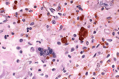 Multinucleate giant cell and
abundant hemosiderin-laden macrophages in an ethmoid hematoma.
(40X, HE, 103K)
Multinucleate giant cell and
abundant hemosiderin-laden macrophages in an ethmoid hematoma.
(40X, HE, 103K)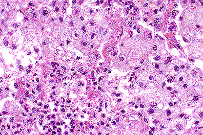 Alveoli filled with characteristic
foamy, eosinophilic surfactant and inconspicuous P. carinii
organisms in a ferret. (40X, HE, 103K)
Alveoli filled with characteristic
foamy, eosinophilic surfactant and inconspicuous P. carinii
organisms in a ferret. (40X, HE, 103K)