Signalment:
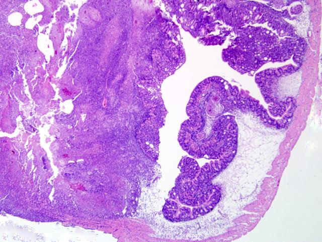
Histopathologic Description:
Morphologic Diagnosis:
1. Stomach: Gastritis, necrotizing and granulomatous, transmural, diffuse, severe, with multifocal granulomas, adjacent granulomatous peritonitis and intralesional protozoa
2. Liver and Kidney (variable between sections): Multifocal necrosis, granulomatous inflammation and epithelioid granulomas with intralesional protozoa
Condition:
Contributor Comment:
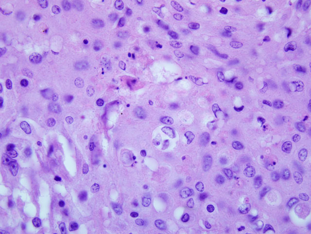
Microscopic findings of severe granulomatous gastritis are consistent with descriptions of Cryptobia iubilans (order Kinetoplastida, family Bodonidae) infection in other cichlid fishes.(1,4,6) The Kinetoplastidea are flagellates with long, tubulous mitochondria curved inside the cell, which contain a kinetoplast, an organized DNA nucleoid. Easily detected by Giemsa and Feulgen stains, it is usually single and located close to the kinetosome. One or two flagella may be present and most parasitic forms are transmitted by a vector.
Although 52 Cryptobia spp. have been tentatively identified in fish, most are leech transmitted hemoparasites and it has been proposed they be assigned to the Trypanoplasma.(2,3) The only fish pathogenic intestinal species, C. iubilans, has a direct life cycle and is common in cichlid fishes. The parasite is ovoid to elongate and averages 19 x 5 μm. The anterior flagellum is 1.5 - 2 times the body length and the recurrent flagellum, attached along the ventral axis, extends 11-19 μm beyond the body. Posterior to the flagellar pocket is a slender triangular kinetoplast. A spherical nucleus lies in the anterior half of the body and the cytoplasm contains large vacuoles filled with glycogen reserves.3 Additional electron microscopic features are available in the literature.(6)
Histopathologic lesions in light infections are confined primarily to the stomach and may range from isolated granulomas to diffuse granulomatous gastritis. In severe cases there can be multi-organ involvement associated with a similar necrotizing and granulomatous response, as seen in the livers and head kidney of some sections. The parasite may occur extracellularly or intracellularly within macrophages, where it is confined to a large parasitophorus vacuole.(2,6)
It has been suggested that C. iubilans may be a part of the normal gastrointestinal fauna, becoming pathogenic only under certain stressful conditions.(6) There are no effective treatments and losses can be acute and severe. Differentials for C. iubilans infection include other flagellates, primarily Spironucleus vortens. This organism is seen typically in the intestinal lumen and while it does not evoke as intense an inflammatory response, systemic spread can occur as well. On wet mounts this diplomonad is differentiated from C. iubilans by its linear movement and 8 flagella.(6) Although mycobacteriosis was considered, acid-fast stains and mycobacterial cultures performed on multiple fish were negative.
JPC Diagnosis:
1. Stomach and intestines: Gastroenteritis, transmural, granulomatous and necrotizing, diffuse, severe with protozoal trophozoites and many bacilli.
2. Liver; pancreas; mesentery; and kidney: Granulomas, with coelomitis.
Conference Comment:
The contributor provides an excellent review of this important disease of cichlids. Clincally, parasitemia results in anemia, exophthalmia, ascites, positive Coombs test, microcytic hypochromic anemia, and anorexia. As a brief additional note, isolation of a cysteine protease and metalloproteinase from the organism recently provided useful information regarding the pathogenesis of the disease; current research is focused on the metalloproteinase that causes lysis of erythrocytes. Additionally, the protein degrades types I, IV and V collagen and laminin, aiding invasion and generation of the observed histologic lesions.(5)
References:
2. Francis-Floyd R, R Yanong. Cryptobia iubilans in Cichlids. University of Florida Institute of Food and Agricultural Sciences Extension Publication VM 104.
3. Gou FC and PTK Woo. Selected parasitosis in cultured and wild fish. Vet Parasitol. 2009;163: 207-216.
4. Lom J, I Dykova. Flagellates (Phylum Mastigophora Diesing, 1866). In: Protozoan Parasites of Fishes. Amsterdam, The Netherlands: Elsevier Science Publishers; 1992:48-53.
5. Woo PTK. Immunological and therapeutic strategies against salmonid cryptobiosis. J Biomed Biotechnol. 2010;10 (Article ID 341783):9 pages.
6. Yanong RP, Curtis E, Russo R, et al. Cryptobia iubilans infection in juvenile discus. J Am Vet Med Assoc. 2004;224:1644-1650.