WSC 22-23:
Conference 8:
CASE I:
Signalment:
10-day-old Red Angus-Simmental calf; Bos taurus, bovine
History:
Over a period of 8 years (2013-2021) a total of six Red Angus-Simmental bull calves were born alive full term with hypotrichosis and oligodontia on a ranch in the Nebraska panhandle, USA. According to the attending veterinarian, the calves appeared almost completely alopecic. Animals tended to die in the first week of life, probably related to being born in spring with associated cold temperatures. The owners kept one calf (current case) alive by housing him indoors, wrapped in an insulated coat. A skin sample was taken to exclude inter-uterine infection with bovine viral diarrhea virus (BVDV), with negative results. The calf was alert and nursed normally. He was euthanized at 10 days of postnatal life in order to characterize cutaneous lesions and to establish whether lesions were present elsewhere, particularly in respiratory tract and oral cavity.
Gross Pathology:
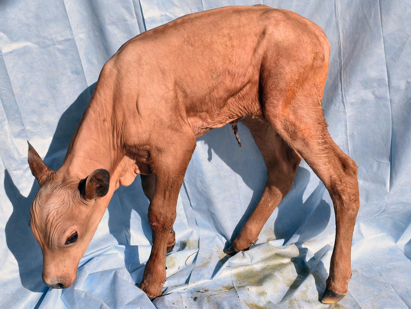
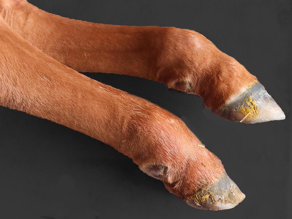
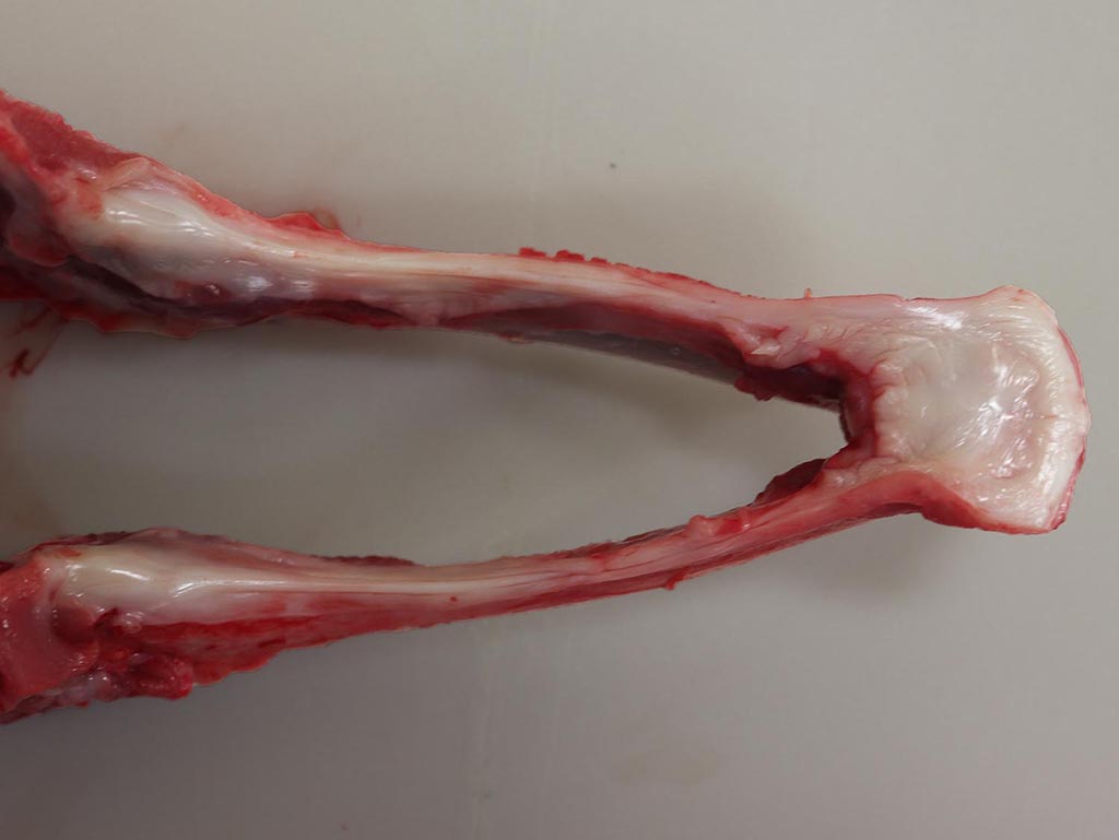
The calf was almost completely devoid of large, guard-type hairs at birth, excepting distal parts of the limbs and tail, eyelids, chin and tragus. Vibrissae were present on the muzzle and chin. Short fine hairs consistent with undercoat were present over the trunk, neck, head and upper two thirds of the limbs. The nasolabial plate was flat and dry. All incisors were absent grossly, a finding that was confirmed radiographically. According to the owner and the attending veterinarian, the other five affected bull calves had a similar phenotype, including the absence of visible teeth. Hooves were unremarkable. Obvious ocular lesions were absent.
Laboratory Results:
A biopsy of aural skin was negative for bovine viral diarrhea virus by antigen ELISA.
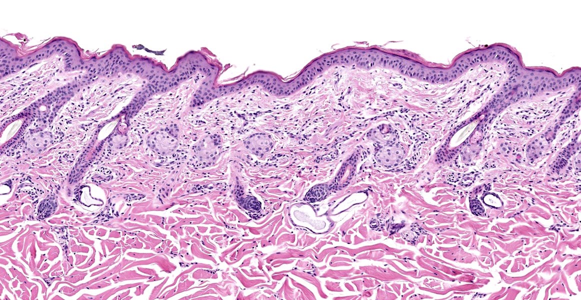
Microscopic Description:
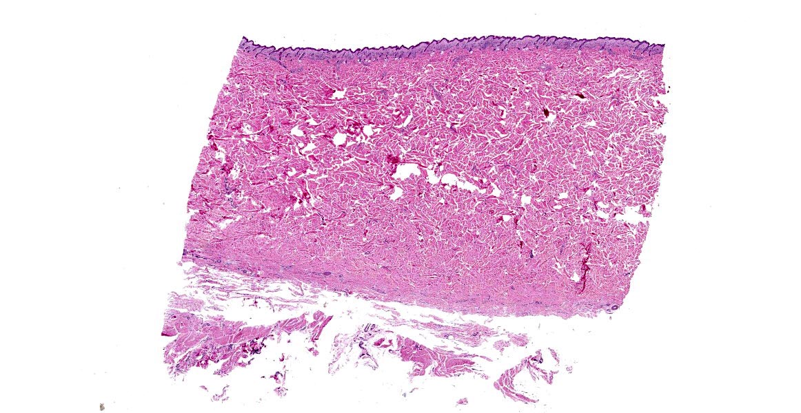
Haired skin-subcutis from flank. There is a complete absence of large caliber hair follicles containing medullated hair shafts. Existing follicles are uniformly small (diameter of 35 - 40 µm) and contain non-medullated, lightly pigmented hairs of unremarkable appearance. Each follicle retains a normal relationship with apocrine glands, arrector pili, and sebaceous glands. There is a mild diffuse lymphohistiocytic inflammation in upper dermis.
Contributor’s Morphologic Diagnoses:
- Severe diffuse congenital hypotrichosis, with absence of primary hair follicles.
- Mild diffuse superficial lymphohistiocytic dermatitis.
Contributor’s Comment:
In addition to hypotrichosis, the affected calf had oligodontia with only 12 deciduous teeth, all of which were unerupted or barely erupted premolars at the time of euthanasia. The calf also lacked nasolabial, tracheal and bronchial-bronchiolar glands. Analysis at the Institute of Genetics in the University of Bern identified a 53 kb deletion in the X chromosome, including part of the EDA gene and all of AWAT2. Partial deletion of EDA is the likely basis for this form of hypotrichosis-oligodontia.8
This combination of lesions involving hair follicles, teeth and aplasia of selected glands indicates an ectodermal dysplasia.5 Mutation in EDA, marked reduction in large (developmentally first-formed) hair follicles containing medullated hairs, restriction of the disorder to bull calves, and the lesions in non-cutaneous tissues is consistent with an X-linked disease. This is reported sporadically in Holstein and Holstein Friesian cattle, Japanese Black cattle, and crossbred cattle. Currently most of the published reports document one or more affected calves in commercial herds in Europe.9 Affected calves die or are killed early in post-natal life. Some can be kept alive when fed a chopped diet after they begin to ruminate, combined with protection from weather extremes. Currently 9 episodes of the bovine condition have been documented.9 The causative abnormality in EDA varies. Investigators report various small or gross deletions, insertions, inversions, splicing, and nonsense (stop-gain) mutations. The resultant phenotype is remarkably uniform, based on published accounts. Similar forms of ectodermal dysplasia due to EDA variants are reported in dogs, mice, and people.3AWAT2 encodes an enzyme in the diacylglycerol acyltransferase family. The enzyme produces wax esters as part of the normal lipid metabolism of skin, primarily in sebocytes. The submitter did not recognize a lesion in sebaceous glands of affected calves.
This bovine ectodermal dysplasia corresponds to X-linked hypohidrotic ectodermal dysplasia-1 in human subjects (ECTD1)(OMIM 305100).10,13EDA codes for ectodysplasin, a member of the TNF-related ligand family which mediate epithelial-mesenchymal interactions during fetal development, including formation of ectodermal appendages.11 Use of the term ECTD has been proposed for calves with analogous mutations in EDA.3
Unlike affected people and mice, there is no simple way to assess the function of sweat glands in neonatal calves. There is disagreement about whether sweat glands in affected calves are histologically normal. The submitter compared sweat glands from affected calves to those in unaffected calves from the source herd and there was no obvious difference.
Affected human patients have a triad of signs comprising sparse hair (hypotrichosis), abnormal or missing teeth (anodontia or hypodontia), and anhidrosis/hypohidrosis. Medical complications in affected people are hyperthermia due to inability to sweat normally, and recurrent respiratory infections. Affected children can be treated successfully using antenatal injections of recombinant ectodysplastin.12 Treatment was also successful two animal models of ECTD-like syndromes (Tabby mice; dogs).4,6
It is diagnostically useful to check for oligodontia in calves with marked congenital hypotrichosis, since its presence narrows differential considerations regarding etiology. Calves that are bald at birth will be obvious to owners. It is common for veterinarians to be contacted about such calves. The first question is generally: could this be inherited? Non-genetic causes of congenital hypotrichosis exist, such as transplacental bovine diarrhea virus infection (excluded here), maternal iodine deficiency, and adenohypophyseal hypoplasia. Neonatal calves that present with hypotrichosis, oligodontia and a smooth dry nasal plate should prompt consideration of an inherited abnormally in the EDA molecular pathway. The defect most commonly involves EDA itself. Mutations may affect other genes in the pathway (i.e., EDAR, EDARADD orTRAF6), resulting in a similar phenotype.11
Mild changes affecting teeth, hair and sweat glands occurs in some mothers of children with ECTD1, but are generally subtle. Investigation of similar changes in carrier carriers and calves is limited.
Contributing Institution:
Wyoming State Veterinary Laboratory; 1174 Snowy Range Road; Laramie; WY 82070; http://www.uwyo.edu/wyovet/
JPC Diagnosis:
- Haired skin: Hypotrichosis, diffuse, severe with trichodysplasia.
- Haired skin: Dermatitis, lymphohistiocytic, superficial and perivascular, mild.
JPC Comment:
This week’s moderator, Dr. Charles Bradley from the University of Pennsylvania, emphasized that it is not possible based on histopathologic examination alone to rule out non-genetic causes of hypotrichosis. Other differentials in calves which be considered include iodine deficiency; toxins (Veratrum album in Japanese cattle), adenohypophyseal hypoplasia in Guernsey and Jersey cattle, and intrauterine infection with bovine pestivirus (bovine viral diarrhea virus; ruled out in this case using ELISA testing).
Currently, there are nine X-linked EDA mutations causing ectodermal dysplasia in various breeds of cattle.1 Additionally, there is one documented autosomal recessive mutation in the EDA receptor gene associated with ectodermal dysplasia in Charolais cattle.8
Humans, mice, and cattle with ectodermal dysplasia may lack of normal glands of the respiratory tract, such as bronchial and bronchiolar glands, which compromises mucociliary clearance and predisposes affected animals to respiratory infections, including sinusitis and pneumonia.1,7,8 Additionally, EDA is crucial for the development and function of lacrimal and Meibomian glands, corneal homeostasis, and corneal wound healing, and recurrent conjunctivitis has been documented in dogs with ectodermal dysplasia and EDA-deficient mice.1,7,8
Recently, it has been reported that several mice and rat strains with EDA deficiency also have significant Zymbal’s gland hypoplasia, with fewer hair follicles and smaller sebaceous glands in the external ear canal.2 Affected rodents have increased prevalence of otitis externa, and the authors concluded this was likely due to sebum deficiency.2 Strains with Zymbal’s gland hypoplasia included EDA-deficient mice, EDA receptor (EDAR) deficient mice, and EDAR-associated death domain (EDARADD) deficient rats.2 The EDA-deficient mice and EDARADD deficient rats were also predisposed to otitis media due to deficiency of nasopharyngeal submucosal glands associated with the auditory tube.2
References:
- Capuzzello G, Jacinto JCP, Hafliger IM, et al. A large deletion encompassing exon 2 of the ectodysplasin A (EDA) gene in a British blue crossbred calf with hypohidrotic ectodermal dysplasia. Acta Veterinaria Scandinavica. 2022; 64(23):1-8.
- Del-Pozo, Headon DJ, Glover JD, et al. The EDA deficient mouse has Zymbal’s gland hypoplasia and acute otitis externa. Dis Model Mech. 2022; 15(3):1-31.
- Drögemüller C, Distl O, Leeb T. X-linked anhidrotic ectodermal dysplasia (ED1) in men, mice, and cattle. Genet Sel Evol. 2003;35: S137-145.
- Gaide O, Schneider P. Permanent correction of an inherited ectodermal dysplasia with recombinant EDA. Nat Med. 2003;9:614-618.
- Hargis AM, Ginn PE. The integument. In: Zachary JF, McGavin MD, eds. Pathologic Basis of Veterinary Disease. 5th Elsevier Inc. 2012.
- Mauldin EA, Gaide O, Schneider P, Casal ML. Neonatal treatment with recombinant ectodysplasin prevents respiratory disease in dogs with X-linked ectodermal dysplasia. Am J Med Genet A. 2009;149A:2045-2049.
- Moura E, Rotenberg IS, Pimap CT. X-Linked hypohydrotic ectodermal dysplasia - General features and dental abnormalities in affected dogs compared with human dental abnormalities. Top Companion Anim Med. 2019, 35, 11-17.
- O’Toole D, Häfliger IM, Leuthard F, et al: 2021, X-linked hypohidrotic ectodermal dysplasia in crossbred beef cattle due to a large deletion in EDA. Animals (MDPI). 2021;11:657.
- OMIA - Online Mendelian Inheritance in Animals database search engine. Accessed 27 May 2021. https://www.omia.org/home/
- OMIM - Online Mendelian Inheritance in Man database search engine. Accessed 27 May 2021. https://www.omim.org/about
- Sadier A, Viriot L, Pantalacci S, Laudet V. The ectodysplasin pathway: from diseases to adaptations. Trends Genet. 2014;30:24-31.
- Schneider H, Faschingbauer F, Schuepbach-Mallepell S, et al. Prenatal correction of X-linked hypohidrotic ectodermal dysplasia. N Engl J Med. 2018;378:1604-1610.
- Wright JT, Fete M, Schneider H, et al. Ectodermal dysplasias: Classification and organization by phenotype, genotype and molecular pathway. Am J Med Genet. 2019, 179, 442-447.