CASE 4: P17-0217 (4100658-00)
Signalment: 1-year old female spayed Beagle
History: The dog was spayed, and the uterus and ovaries were submitted for histopathology.
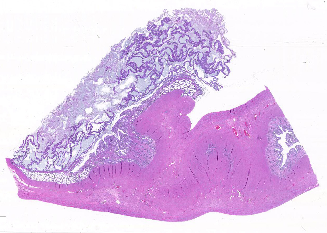
Gross Pathology: The uterus contains a 3 cm diameter well circumscribed mass.
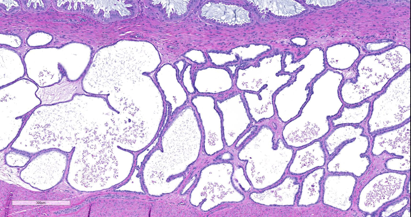
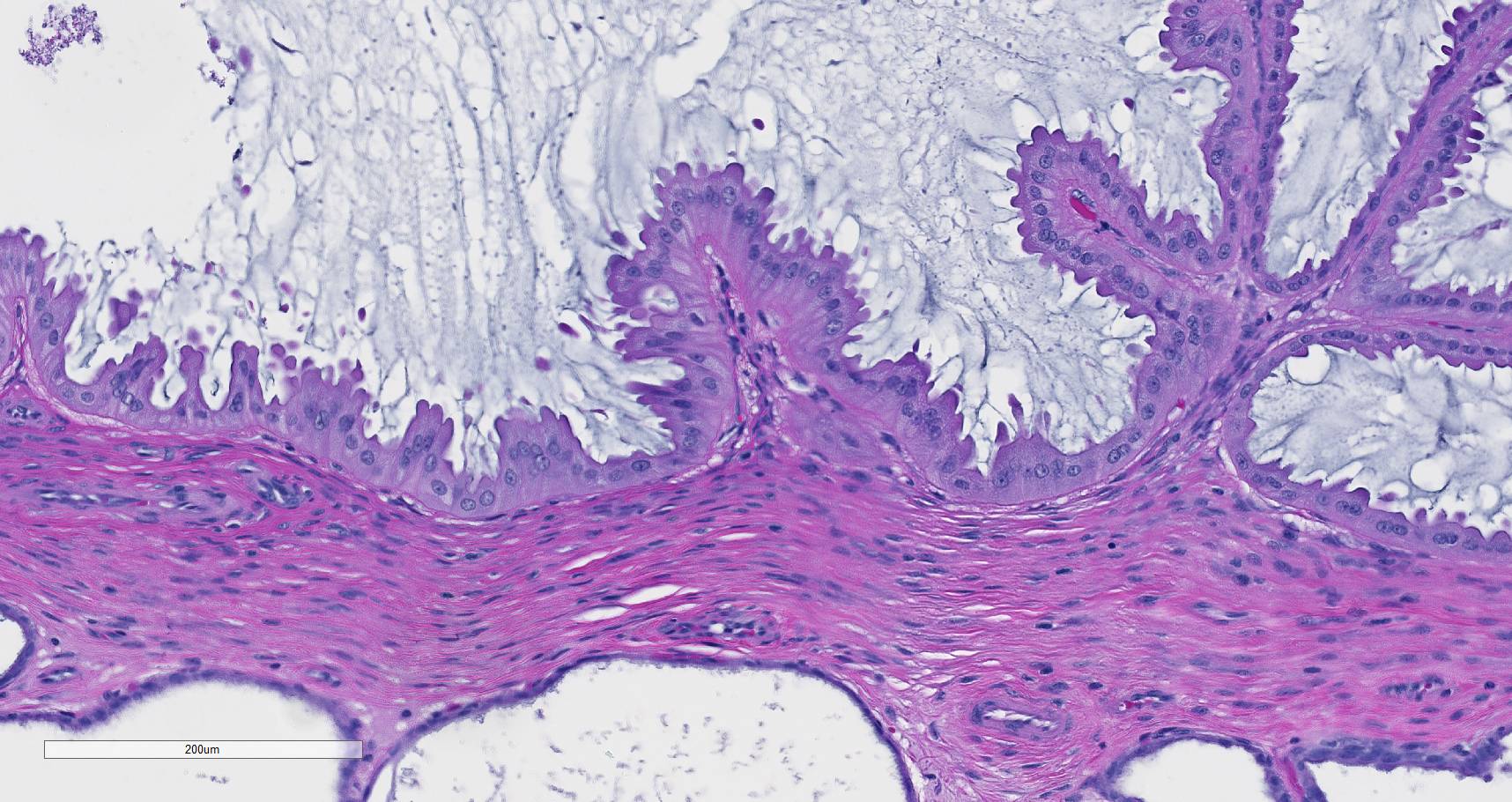
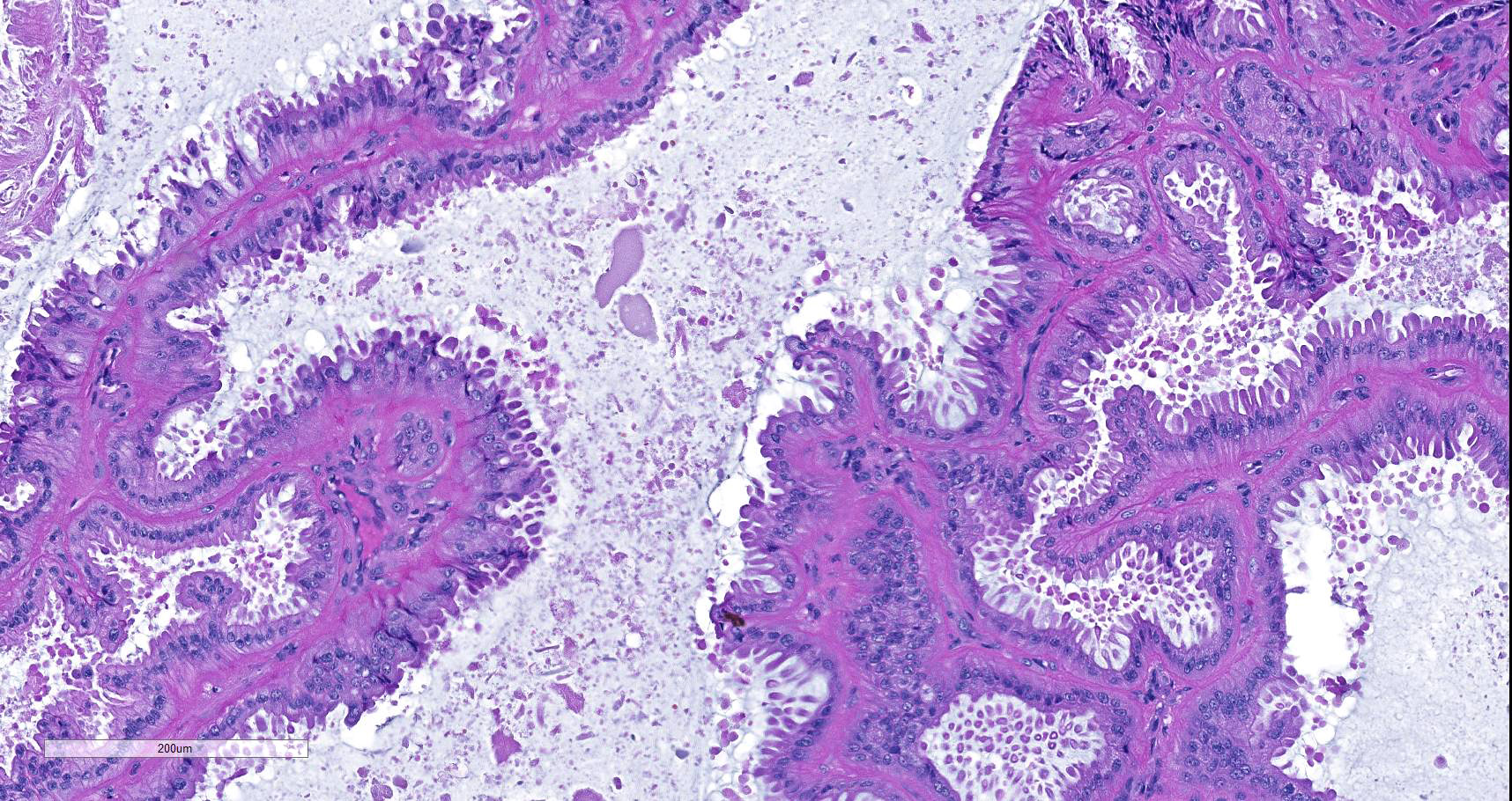
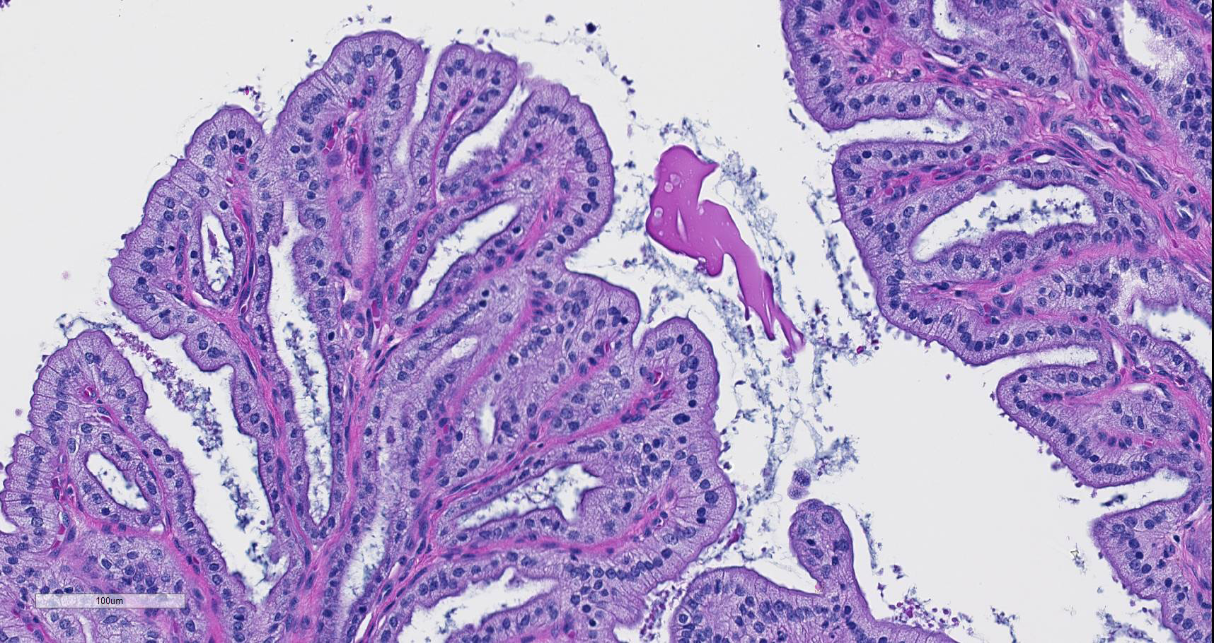
Microscopic description: The endometrium has hyperplasia of the surface epithelium forming long fronds extending into the lumen with secondary branching and papillary projections. The epithelium is a single layer of columnar cells with abundant amounts of eosinophilic cytoplasm and basal-oriented nuclei. The lumen is filled with mucus that permeates between the fronds of epithelium. The endometrial glands have hyperplasia and ectasia. Inflammation is lacking.
Contributor's morphologic diagnosis:
Pseudo-placentational endometrial hyperplasia
Contributor's comment: Endometrial hyperplasia in the dog occurs in two forms. The more common form is called cystic endometrial hyperplasia and is a hyperplasia of the endometrial glands. It is frequently associated with pyometra. The other form occurs uncommonly and is called by various names including pseudo-placentational endometrial hyperplasia, deciduoma, and segmental endometrial hyperplasia. Both forms occur under the influence of progesterone from persistent corpora lutea.
The gross and microscopic appearance of the lesion of pseudo-placentational endometrial hyperplasia resembles the normal placental sites in the dog uterus and is the genesis for the naming of the disease.4,5 The dog with this disease is not pregnant, and the cause of the disease is unknown. The condition has been induced by insertion or injection of a variety of materials within the uterus.
Contributing Institution:
College of Veterinary Medicine
Virginia Polytechnic Institute and State University
Blacksburg, VA 24061
JPC diagnosis:
Uterus: Hyperplasia, endometrial, pseudo-placentational, segmental, Beagle, canine.
JPC comment:
In the bitch, the normal estrous cycle includes differentiation and proliferation of endometrial glands under the influence of estrogen during proestrus, with more extensive proliferation during estrus and metestrus/diestrus under the influence of progesterone. When progesterone levels fall during late diestrus, the endometrium regresses to a quiescent state during anestrus.3
There is debate among authors whether cystic endometrial hyperplasia (CEH) and pyometra are linked, it is currently thought that prolonged exposure to high levels of progesterone cause endometrial gland proliferation which may increase susceptibility of the uterus to infection.1,3 Three forms of endometrial hyperplasia have been described in the bitch, in addition to CEH-pyometra complex: 1) estrogen-induced CEH, 2) focal endometrial polyps, and 3) endometrial hyperplasia associated with pseudopregnancy (PEH, this case entity).
Histologically, PEH is distinguishable from CEH by only involving a focal or multifocal segment of the uterus, mimicking placentation sites. CEH is typically a generalized diffuse reaction involving the entire endometrium.5 PEH masses have the three distinct zone of normal maternal placenta, the stratum spongiosum, a zone of dense connective tissue, and the luminal pseudostratified epithelium with multifocal necrotic syncytia formation.2
References:
1. DeBosschere H, Ducatelle R, Verrneirsch H, et al. Cystic endometrial hyperplasia-pyometra complex in the bitch: should the two entities be disconnected? Theriogenol. 2001;55:1509-1519.
2. Koguchi A, Nomura K, Fujiwara T, et al. Maternal placenta-like endometrial hyperplasia in a Beagle dog (canine deciduoma). Exp Anim. 1995;44:251-253.
3. McKentee K. The uterus: atrophic, metaplastic and proliferative lesions. In: Reproductive Pathology of Domestic Animals. San Diego, CA: Academic Press; 199:171-175.
4. Sato, Y. Pseudo-placentational endometrial hyperplasia in a dog. J Vet Diag Invest. 2011;23(5):1071-1074.
5. Schlafer DH, Giffored AT. Cystic endometrial hyperplasia, pseudo-placentational endometrial hyperplasia, and other cystic conditions of the canine and feline uterus. Theriogenology. 2008;70:349-358.