WSC 2022-2023
Conference 24
Case III:
Signalment:
2 years and 6 months old female Holstein Friesian Cow (Bos taurus).
History:
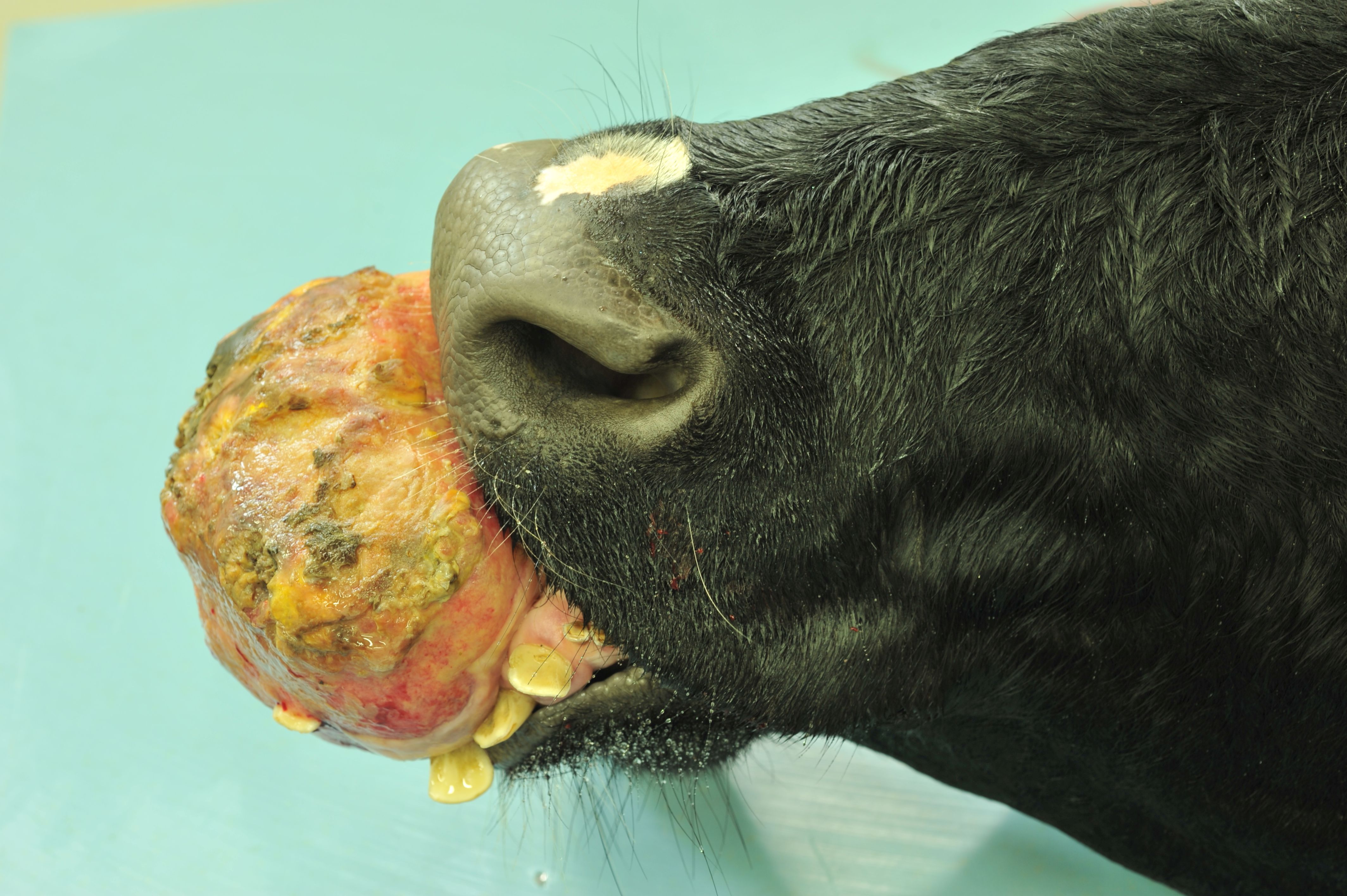
The cow was euthanized due to a large (41.5cm circumference), round, firm, ulcerated mass with embedded incisors at the rostral aspect of the mandible. Whilst the cow was unable to hold saliva, she was still able to eat and drink with some adaptation.
Gross Pathology:
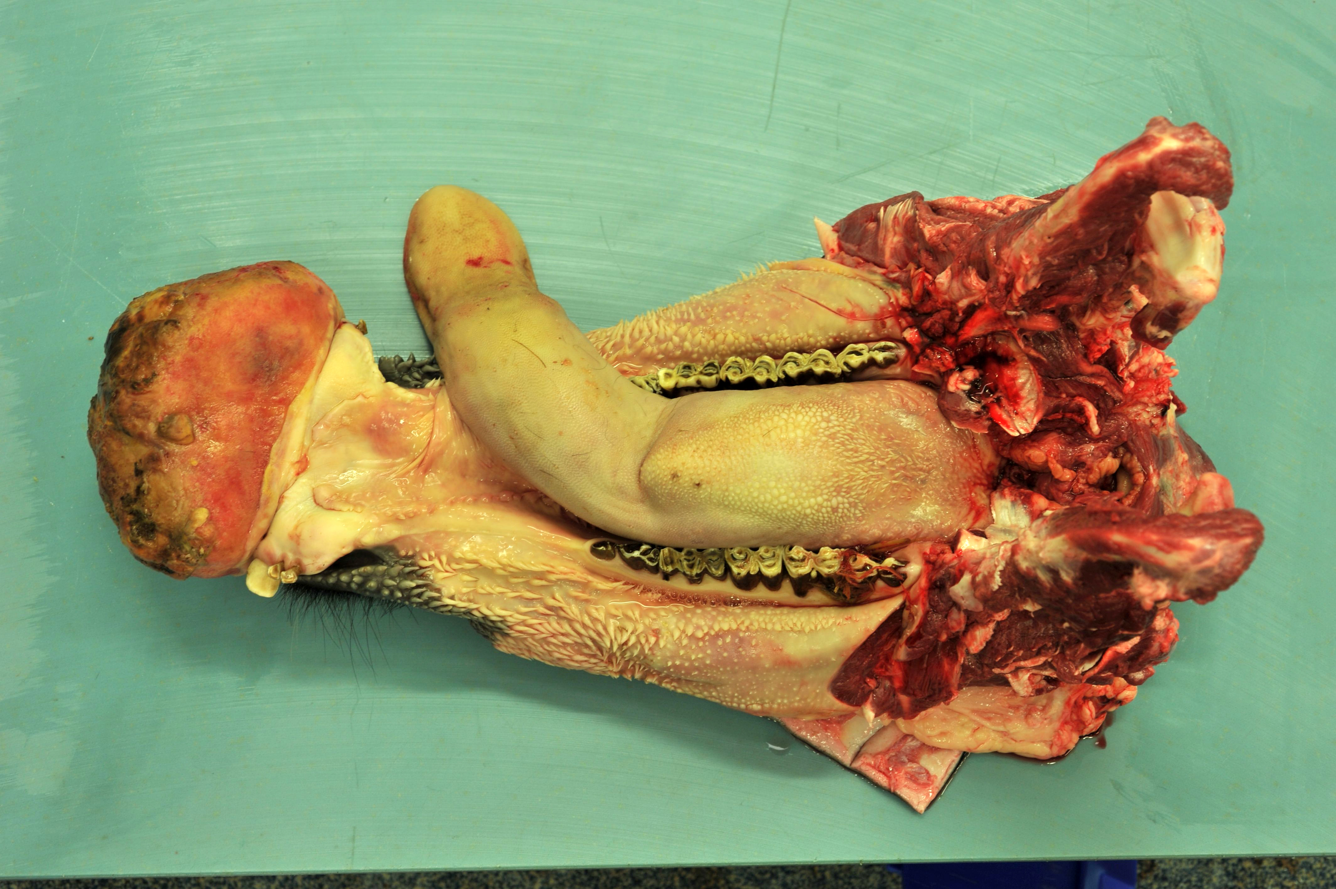
A very large (160x140x160 mm), expansile, broad based, firm, pink to red mass extends from the dorsolingual aspect of the rostral mandible, displacing teeth 301, 302, 401, 402, 403 and 404 laterally. Approximately 70% of the surface of the mass is ulcerated.
On cut surface, the mass exhibits a central 60x24x16 mm cavity which contains approximately 15 ml of clear, yellow tinged, watery fluid surrounded by cream to yellow to pale pink, variably soft to very firm, gelatinous to fleshy tissue. Where the mass merges with regular mandible, the mandible is thickened to a maximum thickness of 69 mm.
The forage in the forestomaches, particularly the rumen, is excessively long.
Laboratory Results: None reported.
Microscopic Description:
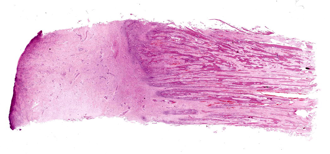
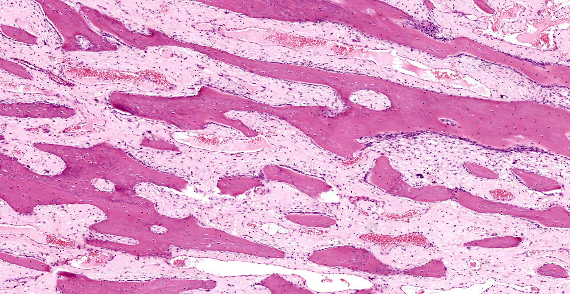
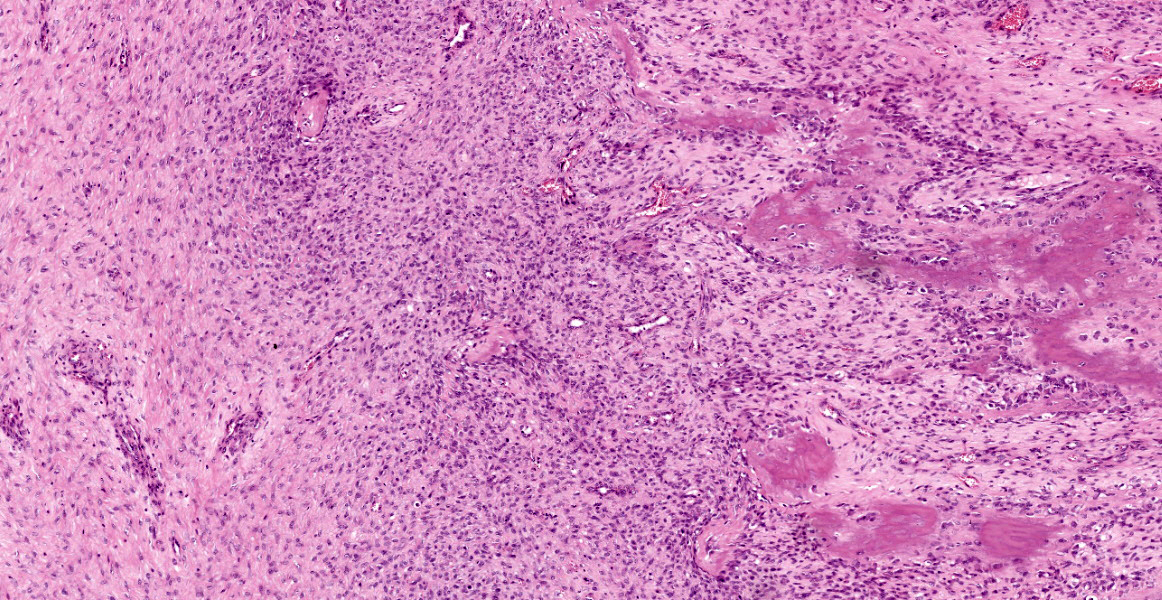
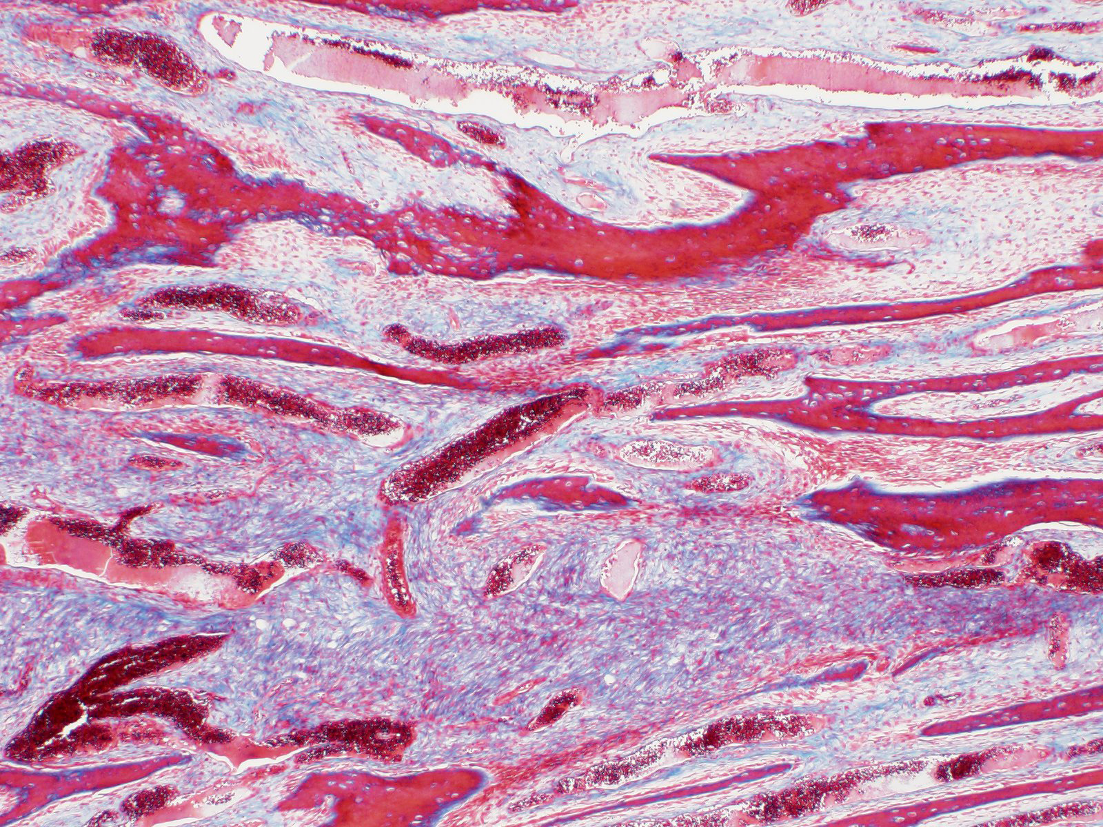
Mandibular mass: The section contains parts of a large and moderately cellular mass which comprises loosely to moderately densely and haphazardly arranged, very vague bundles and streams of neoplastic cells, situated in and supported by a moderate to large amounts of multifocally mildly oedematous, fine collagenous stroma, interspersed with numerous elongated, partially anastomosing bony spicules and trabecules arranged in parallel to one another and perpendicular to the superficial surface. The bone trabecules are lined by a largely one cell (ranging up to three cells) thick layer of polygonal to elongate cells with eosinophilic/mildly basophilic cytoplasm and round to oval nuclei with finely stippled to clumped and marginated chromatin (osteoblasts). The neoplastic cells are spindle-shaped to stellate-shaped with moderately defined cell boundaries and small amounts of eosinophilic cytoplasm. Their nuclei are irregularly oval with finely stippled to mildly clumped, commonly also marginated chromatin and one or two, small basophilic nucleoli. Anisocytosis and anisokaryosis are mild, very small numbers of binucleated cells are present and less than one mitotic figure is seen in 10 high power fields. Mitotic figures, however, are more evident in those regions close to bone formation and at the interface between bone and collagenous tissue, where also a substantial increase in cellularity is evident.
The mucosa is diffusely ulcerated, and covered by fibrin, eosinophilic and basophilic cellular debris and large numbers of viable and degenerate neutrophils, in deeper levels admixed with small numbers of macrophages, lymphocytes and plasma cells which in turn are situated amongst plump fibroblasts vaguely orientated parallel to the surface and small vessels lined by hypertrophied endothelial cells orientated perpendicular to the superficial surface (granulation tissue formation).
Very small numbers of lymphocytes, plasma cells and macrophages are diffusely distributed throughout the neoplastic population and commonly also seen forming very small aggregates in perivascular location.
Contributor’s Morphologic Diagnoses:
Mandible, rostral aspect: Ossifying fibroma, bovine, Bos taurus.
Contributor’s Comment:
Ossifying fibromas (OF) are benign, proliferative, intraosseous lesions with a rapid growth rate and strong predilection for the mandible.3,17 OF are most frequently recognized in human beings and young horses (<1 year old)8, but have also been described in dogs2,3,6,7, cats3, sheep3, and rarely cattle.13,19 Additionally, individual cases of OF are published in a range of other species including a canary14, a rabbit19, a llama5, roe deer21 and a cynomolgus monkey15.
In humans OF are most commonly found in the posterior mandible, although maxillary and zygomatic lesions are also recognized.4 The rostral mandible represents a clear predilection site in young horses (where the condition also is known as equine juvenile mandibular ossifying fibroma)8, and also in ruminants and domestic carnivores3,13,14. Additional reported sites include long bones3,14, equine paranasal sinuses12, the equine proximal phalanx1, the canine os penis7 and the canine zygomatic arch.2 Reported cases in cattle are restricted to the mandible (although paucity of cases may underlie a lack of recognition of atypical locations).13,14
In humans, a predilection for ossifying fibromas has been established in females in their 30s or 40s.4 In contrast to this, equines exhibit such lesions most commonly in young animals (<12 months)8, whilst no clear age-related predilection is reported for other species at present (possibly again due to paucity of cases). No sex predisposition is currently reported in animals.
Human OF are reported to be non-painful. Their most significant effects are of malocclusion and cosmetic impairment.4 Similarly, the primary effects of OF in domestic animals are impaired occlusion and/or prehension as well as a predisposition to pathologic fractures.3,8,13,14 The further effects of OF in atypical locations are largely due to their space-occupying behavior and specific effects dependent on the location in question, for example urethral obstruction in the case of an OF in the os penis.1,2,7,12
Histologically ossifying fibromas are characterized by spindle shaped fibroblasts interspersed with trabeculae of woven bone which are surrounded by a rim of osteoblasts.17 Historical miscategorisation and underreporting of OF in the literature is suspected, owing to the similarity between osteoma and OF and the disagreement about the existence of a disease continuum between fibrous dysplasia (FD) and OF.17,18 Currently, histological differentiation of OF from FD is largely based on the presence of a rim of osteoblasts surrounding foci of woven bone.7,8,10,18,19,20 Osteomas can be distinguished from OF by their grossly sessile or pedunculated appearance arising from the surface of the bone, their containment within the periosteal membrane, relative hypocellularity and the presence of lamellar bone with bone marrow, whilst osteosarcomas can be differentiated from OF by the pleomorphism and a considerably higher mitotic rate of the neoplastic population with osteoblasts typically also filling the intertrabecular spaces rather than only lining the bony trabecules.7,8,12,18,19,20
Contributing Institution:
Division of Pathology, Public Health and Disease Investigation
Veterinary Diagnostic Services
School of Veterinary Medicine
College of Medical, Veterinary and Life Sciences
University of Glasgow (Garscube Campus)
464 Bearsden Road
Glasgow G61 1QH, Scotland
https://www.gla.ac.uk/schools/vet/cad/
JPC Diagnosis:
Bone, mandible: Ossifying fibroma.
JPC Comment:
As the contributor describes, ossifying fibromas are rare in cattle, and since this case was submitted to WSC, another report of three cases of mandibular ossifying fibromas in cattle was published in Journal of Comparative Pathology. All cases were characterized as well demarcated and projected from the rostral mandible, where they replaced or displaced multiple incisors. Histologically, the neoplasms had the characteristic features of ossifying fibroma: trabeculae of bone lined by osteoblasts surrounded by haphazardly arranged neoplastic spindle cells with invasion, degeneration, and necrosis of adjacent bone.11 In one case, the neoplastic spindle cells were confluent with the periodontal ligament, and some have speculated that ossifying fibroma of the mandible may originate from pluripotent stem cells of the periodontal ligament.11
Ossifying fibroma is also rarely reported in marine (striped mullet) and freshwater fish (sauger and walleye). The freshwater fish cases were linked to pollution from a mining operation. A single case of cutaneous osseous fibroma was more recently reported in the caudal peduncle of a tetra.10 The neoplasm contained dysplastic ctenoid scales, and the authors theorize that the neoplasm may have originated from the bone in these scales, a theory which is in line with central ossifying fibromas in humans, which also originate from bone.10
Historically the presence or absence of a rim of osteoblasts around trabeculae of woven bone was used to differentiate ossifying fibroma (OF) from fibrous dysplasia (FD). Recent work, however, has demonstrated that both OF and FD can have osteoblasts around trabeculae. Alternatively, both OF and FD may lack an osteoblastic rim around trabeculae. For these reasons, the presence or absence of an osteoblastic rim is no longer considered diagnostically useful. The degree of bone differentiation histologically, and the sharpness of demarcation radiographically are generally considered the most useful features in distinguishing OF and FD. OF has well differentiated woven bone histologically, and sharply defined margins radiographically. FD may have poorly differentiated bone that is difficult to recognize as bone (ie “proto-bone”) histologically, and is poorly delineated with ill-defined margins radiographically. Both OF and FD arise from the intra-osseous region and can have multinucleated giant cells (osteoclasts). As these represent a spectrum of benign, fibro-osseous lesions, it may be challenging to impossible to definitively differentiate OF from FD without the use of radiographs.8,16
References:
- Balducci J, Selberg K, Pool R and Radue RP. Primary ossifying fibroma of the proximal phalanx in a horse. Equine Vet Educ. 2019;32:40-44.
- Best E. Ossifying fibroma of the zygomatic arch in a 20-month-old male corgi. Vet Times.
- Craig LE, Dittmer KE and Thompson KG. Chapter 2 Bones and Joints. In: Jubb, Kennedy and Palmer’s Pathology of Domestic Animals. Volume 1. Maxie MG. 6th ed. Ames, Iowa; Wiley Blackwell; 2015:109.
- da Silveira DT, Cardoso FO, Silva BJ, Alves Cardoso CA and Manzi FR. Ossifying fibroma: report on a clinical case, with the imaging and histopathological diagnosis made and treatment administered. Rev Bras Ortop. 2015;51(1):100-104.
- McCauley CT, Campbell GA, Cummings CA and Drost WT. Ossifying fibroma in a llama. J Vet Diagn Invest. 2000;12:473-476.
- Miller MA, Towle HAM, Heng HG, Greenberg CB and Pool RR. Mandibular Ossifying Fibroma in a Dog. Vet Pathol. 2008;45(2):203-206.
- Mirkovic TK, Schmon CL and Allen Urinary obstruction secondary to an ossifying fibroma of the os penis in a dog. J Am Anim Hosp Assoc. 2004; 40:152-156.
- Morse CC, Saik JE, Richardson DW and Fetter AW: Equine juvenile mandibular ossifying fibroma. Vet Pathol. 1988;25:415-421.
- Murphy B, Imai DM. Cuatneous Ossifying Fibroma in a Neon Tetra (Paracheirodon innesi). J Comp Pathol. 2016; 155(2-3): 272-275.
- Murphy BG, Bell CM, Soukup JW. Veterinary Oral and Maxillofacial Pathology. Hoboken, NJ: John Wiley & Sons. 2020; 243.
- De Oliveira Freitas DC, Melo FG, de Melo Ocarino N, Abreu DM, Araujo FR, Serakides R. Three Cases of Ossifying Fibroma in Cattle. J Comp Pathol. 2022; 198: 16-21.
- Orsini JA, Baird DK and Ruggles AJ. Radiotherapy of a recurrent ossifying fibroma in the paranasal sinuses of a horse. 2004;224(9):1483?1454.
- Raval SH, Joshi DV, Patel BJ, Patel J, Sutariya P, Soni M and Rathod AS. Bovine dental tumors: A report of four cases. Indian J Vet Pathol. 2017:41:119-122.
- Razmyar J, Dezfoulian O and Peighambari S. Ossifying Fibroma in a Canary (Serinus canaria). J Avian Med Surg. 2008;22(4):320-322.
- Schmelting B, Zöller M, Kaspareit J. Peripheral ossifying fibroma and juxtacortical chondrosarcoma in cynomolgus monkeys (Macaca fascicularis). J Am Assoc Lab Anim Sci. 2011;50(1):98-104.
- Soltero-Rivera M et al. Benign and malignany proliferative fibro-osseous and osseous lesions of the oral cavity of dogs. Vet Pathol. 2015; 52(5):894-902.
- Thompson KG, Dittmer Tumors of Bone. In:Meuten DG, ed. Tumors in Domestic Animals. 5th ed. Ames, Iowa: Wiley Blackwell; 2017:362-363.
- Toyosawa S, Yuki M, Kishino M, Ogawa Y, Ueda T, Murakami S, Konishi E, Iida S, Kogo M, Komori T and Tomita Y. Ossifying fibroma vs fibrous dysplasia of the jaw: molecular and immunological characterization. Mod Pathol. 2007;20:389-396.
- Whitten KA, Popielarczyk MM, Belote DA, McLeod GC and Mense MG. Ossifying fibroma in a miniature rex rabbit (Oryctolagus cuniculus). Vet Pathol. 2008;43:62-64.
- Yayla S, Kiliç E, Özen H, Da? S, Baran V and Aydin U. A case of osteofibroma on the symphysis mandible in a cow. J Hell Vet Med Soc. 2018;69(1):879-882.
- Zürcher-Giovannini S, Ruder TD, Pool R, Erdelyi K and Origgi FC. Mandibular Ossifying Fibroma and Multiple Oral Papillomas in a Roe Deer (Capreolus capreolus). Front Vet Sci. 2020;7:166.