Signalment:
Nine-year-old, female, golden retriever, (
Canis familiaris).The dog was
brought to the clinic with a history of intermittent rectal prolapse. Clinical
examination revealed a rectal mass (5 cm cranial from the anus), and the dog
was referred to the surgical excision of the mass.
Gross Description:
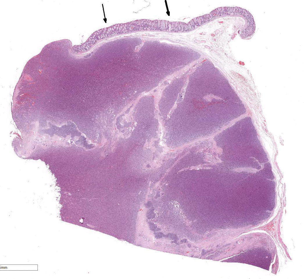
Rectal
mass: an oval 3x5 cm mass, on cross section light brown in color and firm
consistency.
Histopathologic Description:
Rectum:
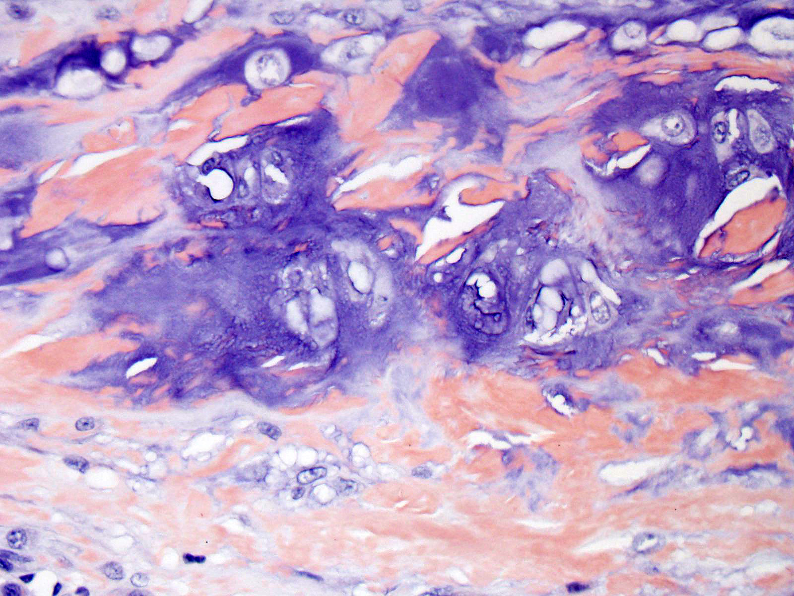
Expanding the submucosa and elevating the overlying ulcerated mucosa is a
well circumscribed, partially encapsulated, densely cellular neoplasm composed
of sheets and packets of round cells
separated by a fine fibrous stroma.
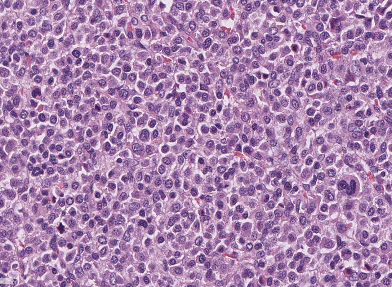
Neoplastic cells have variably distinct cell borders and moderate amounts of
eosinophilic granular cytoplasm. Nuclei are round, rarely oval, usually
eccentrically located with finely stippled to finely clumped chromatin and
usually one indistinct nucleolus. There is moderate anisokaryosis, multifocal
karyo-megaly and a moderate number of multinucleate cells. Mitotic figures
average 10 per 10 HPF. Multifocally between neoplastic cells or at the
periphery of the tumor are variably sized and shaped, extracellular deposits of
amorphous, homogenous, eosinophilic material (amyloid) with multifocal islands
of cartilage formation (cartilaginous metaplasia) that is occasionally
mineralized.
Morphologic Diagnosis:
>Rectum:
Plasmacytoma (or Extramedullary Plasmacytoma), golden retriever, canine.
Lab Results:
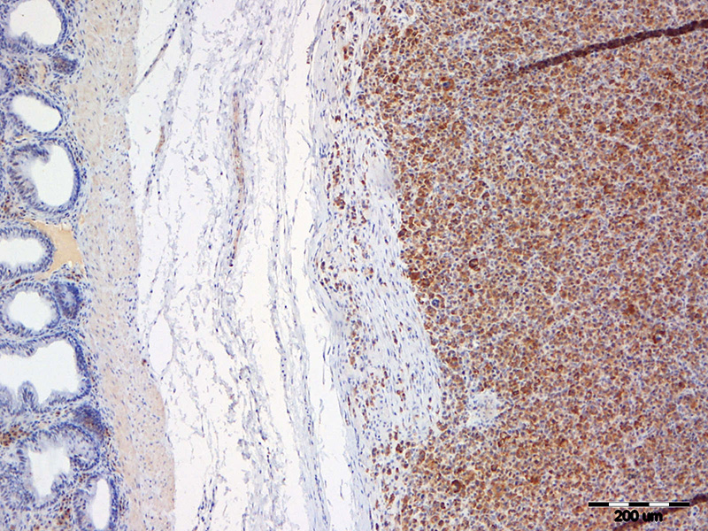
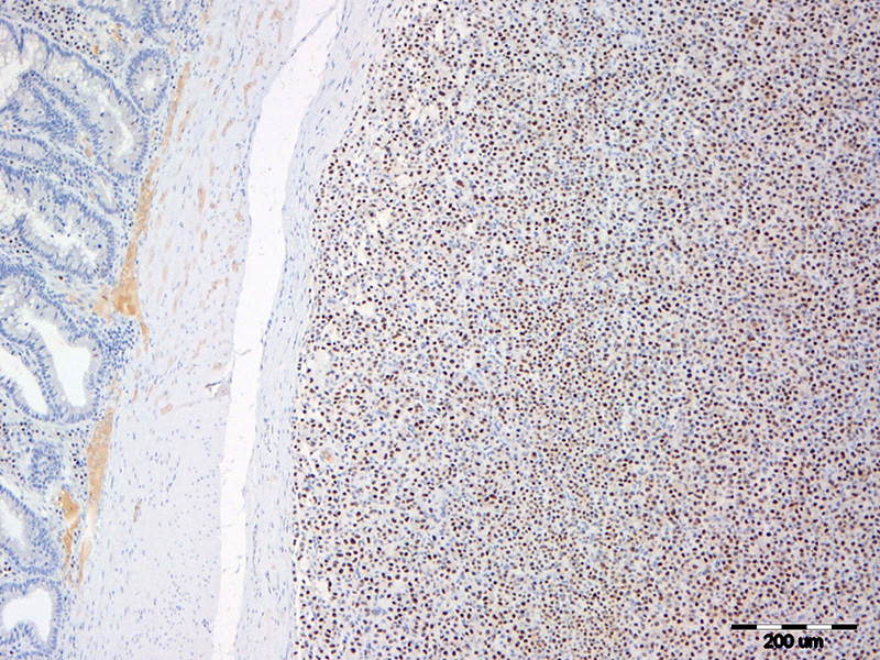
Immunohisto-chemistry antibodies
against CD 3, CD79 and MUM1 were applied. The tumor was CD79 and MUM1 positive,
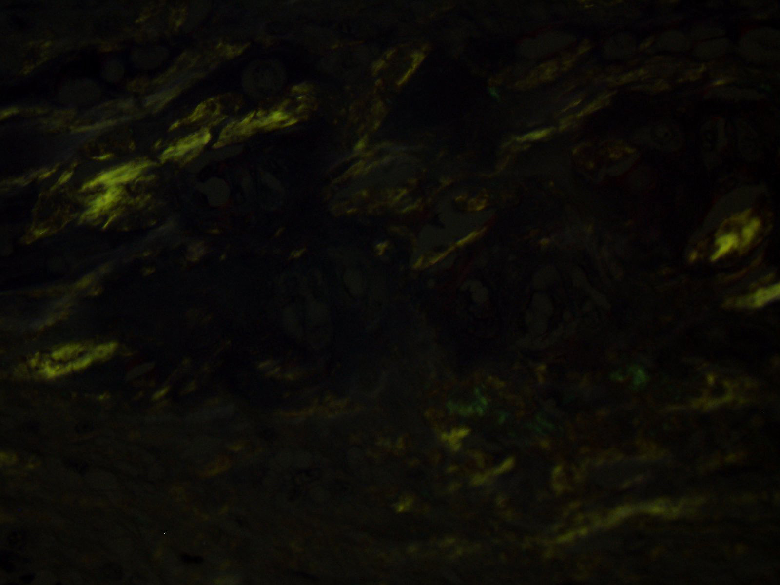
and CD3 negative. Congo red stain revealed amyloid deposits at the periphery of
the tumor.
Condition:
Extramedullary plasmacytoma
Contributor Comment:
Extramedullary
plasmacytoma (EMP) is a relatively common tumor in older dogs and occur most
frequently on the skin and mucous membranes, but has been reported in other
areas such as the brainstem, spinal cord, lymph nodes, abdominal viscera,
genitalia, and eyes.
2,9
Plasmacytomas of
the lower gastrointestinal tract are uncommon neoplasms in dogs, and rare in
cats and other species. They are encountered most frequently in the submucosa
of the distal colon and rectum of dogs, where they are associated with signs of
large bowel diarrhea and bleeding
5. Gastrointestinal EMP has also
been reported in other sites, including the esophagus, stomach and small
intestine.
10
Histologically
they resemble plasmacytomas of the skin, oral cavity or larynx. The tumor is
formed by solid packets of pleomorphic round cells with various degrees of
plasmacytoid maturation, especially at the periphery of the tumor. There is a
frequent nuclear hyperchromasia and convolution. The cells are typically
arranged in solid endocrine-like packets and there may be AL amyloid deposition
among the tumor cells. The majority of the tumor growth is submucosal. A small
proportion exhibit more aggressive behavior, including invasion of tunica
muscularis, and some spread to regional lymph nodes and spleen.
9
Canine EMP histological typing system has been established and is helpful
diagnostic tool, although the types cannot be used for a tumor grading system.
4
An unusual
finding in plasmacytomas is the presence of cartilage and bone in close
association with amyloid deposits. Meta-plastic bone and cartilage have been
described in two canine intestinal EMPs
5 and in human amyloidosis of
tongue
11 and myelomas.
1 The exact pathogenesis of
chondroid metaplasia in amyloidosis is not clearly described. Ramos Vara and
colleagues hypothesized that in amyloidosis the inducing stimulus for bone
formation is the amyloid deposits, with resultant stimulation of mesenchymal
precursor cells to
differentiate into cartilage- and bone-forming cells under the influence of
soluble factors liberated by histiocytic and other cells.
5
JPC Diagnosis:
Colon: Plasmacytoma, extramedullary, golden retriever,
Canis
familiaris.
Conference Comment:
The contributor provides a great example of a relatively common
neoplasm in the skin or mucus membrane of dogs in an uncommon location within
the abdominal viscera. Extra-medullary plasmacytomas of the lower
gastrointestinal tract have been reported in dogs to occur most frequently in
the submucosa of the distal colon and rectum, as in this case, and are
considered a benign neoplastic proliferation of monoclonal B cells.
9
Rectal and colonic plasmacytomas represent only 4% of all extramedullary
plasmacytomas in dogs.
6 Despite the uncommon location, conference
participants identified solid packets of pleomorphic plasmacytoid round cells
with a perinuclear clearing (Golgi complex) admixed with abundant homogenous
smudgy material recognized as amyloid, commonly deposited in plasma cell
tumors. The contributor confirmed the presence of amyloid via Congo-red
staining and apple-green birefringence with polarized light. The conference
moderator also pointed out the distinctive clumping pattern of the hetero-chromatin,
giving the neoplastic cells the classic clock face histomorphology of plasma
cells.
9
The most striking feature of
this case is the prominent streaks of amyloid within and surrounding the neoplasm,
often admixed with foci of chondroid metaplasia. Amyloid is a proteinaceous
substance composed of polypeptides arranged in beta-pleated sheets and is
deposited in tissues in response to a number of pathogenic processes. Primary
systemic or immunoglobulin associated amyloid (AL) is associated with light
chains derived from plasma cells in monoclonal B cell proliferations of
extramedullary plasmacytomas or multiple myelomas. The light chains are then
converted to amyloid fibrils by proteolytic enzymes in macro-phages and
deposited within tissues.
8 As mentioned by the contributor,
chondroid metaplasia has been uncommonly reported to occur with amyloidosis
secondary to a variety of conditions, although the exact pathogenesis is
currently unknown.
3,11
Another type of amyloid is
formed secondary to a variety of chronic inflammatory conditions inciting
excessive release of serum amyloid associated (AA) acute phase protein,
predominantly by the liver under the influence of interleukin-6 (IL-6) and
IL-1. AA is then deposited in multiple organs including the spleen, pancreas,
intestinal lamina propria, lymph nodes, kidneys and liver.
7,8,9
Amyloid-beta (AB) has been found in cerebral plaques of aging humans and rhesus
macaques and is associated with Alzheimers disease. Interestingly,
neurofibrillary tangles, another feature of Alzheimers disease in humans, have
not been found in aging rhesus macaques.
7
References:
1. Karasick
DV, Schweitzer ME, Miettinen M, OHara BJ. Osseous metaplasia associated
with amyloid-producing plasmacytoma of bone: A report of two cases.
Skeletal Radiol. 1996; 25:263267.
2. Jacobs JM, Messick JB, Valli VE.
Lymphoid Tumors. In: Meuten DJ, ed.
Tumors in Domestic Animals. 4
th
ed. Iowa State Press; Blackwell Publishing; 2002: 163.
3. Gross TL, Ihrke PJ, Walder EJ,
Affolter VK.
Skin Diseases of the Dog and Cat. 2nd ed. Ames, IA:
Blackwell; 2005: 870-871.
4. Platz SJ, Breuer W, Pfleghaar S,
Minkus G, Hermanns W. Prognostic value of histopathological grading in
canine extramedullary plasma-cytomas.
Vet Pathol. 1999; 36: 23-27.
5. Ramos-Vara JA, Miller MA, Pace LW,
Linke RP, Common RS, Watson GL. Intestinal multinodular AL deposition
associated with extramedullary plasmacytoma in three dogs:
Clinicopathological and immunohistochemical Studies.
J Comp Path.
1998; 119: 239-249.
6. Rannou B, Helie P, Bedard C. Rectal
plasmacytoma with intracellular hemosiderin in a dog.
Vet Pathol.
2009; 46:1181-1184.
7. Simmons HA. Age-associated
pathology in rhesus macaques (
Macaca mulatta).
Vet Pathol.
2016; 53(2):399-416.
8. Snyder PW. Diseases of immunity.
In: McGavin MD, Zachary JF, ed.
Pathologic Basis of Veterinary Disease,
6th ed. St Louis, MO: Elsevier Mosby; 2017:285.
9. Uzal FA, Plattner BL, Hostetter JM.
Alimentary system. In: Maxie MG, ed.
Jubb, Kennedy, and Palmer's
Pathology of Domestic Animals. 6th ed. Vol 2. St. Louis, MO: Elsevier;
2016:109.
10. Vail DM. Plasma Cell Neoplasms. In:
Withrow SJ, Vail DM, eds.
Small Animal Clinical Oncology. 4
th
ed. Philadelphia, PA: Elsevier Saunders; 2007: 769-784.
11. Vasudevan JA, Somanathan T, Patil
SA, Kattoor J. Primary systemic amyloidosis of tongue with chondroid
metaplasia.
J Oral Maxillofac Pathol. 2013; 17(2): 266-268.