Signalment:
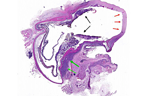
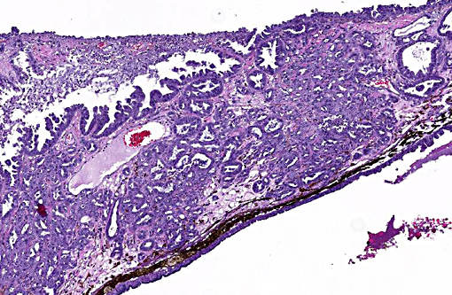
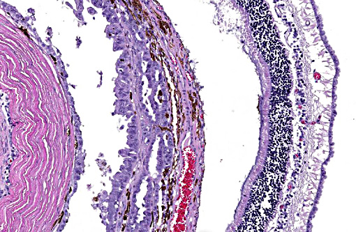
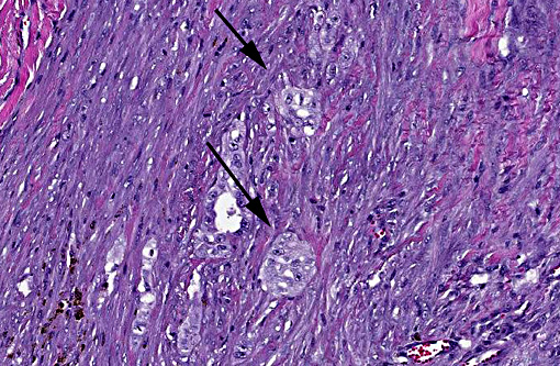
Histopathologic Description:
Morphologic Diagnosis:
Lab Results:
Condition:
Contributor Comment:
Carcinomas reported as metastasizing to the feline eye have been of pulmonary, sweat gland, mammary, uterine, and squamous cell origin.(1,2) In this case, the metastatic carcinoma in the eye was presumed to be of pulmonary origin based on the radiographic findings of a tissue opacity in the caudal lung, the most commonly affected lung field by primary lung tumors in cats.(4) CT scan and fine needle aspirate of this lung lesion with follow-up with the oncology team was recommended. The owners declined any further workup, however. The cat was euthanized three months later, at which point it was emaciated and unresponsive. No necropsy was performed.
Primary lung tumors are uncommon in cats, with older animals more commonly affected (mean age 12 years).(3,4) Lesions tend to be malignant and with a grave prognosis. When diagnosed early, with no evidence of metastatic disease, solitary lung tumors may be surgically resected. The degree of differentiation of the pulmonary neoplasm has been suggested to be a prognostic indicator, with cats with moderately differentiated primary lung tumors having reportedly a significantly longer survival time (median 698 days) than cats with poorly differentiated primary lung tumors (median 75 days).(5) However, early diagnosis is difficult since most cats initially present with nonspecific clinical signs or signs related to metastases.3 Lameness, associated with metastasis to the bone and skin of the digits,(3) or visual deficits (associated with intraocular metastasis),(1) may be the only initial presenting sign. Intraocular metastasis may be underestimated since ophthalmoscopic and microscopic examinations are not performed on a routine basis in many cases. Other sites of metastasis of primary lung tumors in cats include skeletal muscle, and multiple thoracic and abdominal organs.(4) Thorough clinical examination and thoracic radiography can provide a high index of suspicion of the primary neoplasm.(4) Cytologic evaluation of cells obtained by fine-needle aspiration of the lung mass, as well as of cells collected from thoracic fluid or bronchoalveolar lavage can help determine the diagnosis.(3,4) Effective treatment has yet to be demonstrated for metastatic lung carcinomas in cats. As a result, most cats with metastatic lung carcinoma die or are euthanized within 6 weeks of diagnosis.(5)
JPC Diagnosis:
Conference Comment:
Primary uveal neoplasms are common in dogs and cats and considerably more common than metastatic tumors, with the exception being metastatic uveal lymphoma in cats. Among primary ocular tumors, melanoma, lymphoma, posttraumatic sarcoma and iridociliary adenocarcinoma are most common in cats. Iridociliary adenocarcinoma is a reasonable differential in this case, but these typically benign neoplasms tend to infiltrate and expand the posterior chamber, often in solid sheets, in contrast to this case which filed along the uvea and choroid forming tubules.(6) Additionally, a PAS-positive basement membrane is highly conserved in these neoplasms and can aid in distinguishing from metastatic disease.(7) Extraocular metastasis from these primary tumors is rare or nonexistent; however, secondary complications are often significant, with glaucoma or intractable hyphema occurring most commonly.(6)
Of greater significance in cats are posttraumatic sarcomas, which are usually high-grade neoplasms of lens epithelial origin following lens rupture.(6) The majority of these are of the spindle cell variant and are often not recognized for years following a traumatic event. They are highly invasive within the globe though rarely metastasize. A round cell variant resembling lymphoma and osteo- or chondrosarcoma also occur following lens trauma, albeit at a much lower frequency.(2)
References:
1. Cassotis NJ, Dubielzig RR, Gilger BC, Davidson MG. Angioinvasive pulmonary carcinoma with posterior segment metastasis in four cats. Vet Ophthalmol. 1999;2(2):125131.
2. Dubielzig RR, Ketring KL, McLellan GJ, Albert DM. Metastatic neoplasia. In: Veterinary ocular pathology: A comparative review. New York: Saunders Elsevier. 2010; 97-103,309-315.
3. Goldfinch N, Argyle DJ. Feline lung-digit syndrome: unusual metastatic patterns of primary lung tumours in cats. J Feline Med Surg. 2012;14(3):202-8.
4. Hahn KA, McEntee MF. Primary lung tumors in cats: 86 cases (1979-1994). J Am Vet Med Assoc. 1997;211(10):1257-60.
5. Hahn KA, McEntee MF. Prognosis factors for survival in cats after removal of a primary lung tumor: 21 cases (1979-1994). Vet Surg. 1998;27(4):307-11.
6. Njaa BL, Wilcock BP. The ear and eye. In: Zachary JF, McGavin MD, eds. Pathologic Basis of Veterinary Disease. 5th ed. St. Louis, MO: Elsevier Saunders; 2012:1228-1230.
7. Wilcock B, Dubielzig RR, Render JA. Histological classification of ocular and otic tumors of domestic animals. Second series. Vol IX. Washington, D.C.: Armed Forces Institute of Pathology; 2002:26.