Signalment:
1.5-year-old female New Zealand white rabbit,
Oryctolagus cuniculus.In August of 2007, a pet rabbit was presented to an Ontario veterinary clinic with a 3-day history of lethargy, anorexia and facial swelling. The affected animal was one of a group of six housed in a large hutch on a grassy enclosure surrounded by a chain link fence. Chipmunks, other small mammals and occasionally birds were seen within the enclosure on a number of occasions and direct contact with other forms of wildlife could occur across the fence; two dogs also resided on the property.
During the examination, the rabbit was noted to be tachypneic. A 1-cm crust was noted on the nasal planum and skin over the right nares and upper lip was swollen. Excessive waxy debris was present in the left ear canal.
A presumptive diagnosis of
Pasteurella multocida-induced pneumonia was made and oral antibiotic therapy (chloramphenicol palmitate) was prescribed and initiated. Mineral oil was infused in the left ear and when the luminal debris was removed, mild bleeding and ulceration of the epithelium was noted. Antibiotic ointment was instilled into the ear canal and the animal was discharged. Approximately 30 minutes after leaving the clinic, the owner reported that the animal began choking and bleeding from the nose. Shortly afterwards, the rabbit died and the body was submitted for postmortem examination.
Gross Description:
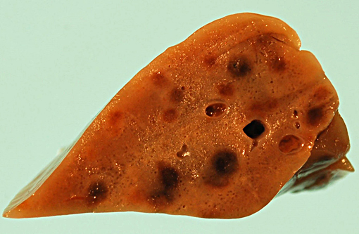
The rabbit was in good body condition with abundant internal fat stores. Blood stained the inside of the left pinna and a blood clot was present at the base of the ear canal. A 1 cm round raised reddened area was noted on the nasal planum. Blood stained the perinasal and dewlap fur. The tracheal mucosa was mildly congested and there was generalized purple red mottling of the lungs with numerous, up to 3 mm, foci of hemorrhage on the pleural surface and in the parenchyma. Within the abdomen, the spleen was enlarged and congested. There was transmural reddening of the caudal 2 cm of the ileum and the lumen contained a blood clot.Â
Histopathologic Description:
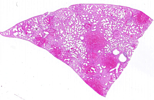
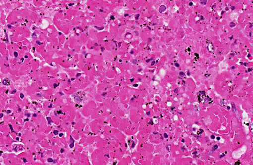
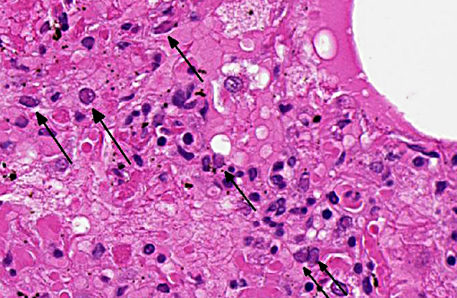
There is marked generalized acute necrotizing and hemorrhagic bronchopneumonia with segmental bronchial epithelial proliferation and necrosis, large areas of alveolar necrosis and hemorrhage and flooding of alveoli by edema fluid, fibrin, heterophils and large macrophages. Numerous bronchiolar epithelial cells, bronchiolar epithelial syncytia, pneumocytes, endothelial cells and macrophages contain prominent glassy eosinophilic intranuclear viral inclusions. Several vessels have perivascular hemorrhage, edema, mural fibrinous necrosis, vasculitis and thrombosis. There is patchy alveolar overinflation and emphysema.
Morphologic Diagnosis:
Marked acute generalized necrotizing and hemorrhagic bronchopneumonia with syncytia containing numerous eosinophilic intranuclear viral inclusions.
Lab Results:
Low numbers of
E. coli were isolated from the lung. An alphaherpesvirus isolated from the lung and skin by cell culture was further identified by electron microscopy and ribonucleotide reductase gene sequencing as leporid alphaherpesvirus-4 (LHV-4).1
Condition:
Leporid herpesvirus-4
Contributor Comment:
In the early 1990s, two herpes-virus like outbreaks with high mortality characterized by ulcerative dermatitis, pneumonia, splenic necrosis, and gastrointestinal hemorrhage were reported from commercial rabbitries in Alberta(4,6) and British Columbia.(4) In both cases, herpes-like viral particles were identified in formalin-fixed, paraffin-embedded tissue sections and herpesvirus was isolated from affected tissues, but neither isolate was further characterized. However, the disease was experimentally reproduced in meat-type rabbits using one of the isolates. In the summer of 2006, a commercial pet and agricultural rabbitry in Alaska also reported high morbidity and mortality associated with systemic herpesvirus infection.(3) Rabbits were housed outside in open-sided hutches, where mosquito and biting fly activity was high. Snowshoe hares were present in the surrounding area and feral domestic rabbits had been in close proximity to the hutches earlier in the spring. In the following spring and summer, several rabbits from this same rabbitry developed conjunctivitis and skin lesions; and one breeding rabbit that had recovered from clinical infection in the previous year experienced perinatal mortality. The herpesvirus was isolated and characterized as leporid herpesvirus-4.(2)
The case presented here is the first documented and characterized case of leporid herpesvirus-4 infection in a pet Canadian rabbit.(1) This viral disease should be included in the list of differential diagnoses for acute morbidity and mortality in domestic rabbits presenting with epistaxis or respiratory distress, which presently includes rabbit hemorrhagic disease virus (RHDV), per acute
Pasteurella multocida septicemia or chronic
Pasteurella multocida pleural/pulmonary abscessation with rupture.
JPC Diagnosis:
Lung: Pneumonia, bronchointerstitial, necrohemorrhagic, multifocal to coalescing, acute, severe, with intranuclear viral inclusions and syncytia.Â
Conference Comment:
There are four known herpesviruses of rabbits, two of which are gammaherpesviruses that produce lymphoproliferative disease and neoplasia (
Leporid herpesvirus 1 and
Leporid herpesvirus 3).Â
Leporid herpesvirus 2 is also a gammaherpesvirus, but is capable of inducing encephalitis. Additionally, natural infections of
Human herpesvirus 1 (herpes simplex) have been reported in rabbits causing a fatal encephalitis.(7) The virus demonstrated in this case,
Leporid herpesvirus 4 (LHV4), is a novel herpesvirus with rare reports in the literature as the contributor highlighted.Â
LHV-4 is classified as an alphaherpesvirus on the basis of its rapid growth and cytopathic effect in cell culture.(5) This case illustrates the severity of bronchopneumonia which results from infection and leads to the reported 50% morbidity and 29% mortality rates.(6) Additionally, ulcerative rhinitis and splenic necrosis has been observed in experimental infections, and hemorrhagic dermatitis and myocarditis in natural infections.(5)
References:
1. Brash ML, Nagy E, Pei Y, Carman S, Emery S, Smith AE, Turner PV. Acute hemorrhagic and necrotizing pneumonia, splenitis, and dermatitis in a pet rabbit caused by a novel herpesvirus (leporid herpesvirus-4). Can Vet J. 2010;51:1383-1386.
2. Jin L, Lohr CV, Vanarsdall AL, Baker RJ, Moerdyk-Schauwecker M, Levine C, et al. Characterization of a novel alphaherpesvirus associated with fatal infections of domestic rabbits. Virol. 2008;378:13-20.Â
3. Jin L, Valentine BA, Baker RJ, Lohr CV, Gerlach RF, Bildfell RJ, Moerdyk-Schauwecker M. An outbreak of fatal Herpesvirus infection in domestic rabbits in Alaska. Vet Pathol. 2008;45:369-374.
4. Onderka DK, Papp-Vid G, Perry AW. Fatal herpesvirus infection in commercial rabbits. Can Vet J. 1992;33:539-543.
5. Sunohara-Neilson JR, Brash M, Carman S, Nagy E, Turner PV. Experimental infection of New Zealand White rabbits (Oryctolagus cuniculi) with Leporid herpesvirus 4. Comp Med. 2013;63(5):422-431.Â
6. Swan C, Perry A, Papp-Vid G. Herpesvirus-like viral infection in a rabbit. Can Vet J. 1991;32:627-628.
7. Weissenbock H, Hainfellner JA, Berger J, Kasper I, Budka H. Naturally occurring herpes simplex encephalitis in a domestic rabbit (Oryctolagus cuniculus). Vet Pathol.1997;34(1):44-47.