CASE 1: Case 2 N-169/20 (4153943-00)
Signalment:
1-day old foal, male, Pure Spanish Breed, Equine (Equus ferus caballus).
History:
The physical examination reveals hyperthermia and weakness. Supporting treatment consisting of IV fluid therapy, antibiotic (cefquinoma) and NSAID (flunixin). The animal died 12 hours later.
Gross Pathology:
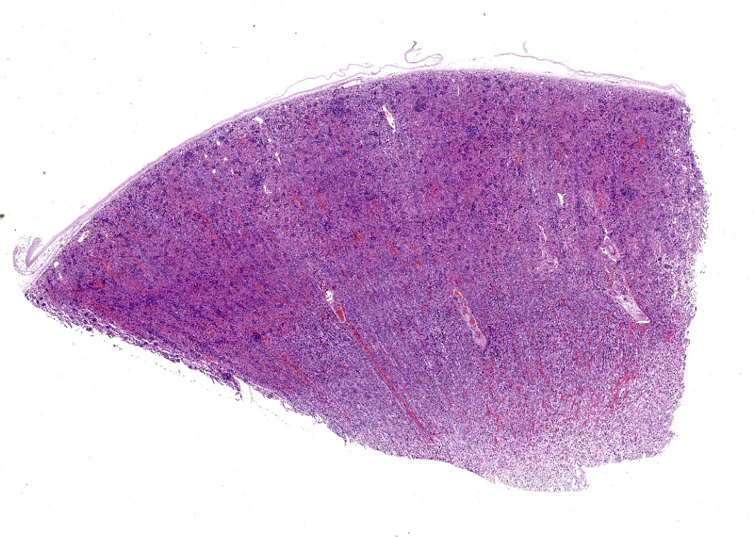
The kidneys were swollen, diffusely congested and up to 10% of the renal cortex had multiple, 1-2 mm diameter, well demarcated, round, white foci (embolic nephritis).
Laboratory results:
None submitted
Microscopic description:
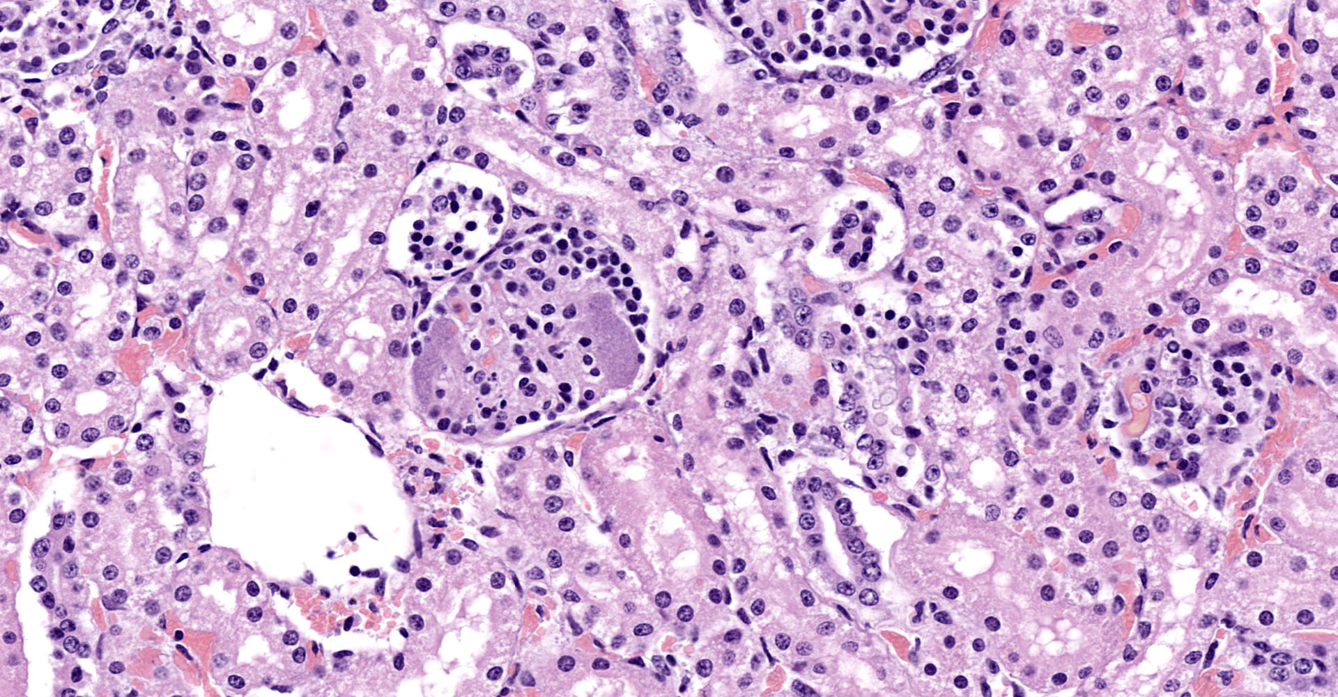
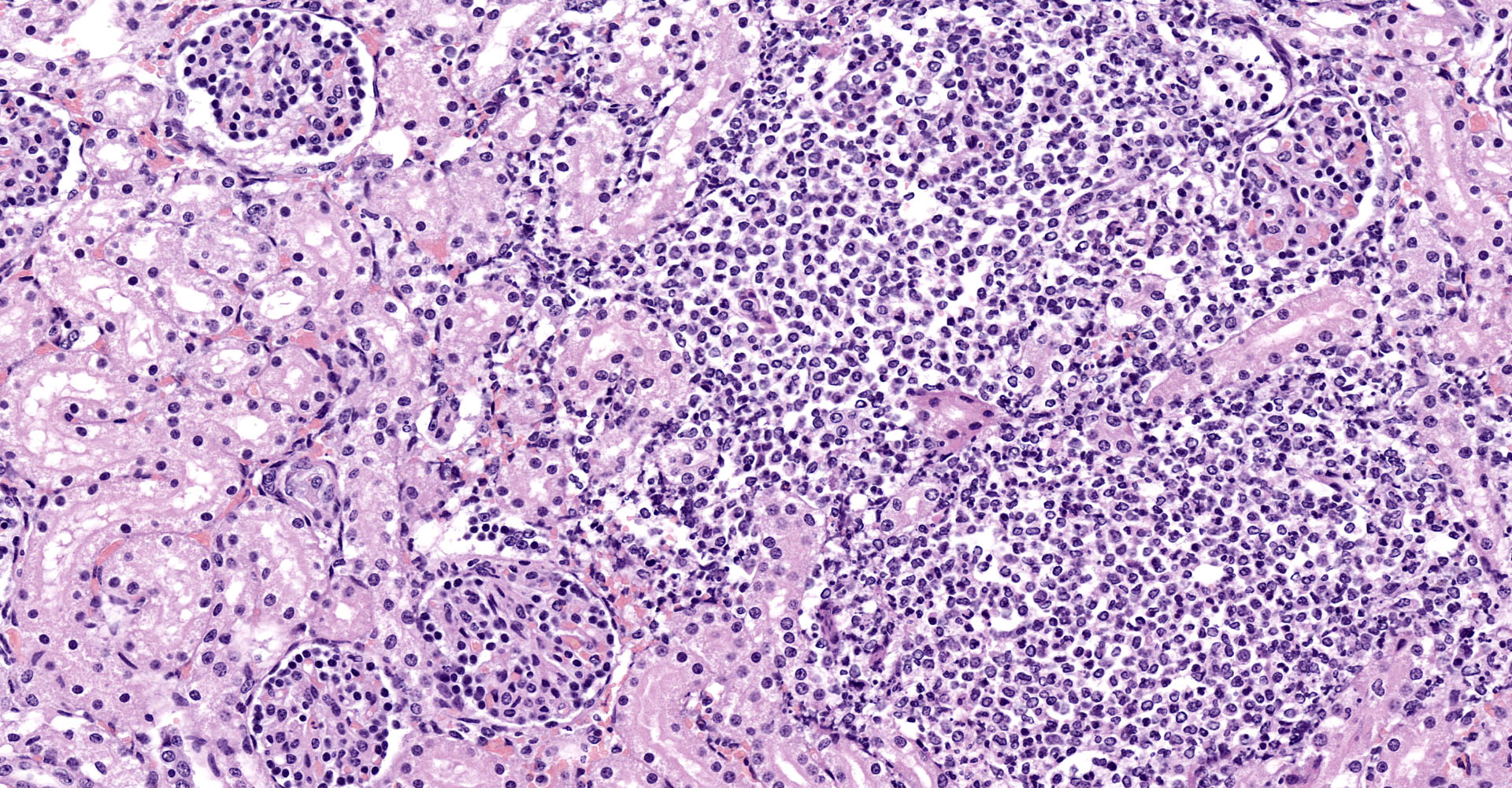
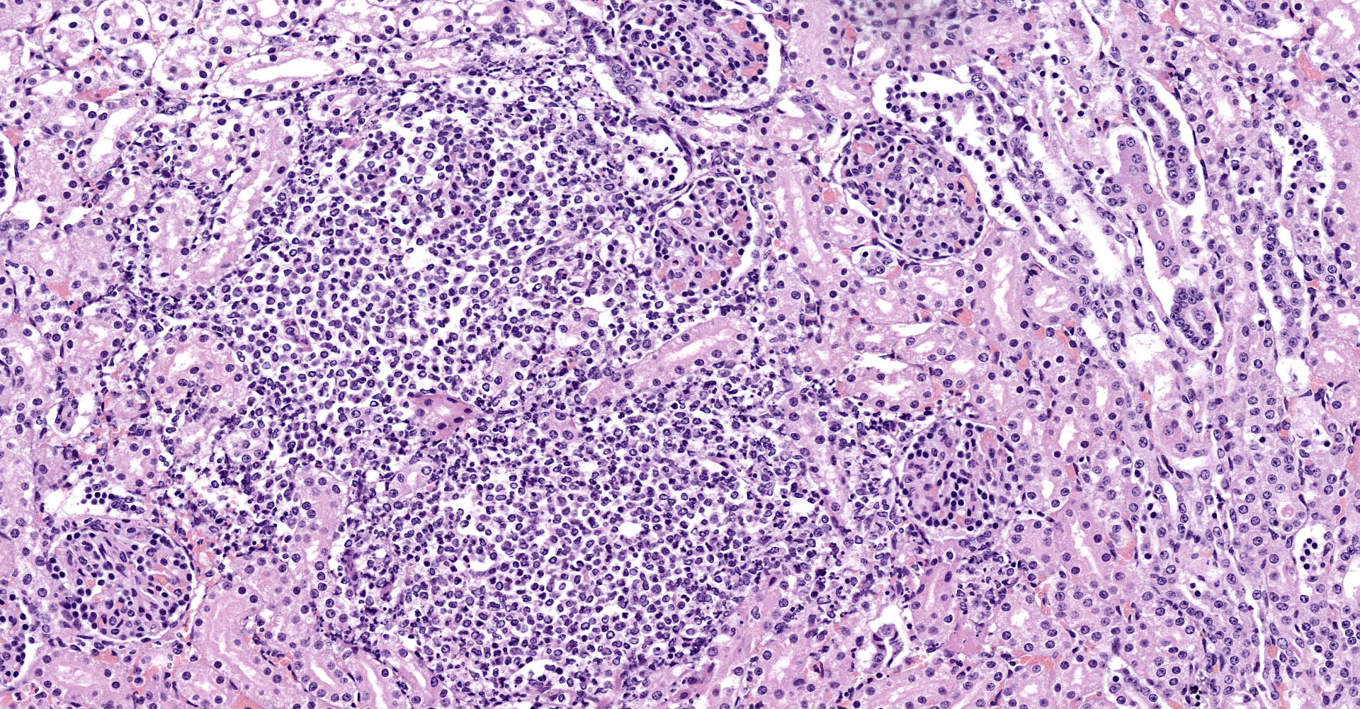
Kidney: Multifocally, up to 20% of the renal cortex is affected by an inflammatory and necrotizing process. Diffusely, expanding and effacing the glomeruli and peripheral tissue, there are abundant viable and degenerated neutrophils, moderate number of macrophages, few lymphocytes and plasma cells, admixed with abundant eosinophilic cellular debris with pyknosis and karyolysis with loss of cellular outlines (lytic necrosis), moderate amount of fibrin, hemorrhage, edema and small to medium extracellular basophilic colonies of 1-2µm coccobacilli (microabscesses). Adjacent tubular epithelium variably presents one of the following changes: shrunken hypereosinophilic cytoplasm with pyknosis (coagulative necrosis) or swollen pale eosinophilic vacuolated cytoplasm (tubular degeneration). Diffusely, the interstitium shows moderate congestion. Remaining tubules present minimal tubular degeneration with few intraluminal, pale round eosinophilic material (protein cast).
Contributor's morphologic diagnosis:
1. Kidney: Multifocal suppurative glomerulonephritis diffuse, global, subacute, moderate with intralesional small colonies of coccobacilli.
2. Kidney: Tubular degeneration and necrosis, acute, diffuse, mild.
Contributor's comment:
Actinobacillus equuli, a Gram-negative pleomorphic rod, is the causative agent of several diseases in horses. The most serious is an acute, usually fatal septicemia of newborn foals known as: "sleepy foal disease",4 which causes lesions at several organs, including the kidneys, joints, lungs and intestine. The most likely source of infection for the neonate is the mare, where the organism is present in the normal oral flora.4
A. equuli affects more the newborn foals, in adult horses are less common and generally more localized, causing acute and chronic peritonitis.11
Rarely, A. equuli has been reported to be an opportunistic pathogen of pigs, with these infections typically associated with abortion, septicemia, and polyarthritis.11
Fecal contamination or extension from oral mucous membranes is the method of inoculation, is acquired in utero during parturition, or shortly after birth as an umbilical infection, the last one is the most common route of infection in foals, resulting in septicemias.1,5
Two subspecies of Actinobacillus equuli, subsp equuli and haemolyticus are normal inhabits of mucous membranes of the alimentary tract. Subspecies are based on the repeats-in-toxin (RTX), toxin genotype (Aqx-equuli toxin).1,7 The higher isolation rates of A. equuli subsp equuli over A. equuli subsp haemolyticus from septicemic cases indicates that other virulence factors play a major role in pathogenesis. Toxins belonging to RTX family are produced by some species of Actinobacillus, and they exert hemolytic, cytotoxic activity and host specificity.7,8,12
The foal is born without anti-Aqx antibodies but acquires them by passive transfer from the mare's colostrum within an hour of ingesting it. The complete failure of passive transfer caused by the delay in the colostrum supply, or by a poor-quality colostrum predisposes foals to infection and sepsis.2,4
A. equuli is of clinical importance, since it can cause disease in horses and is found in infected wounds of humans bitten by horses. Hemolytic strains of A equuli are also isolated from tracheal washes, indicating a preference for the respiratory tract. These strains are opportunistic pathogens and can cause respiratory infections, septicemia, metritis, mastitis, arthritis, endocarditis, meningitis and peritonitis. Non-hemolytic strains is well known to cause the clinical condition: 'sleepy foal disease.'4
A. equuli is a well-known cause of embolic nephritis in young horses. At gross examination small abscesses occur in a variety of organs, including the liver, adrenal gland, joints, and the kidney.1 Microscopically, glomerular capillaries and to a lesser extent interlobular arterioles contain microabscesses. If the animal survives the infiltrate can persist as focal residual abscesses or coalescing scar.1
Embolic nephritis also occurs commonly in the bacteremia of pigs infected with Erysipelothrix rhusiopathiae, sheep and goats infected with Corynebacterium pseudotuberculosis, Trueperella pyogenes is the most common isolate in cattle, but Staphylococcus aureus, Mannheimia haemolytica and Streptococcus bovis were also present.1
Contributing Institution:
Universidad de Zaragoza
Departamento de Patología Animal
https://patologiaanimal.unizar.es
JPC diagnosis:
Kidney: Nephritis, suppurative, embolic, with mild fibrinosuppurative glomerulitis and rare large colonies of bacilli.
JPC comment:
The contributor provides a concise summary of this disease. In addition to the described diseases in horses, Actinobacillus equuli ssp. equuli is one of the most commonly isolated agents causing osteomyelitis in foals, while Actinobacillus equuli ssp. haemolytica is a common cause of bronchopneumonia and pleuritis in horses.6
A number of species within the Actinobacillus genus are closely related, and A. equuli is difficult to differentiate from A. suis using even 16S rRNA sequencing. Recent whole genome sequencing of the Actinobacillus genus phylogenetic tree shows that a number of species capable of causing invasive disease cluster closely by surface antigen type, and include A. suis, A. ureae, A. equuli equuli, and A. capsulatus.3
As stated previously, A. equuli also occasionally affects pigs and piglets. Recently, an outbreak of A. equuli caused swollen joints and moderate to severe lameness in 6-8-hour old piglets. They experienced lethargy and a subset became non-ambulatory. The primary findings in the affected piglets included purulent polyarthritis, tendovaginitis, and purulent inflammation in the brain and kidneys of one animal.8
During the conference, the moderator emphasized how glomerular morphology in this case allows for some age determination of the animal. The glomerular visceral epithelial cells are plump podocyte precursor cells, consistent with fetal glomerular morphology of a young animal. It was also emphasized that this agent, as well as other possible bacterial agents, will often be found in the peritubular capillaries.
The moderator reminded all participants that the vessels in the kidney are arteries, with the term "arteriole" being reserved for the afferent and efferent vessels immediately arriving and departing the glomerulus, respectively. Arterioles lack the internal elastic lamina, allowing for direct and immediate control of lumen diameter and blood pressure.
References:
1. A, Breshears Melanie CAW. The Urinary System. In: Zachary F J, ed. Pathologic Basis of Veterinary Disease. Elsevier Inc; 2017:617?680.
2. Berthoud H, Frey J, Sternberg S, Straub R, Kuhnert P. Antibodies to Aqx toxin of Actinobacillus equuli in horses and foals. Vet Rec. 2004 Aug 21 [cited 2020 Jun 22];155:231?233.
3. Bujold AR, Shure AE, Liu R, Kropinski AM, MacInnes JI. Investigation of putative invasion determinants of Actinobacillus species using comparative genomics. Genomics. 2019;111(1):59-66.
4. Castagnetti C, Rossi M, Parmeggiani F, Zanoni RG, Pirrone A, Mariella J. Facial cellulitis due to Actinobacillus equuli infection in a neonatal foal. Vet Rec. 2008 Mar 15 [cited 2020 Jun 19];162:347?349.
5. Cianciolo E, Rachel. Mohr CF. Urinary System. In: Grant Maxie M, ed. Pathology of Domestic Animals. Elsevier Inc.; 2016:377?463.
6. Fenwick BW, Woolums AR. Pasteurellaceae: Actinobacillus. In: McVey DS, Kennedy M, Chengappa MM, eds. Veterinary Microbiology, 3rd Ed. Ames, IA:Wiley-Blackwell. 2013;108-114.
7. Kuhnert P, Berthoud H, Straub R, Frey J. Host cell specific activity of RTX toxins from haemolytic Actinobacillus equuli and Actinobacillus suis. Vet Microbiol. 2003 Mar 20;92:161?167.
8. Löhr C V., Polster U, Kuhnert P, Karger A, Rurangirwa FR, Teifke JP. Mesenteric Lymphangitis and Sepsis Due to RTX Toxin-Producing Actinobacillus spp in 2 Foals With Hypothyroidism-Dysmaturity Syndrome. Vet Pathol. 2012 Jul;49:592?601.
9. Maul C, Suchowski M, Klose K, Antov V, Pfeffer M, Schwarz BA. Detection of Actinobacillus equuli ssp. equuli in piglets with purulent polyarthritis and tendovaginitis. Tierarztl Prax. 2020;48(1):51-58.
10. Patterson-Kane JC, Donahue JM, Harrison LR. Septicemia and Peritonitis Due to Actinobacillus equuli Infection in an Adult Horse. Vol. 38, Vet Pathol. 2001.
11. Thompson AB, Postey RC, Snider T, Pasma T. Actinobacillus equuli as a primary pathogen in breeding sows and piglets. Can Vet J = La Rev Vet Can. 2010 Nov [cited 2020 Jun 22];51:1223?1225.
12. Uchida-Fujii E, Niwa H, Kinoshita Y, Nukada T. Actinobacillus species isolated from Japanese Thoroughbred racehorses in the last two decades. J Vet Med Sci. 2019 Sep 3 [cited 2020 Jun 19];81:1234?1237.