Joint Pathology Center
Veterinary Pathology Services
Wednesday Slide Conference
2019-2020
Conference 18
12 February, 2020
Timothy Cooper, DVM, DACVP
Pathology Department
NIH/NIAID
Integrated Research Facility
Frederick, MD
CASE II: 17/1060 Z (JPC 4032584).
Signalment: 4 year old, female spayed, Bull Arab (canine)
History: Sudden death. The week before death the animal was showing signs of hind limb/spinal pain. Radiographs at that time revealed a sublumbar mass and fluid in the caudal abdomen, fluid PCV/TP 15/70.
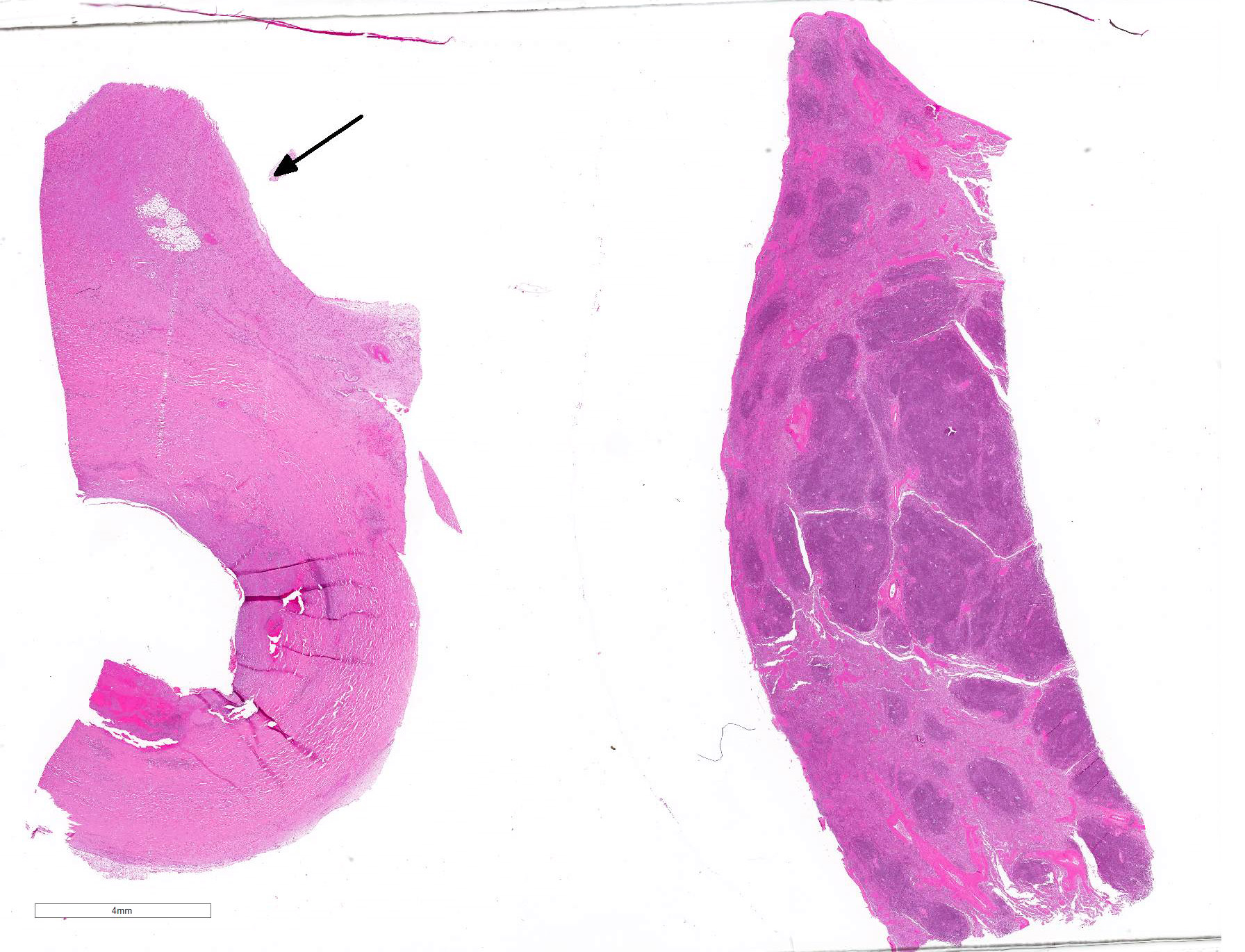
Gross Pathology: Abdominal cavity: The abdomen contains approximately 1300ml of dark red, slightly turbid fluid (blood) admixed with tens of variably sized, friable, soft dark red material (blood clots). The dorsocaudal abdomen, ventral to lumbar vertebrae 6 and 7 and the iliopsoas muscles, contains a poorly demarcated, approximately 11 x 5 x 4 cm, firm mass which surrounds and replaces approximately 7cm of the aorta. On cut section, the mass is mottled dark pink to dark red and cavitated. The caudal aspect of the mass is friable with roughened edges and contains approximately 10ml of clotted blood (site of rupture).
Spleen: The tail of the spleen contains a focal, poorly demarcated, approximately 1cm in diameter, raised, irregular, soft mass which is mottled dark red to tan on cut surface.
Kidney: The medulla of the right kidney contains a focal, 0.9 x 0.5 x 0.4cm depressed area, which contains less than 1ml of turbid, mucoid, viscous, yellow fluid (granuloma).
Gross morphological diagnoses:
Abdominal cavity: Aortic arteritis, aneurysm with rupture and haemoabdomen
Spleen: Granulomatous splenitis, focal, moderate, chronic
Kidney: Granulomatous nephritis, focal, moderate, chronic
Laboratory results:
Fungal culture:
Spleen: Very small fungal colonies isolated following 48 hours incubation. Growth identified as Aspergillus terreus
Microscopic Description:
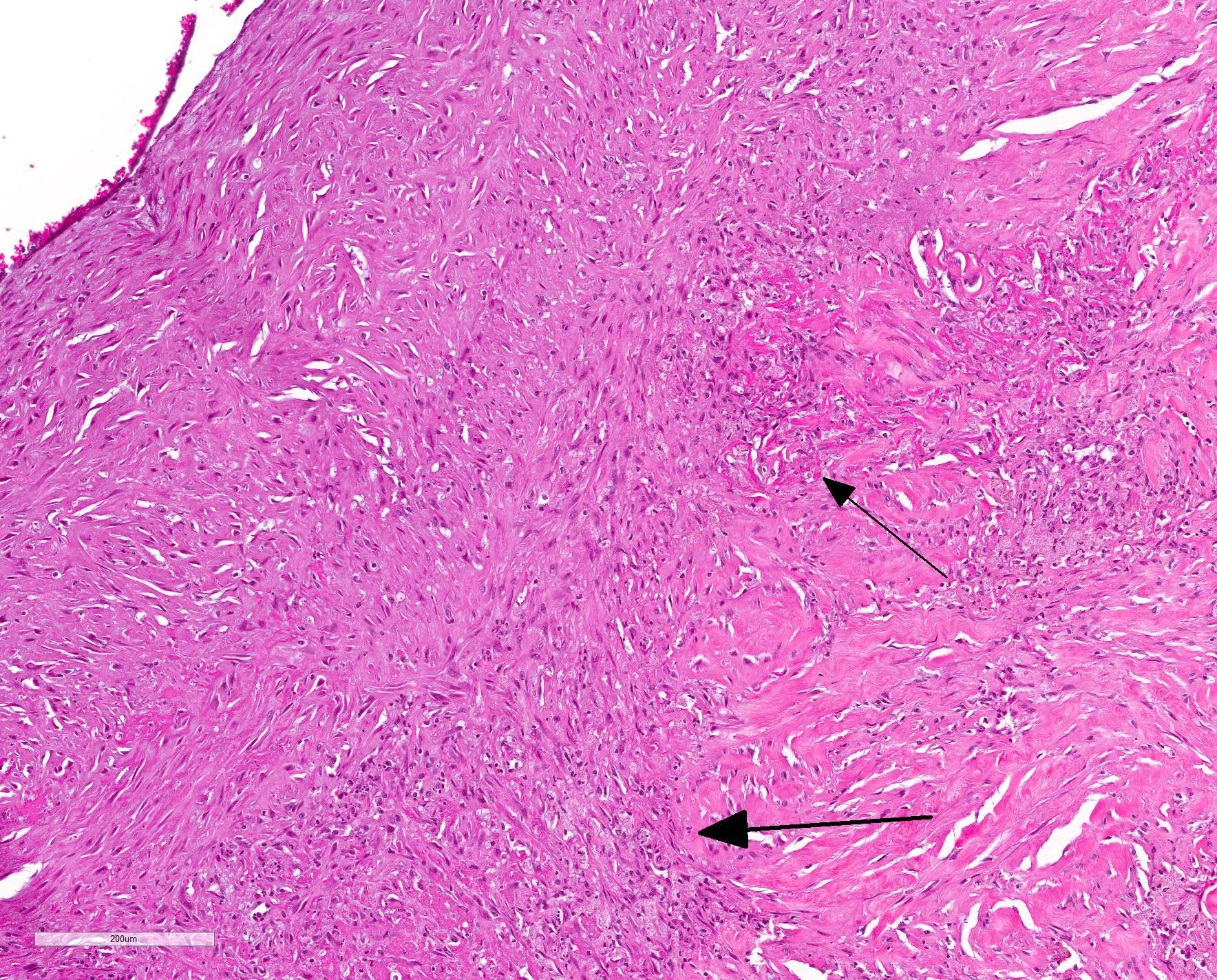
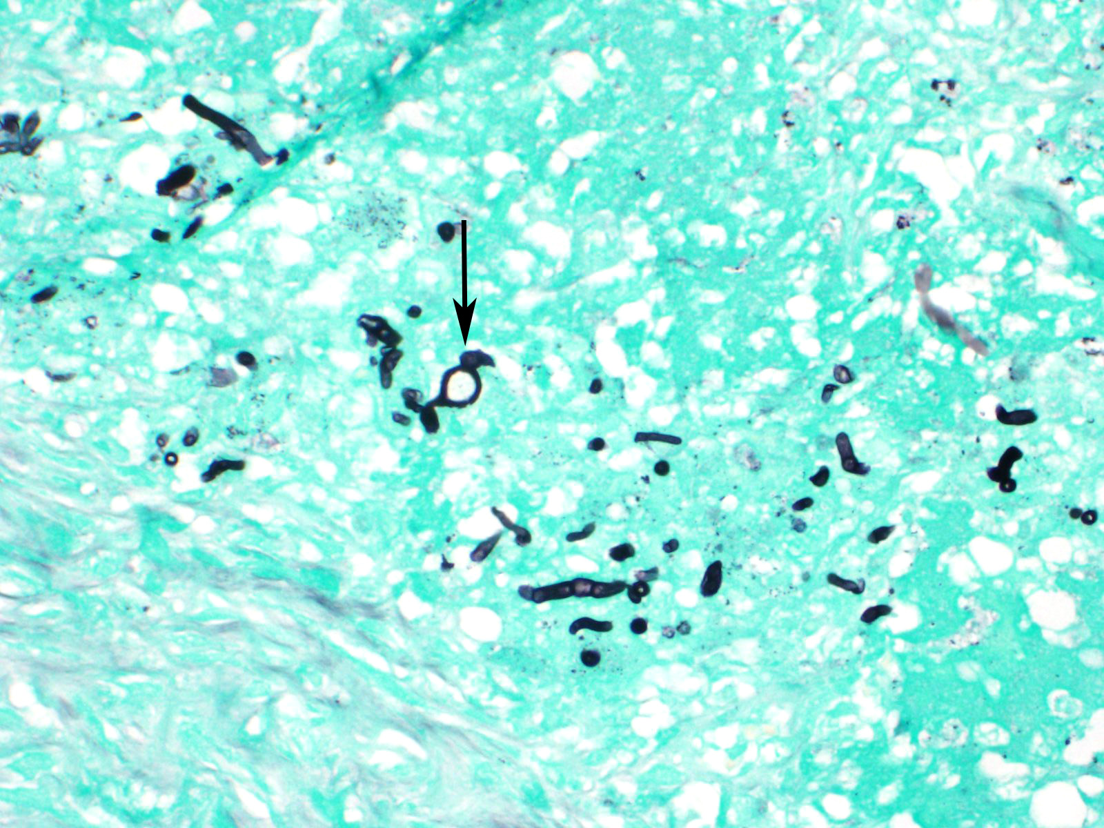
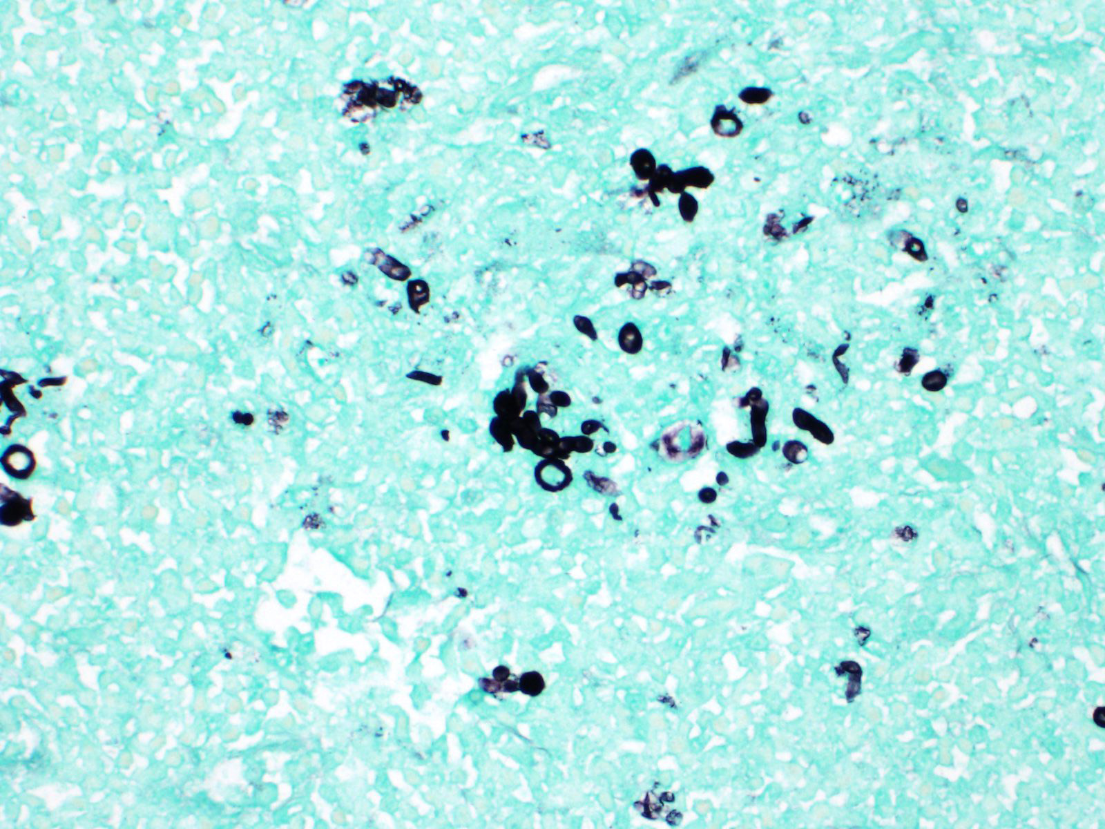
Aortic mass: Generally, approximately 20% of the aorta is expanded up to three-fold with inflammation, necrosis and haemorrhage. The tunica intima, and to a lesser extent, the tunica media contain multifocal to coalescing areas of dense aggregates of degenerate neutrophils and macrophages admixed with large amounts of eosinophilic fibrillary material (fibrin). Macrophages are often epithelioid or forming multinucleated giant cells which engulf or surround hundreds of 3-5 micron thick, 5-100 micron long, branching fungal hyphae which are septate with conidial heads and are GMS positive (Figs. 1 and 2, morphology consistent with Aspergillus terreus). The tunica media contains large, up to 2000 micron in diameter lakes of extravasated erythrocytes (haemorrhage) admixed with degenerate neutrophils, hyperesinophilic material, pyknotic and karyorrhectic debris (necrosis). No bacteria are observed on gram stain.
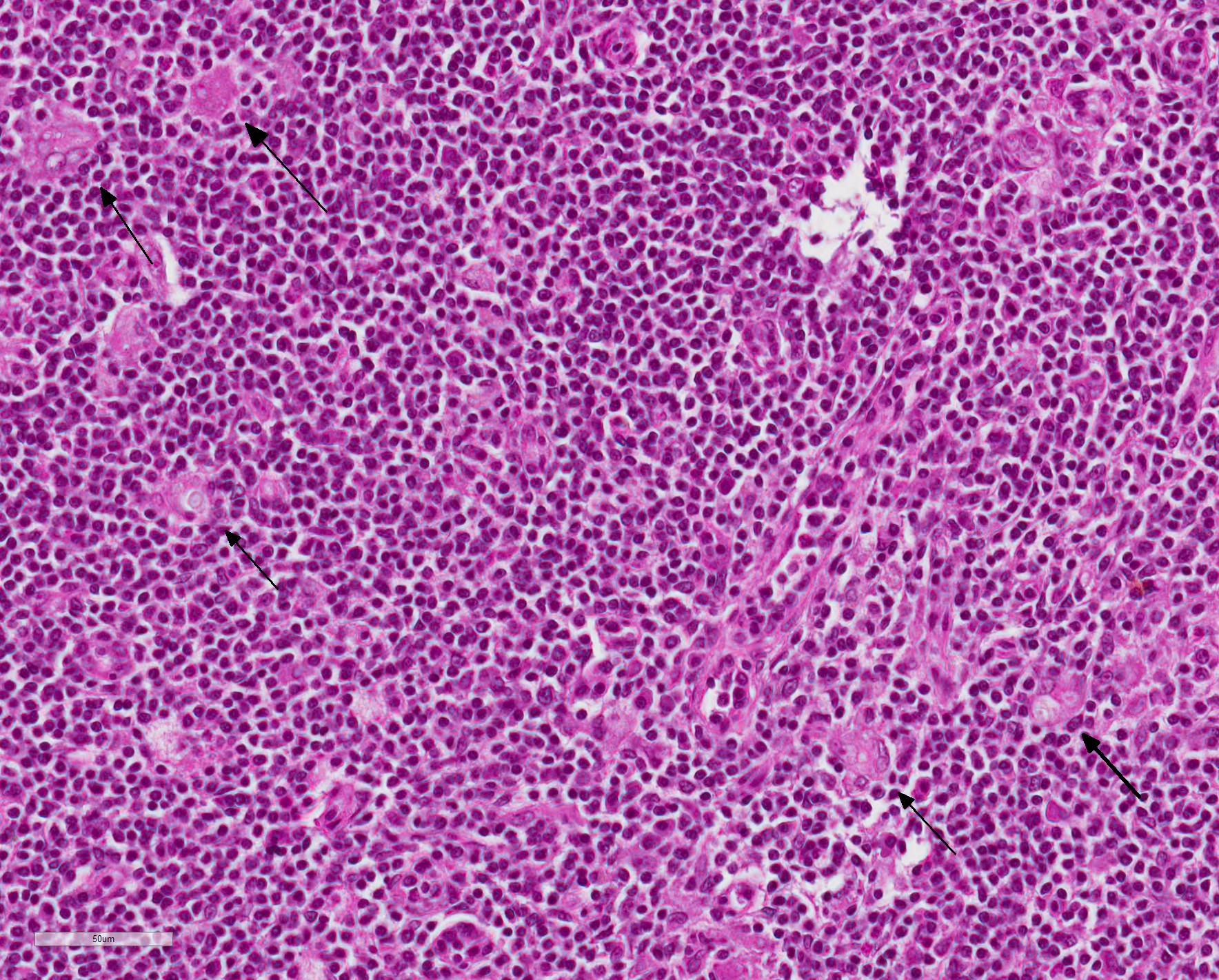
Spleen: In approximately 60% of the tissue section, the white and red pulp are completely effaced by dense infiltrates of lymphocytes and plasma cells which surround hundreds of multifocal aggregates of macrophages, multinucleated giant cells and lesser numbers of neutrophils. Macrophages and giant cells often contain or are associated with moderate numbers of fungal organisms as described for the aortic mass (Fig. 3). The remaining parenchyma contains a reduction in lymphocyte density (depletion) with increased numbers of randomly scattered neutrophils admixed with necrotic debris.
Contributor Morphologic Diagnosis:
Aortic mass: Necrotising granulomatous arteritis, severe, generalised, chronic with fungal hyphae (morphology consistent with Aspergillus terreus).
Spleen: Granulomatous splenitis, focal, severe, chronic with intralesional fungal hyphae
Contributor Comment: Aortic aneurysm is a localised dilation of the vessel which may occur secondary to loss of vessel wall integrity. Aneurysms are rare in domestic animals, and may occur secondary to atherosclerosis, medial degeneration, trauma, infection or arterial dissection.5 In this animal, large amounts of necrosis and inflammation which contain fungal organisms are consistent with a mycotic (fungal) aneurysm. In this case, this infection has caused a weakening of the vessel wall and subsequent rupture, leading to sudden death from acute and massive haemorrhage into the abdomen.
Aspergilli are ubiquitous and can be isolated from soil, air and decomposing organic matter. Infection is acquired from environmental sources, generally by inhalation or ingestion. It is an opportunistic pathogen depending on impaired, overwhelmed or bypassed host defences. Infection may cause local disease, or disseminate to other parts of the body. Evidence of infection and inflammation within the spleen and kidney in this animal is consistent with disseminated disease, however the original route of infection is undetermined.
Fungal aortic aneurysms have been reported twice previously in dogs.1,3 In one case, Graphium species was isolated from the aneurysm, and Candida infection was suspected in the second. In humans, Aspergillus species are the most common mycotic infections associated with aneurysms.
A variety of risk factors may be associated with the development of these infections.1 Reported risk factors in humans include broad-spectrum antibiotic use, mechanical ventilation, parenteral nutrition, renal failure, prior surgery, neutropenia, chemotherapy, severe illness, age, and indwelling catheters (especially central venous catheters). Although less commonly reported, veterinary patients appear to have similar risk factors. In one of the case studies cited, however, no predisposing factors were able to be identified.
Contributing Institution:
School of Veterinary Science
University of Queensland Gatton Campus
Gatton, Qld, 4343 Australia
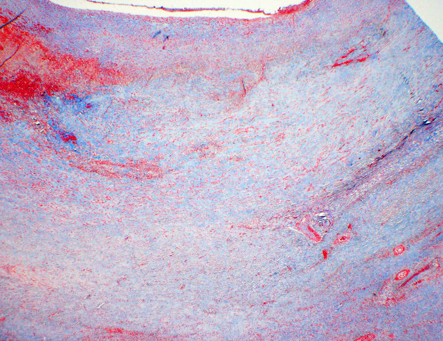
JPC Diagnosis: 1. Aorta: Elastolysis and fibrosis, transmural, diffuse, severe, with vascularization and dissection.
2. Aorta: Aortitis, pyogranulomatous, focally extensive, severe, with transmural fibrosis and numerous fungal hyphae.
3. Spleen: Splenitis, granulomatous, diffuse, marked with numerous fungal hyphae and lymphoid depletion.
JPC Comment: A caution - the term ?mycotic aneurysm? (which may be found repeatedly in Pubmed) is itself a misnomer, as this term, coined by Osler in 1885, refers to a focal aneursym of an arterial wall due to any infectious cause, not just a fungal one.6 The term, as applied by Osler, was used to describe the ?mushrooming? appearance of the lesion, rather than any particular fungal origin.
True fungal infections of the aorta are uncommon in all species. They are most commonly dommented in humans, where the may be the result of mycotic infection from other sites (especially in immunocompromised individuals) as well as seen as a devastating complication from a variety of surgical interventions, including valvular replacement surgery, aortocoronary bypass, and aortic segment replacement usually as a result of improper sterilization.8
Documented manifestations of aortic aspergillosis include ascending aortic pseudoaneurysm ansd aspergilloma with supravalvular aortic stenosis.9 Reports of mycotic aneurysms in animals are even less common. A previous report of Basidiobolus infection in a sooty mangabey was published7 shortly prior to its usage in the Wednesday Slide Conference (WSC Conf. 4, Case 1). The animal was found dead with no premonitory sign following spontaneous rupture of the aneurysm.7 Aortic aneurysms are well-known in horses, both verminous and spontaneous, but only one report of an aortic aneurysm arising from a fungal agent exists.4
Few reports of aortic aneurysms due to Aspergillus terreus exist for any species (including humans); approximately 90% of reports are the result of infection with A. fumigatus. A terreus species complex infections are about 4%. 2 Two cases of aortic aneurysm as a result of A. terreus infection have been reported, both arising in sites of previous surgery in patients undergoing cancer chemotherapy.8
The most common pathogenic species of Aspergillus include the members of the A. fumigatus species complex, the A. flaus species complex, the A. niger species complex, and the A terreus species complex. The A. terreus species complex is composed of 16 related species which are found worldwide. The are common soil-borne fungi which can be found in arable soil, compost, and even deserts. They produce light-brown colonies on Sabouraud agar, with characteristic lateral aleurioconidia which attach directly to hyphae. It is used in the pharmaceutical industry to produce lovastatin. 2
In humans, most A. terreus infections are seen in immunocompromised hosts, but chronic pulmonary disease may also be a precipitating factor.2r In animal species, A. terreus has been well documented as a cause of disseminated aspergillosis in the dog,1,5 with German Shepherd dogs being overrepresented. Breed-associated abnormalities in IgA levels have been postulated as a cause, but not definitively proven. Granulomatous inflamation with fungal hyphae is seen in a number of organs; renal involvement is common (seen in this case, but not submitted) as is myocardial involvement.1,5 Aspergillus terreus is known for a resistance to amphotericin B; 98% of clinical isolates in humans demonstrate a resistance to the drug.2,8 Luckily, it is the species of Aspergillus most susceptible to itraconazone and related antifungals.2,8
The
moderator noted the presence of vessels within the wall of the aorta ? as this
is a section of abdominal aorta which should normally be avascular. As a
general rule, the walls elastic arteries of small and laboratory animals should
not have vasa vasorum; those of ruminants and horses do. The moderator said
that he suspected the aneurysm to be the pre-existent lesion and the fungal
infection to be secondary.
References:
1. Gershenson RT, et al. Abdominal Aortic Aneurysm Associated with Systemic Fungal Infection in a German Shepherd Dog. J Am Anim Hosp Assoc. 2011;47(1):45-49.
2. Lass-Florl, C. Treatment of infections due to Aspergillus terreus species complex. J Fungi 2018: 4:83-92.
3. Murata Y, et al. Mycotic aneurysm caused by Graphium species in a dog. J Vet Med Sci. 2015; 77(10):1285-1288.
4. Okamoto M, Kamitani M, Tunoda N, Tagami M, Nagamine N, Kawata K, Itoh H, Kawasako K, Komine M, Akihara Y, Shimoyama Y, Miyasho T, Hirayama K, Kikuchi N, Taniyama H. Mycotic aneurysm in the aortic arch of a horse associated with invasive aspergillosis. Vet Rec 2007; 160:268-270.
5. Robinson WF and Robinson NA. Cardiovascular system. In: Grant Maxie M, ed. Jubb, Kennedy and Palmer's Pathology of Domestic Animals. vol 3, 6th ed. Elsevier, Missouri. 2016:68-69.
6. Salzberger LA, Cavuoti D, Barnard J. Fatal Salmonella aortitis with mycotic aneurysm rupture. Am J Forens Med Path 2002; 23(4):382-385.
7. Sharma P, Cohen JK, Lockhart SR, Hurst SF, Drew CP. Ruptured mycotic aortic aneurysm in a sooty mangabey (Cercocebus atys).
8. Silva ME, Malagolowkin MH, Hall TR, Sadeghi AM, Krogstad P. Mycotic aneurysm of the thoracic aorta due to Aspergillus terreus: case report and review. Clin Inf Dise 200; 31:1144-1148.
9. Watanabe,
I, Nakayama T, Yamada E, Tsukino M, Hayashi E. Invasive aspergillosis in the
aortic arch with infectious Aspergillus lesions in pulmonary bullae. Med
Mycol Case Rep 2015; 7:15-19.