Signalment:
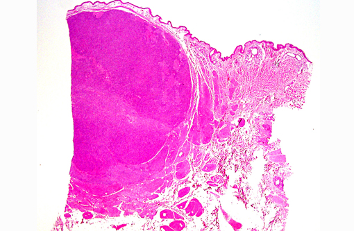
Gross Description:
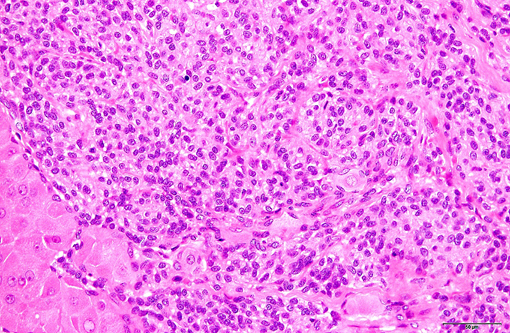
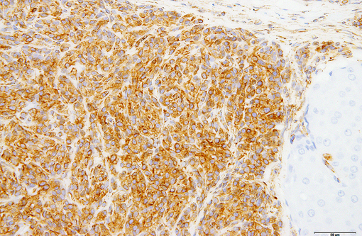
Histopathologic Description:
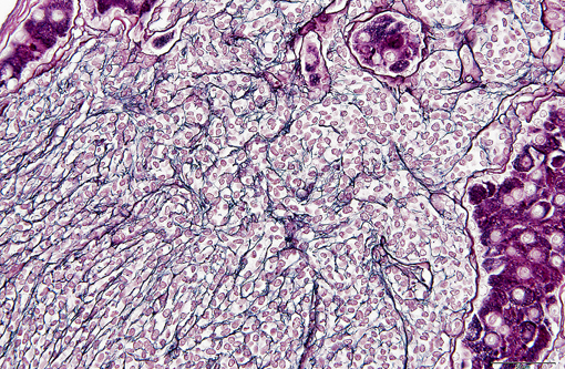
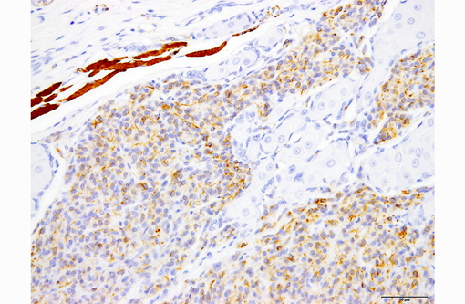
Immunohistochemically, neoplastic cells are diffusely, strongly positive for vimentin, multifocally, weakly positive for desmin, and negative for alpha-smooth muscle actin, calponin, cytokeratins (AE1/AE3, 7, 14, CAM5.2 and CK-MNF), chromogranin A, synaptophysin, PGP9.5, NSE, CD31, Factor VIII, melan-A, PNL2, S-100, MHC Class II, Iba-1and CD18. The cytoplasmic border of each neoplastic cell is strongly positive for type IV collagen.
Morphologic Diagnosis:
Condition:
Contributor Comment:
Glomus tumors are rare in animals, though there are a few case reports in dogs and cats.(2,3,8,9) Similarly to humans, most tumors in dogs and cats arise on the lower extremities and the digit.(2,3,8,9) Although glomangioma has been reported n a cow and a dog,(4,7) most glomus tumors in animals are most consistent with the classical glomus tumor (solid type) in humans.(2,3,8,9) Histopathologically, glomus tumors are composed of small round cells with round nucleus and eosinophilic cytoplasm, arranged in sheets, nests, cords, ribbons or duct like structures.(2,3,8-10) Nests and individual tumor cells are surrounded by a basement membrane.(1,9,10) Variably sized vascular structures are present within the neoplasm, and tumor cells often palisade along vessel walls.(1-3,6-8,10) Immunohistochemically, nearly all glomus tumors express vimentin and alpha-smooth muscle actin. Desmin is variably expressed.(1-3,8-10) The basement membrances surrounding neoplastic cells typically express type IV collagen and laminin.(1,10)
All glomus tumors reported in dogs and cats demonstrate palisading of tumor cells along vessel walls, as well as positive immunoreactivity for vimentin and alpha-smooth muscle actin.(2,3,8,9) Although the present case does not display all of these features, the morphological and immunohistochemical characteristics are most consistent with a glomus tumor. In particular, the basement membrane around each tumor cell is strongly suggestive of a glomus tumor. Glomus tumors in humans and animals must be distinguished from epithelial tumors (such as trichoblastoma), canine hemangiopericytoma, synovial sarcoma and epithelial leiomyoma.(2,3,8-10) In this case, all of these tumors were ruled out based on the histopathological and immunohistochemical features of the neoplastic cells.
JPC Diagnosis:
Conference Comment:
This case is challenging, in that it demonstrates some, but not all of the classic histomorphologic and immunohistochemical features of a glomus tumor. After extensive debate within conference and consultation with pathologists from the Join Pathology Center soft tissue subspecialty, we are unable to definitively diagnose a glomus tumor; however we concur with the contributors conclusion that the microscopic and staining characteristics are most consistent with this entity.
References:
2. Dagli ML, Oloris SC, Xavier JG, dos Santos CF, Faustino M, Oliveira CM, Sinhorini IL, Guerra JL. Glomus tumour in the digit of a dog. J Comp Pathol. 2003;128:199-202.
3. Furuya Y, Uchida K, Tateyama S. A case of glomus tumor in a dog. J Vet Med Sci. 2006;68:1339-1341.
4. Galofaro V, Rapisarda G, Ferrara G, Iannelli N. Glomangioma in the prepuce of a dog. Reprod Domest Anim. 2006;41:568-570.
5. Rosai J. Rosai and Ackermans Surgical Pathology. Vol. 2. 9th ed. St Louis MO: Mosby; 2004:2288-2290.Â
6. Park CH, Kozima D, Tsuzuki N, Ishi Y, Oyamada T. Malignant glomus tumour in a German shepherd dog. Vet Dermatol. 2009;20:127-130.
7. Roperto S, Borzacchiello G, Brun R, Perillo A, Russo V, Urraro C, et al. Multiple glomus tumors of the urinary bladder in a cow associated with bovine papillomavirus type 2 (BPV-2) infection. Vet Pathol. 2008;45:39-42.
8. Shinya K, Uchida K, Nomura K, Ozaki K, Narama I, Umemura T. Glomus tumor in a dog. J Vet Med Sci. 1997;59:949-950.
9. Uchida K, Yamaguchi R, Tateyama S. Glomus tumor in the digit of a cat. Vet Pathol. 2002;39:590-592.
10. Weiss SW, Goldblum JR. Enzinger and Weisss Soft Tissue Tumors. 5th ed. St. Louis, MO: Mosby; 2008:751-754.Â
11. Young B, Lowe JS, Stevens A, Heath JW, eds. Wheaters Functional Histology: A Text and Colour Atlas. 5th ed. Philadelphia, PA: Elsevier Limite; 2006:183.