Signalment:
13-week-old male Sprague-Dawley CD/IGS rat, (
Rattus norvegicus).Rats were necropsied 24 hours following two daily oral gavage doses of a test article.
Gross Description:
There were no notable gross observations.
Histopathologic Description:
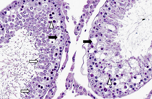
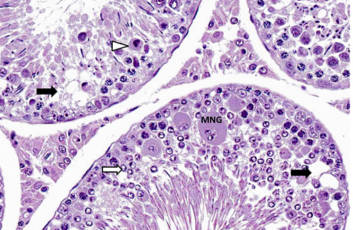
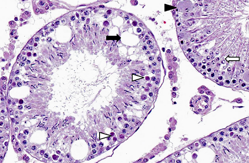
Many seminiferous tubule profiles, both early-stage and late-stage, have abnormal features including Sertoli cell vacuolation and germ cell degeneration, disorganization and depletion. Features of germ cell degeneration/necrosis include individual necrotic/apoptotic cells (especially spermatocytes); round spermatids with condensed and marginated nuclear chromatin and sometimes with excessive lightly basophilic granular cytoplasm or forming multinucleated giant cell syncytia. Some late-stage tubules have elongated spermatids with wavy, bent, or folded heads.Â
Morphologic Diagnosis:
Testis, seminiferous tubule: Germ cell degeneration/ necrosis and Sertoli cell vacuolation, acute, diffuse, marked.
Condition:
Testicular toxicity; 1,3-dinitrobenzene
Contributor Comment:
The case provides an example of testicular injury 48 hours following treatment with 1,3-dinitrobenzene (1,3-DNB), a chemical intermediate formed during manufacture of many chemical compounds and a robust testicular toxicant in rodents.(3) Testicular toxicity is thought to be mediated by a reactive intermediate, 3-Nitrosonitrobenzene, formed during the reduction of 1,3-DNB to nitroaniline.(1,3) The Sertoli cell is thought to be the primary target cell of toxicity since ultrastructural changes in Sertoli cells have been shown to precede germ cell changes.(3) Sertoli cell vacuolation followed by degeneration of pachytene spermatocytes occur within the first 12 to 24 hours after administration of toxic doses and are reported to initially affect selected late stage tubules preferentially.(3,4) In the submitted case, 48 hours following exposure to the toxin, damage is more widespread across stages of the spermatogenic cycle and includes changes to early-stage as well as late-stage tubules. Tubular atrophy is a common sequel to the acute degenerative changes resulting from a single oral toxic dose of 1,3-DNB and may become apparent within about three weeks, though tubular regeneration and recovery may occur.(4)
The severity of testicular damage induced by 1,3-DNB increases with age in rodents, and the proportion of early-stage tubules exhibiting significant injury was shown to be notably higher in 120 day-old compared to 75 day-old rats.(1) Differences in susceptibility to testicular toxicity are thought to be related to differences in rates of hepatic clearance and intratesticular metabolism.(1) Methemoglobinemia, anemia, and liver injury are features of 1,3-DNB toxicity in humans; a literature search did not identify citations confirming that testicular injury is recognized in humans.Â
JPC Diagnosis:
Testicle, seminiferous tubules: Degeneration, multifocal, moderate, with Sertoli cell vacuolation, spermatocyte degeneration and necrosis and multinucleate spermatid formation.
Conference Comment:
Spermatogenesis results from a complex interaction between various hormones, germ cells, Sertoli cells and interstitial (Leydig) cells. Luteinizing hormone (LH) released from the pituitary gland binds LH receptors on Leydig cells, inducing the production of testosterone, which binds to receptors on Sertoli cells (and possibly germ cells). Testosterone has also been shown to inhibit germ cell apoptosis, a normal method of regulating the cell population. Follicle-stimulating hormone (FSH) from the pituitary gland directly stimulates Sertoli cells (and possibly germ cells), while inhibin, produced by Sertoli cells, is a negative feedback mechanism that inhibits FSH production. During spermatogenesis, germ cells pass through spermatogonia, spermatocyte and spermatid stages.(2) In the rat, spermatogenesis is divided into 19 stages.(5) Spermiation is the active release of spermatozoa by Sertoli cells into the lumen. Sertoli cells, the support cells of the seminiferous tubule, maintain tubular epithelial integrity, phagocytose apoptotic germ cells, secrete fluid and proteins, regulate spermatogenesis, metabolize steroids, provide nutrients to germ cells, and mediate hormonal effects on the germ cells. Furthermore, occluding junctions of Sertoli cells form an important part of the blood-testis barrier.(2) Thus, damage to Sertoli cells can also result in degenerative changes in the germ cell line, as demonstrated in this case.Â
The moderator additionally offered several recommendations for performing testicular and epididymal evaluation in the rat, including: a) using Bouins or Modified Davidsons solution for fixation; histochemical staining with PAS-Hematoxylin to highlight the acrosome (this does not work in dogs); and performing a stage-aware assessment of seminiferous tubules using species-specific spermatogenesis chart.
References:
1. Brown CD, Forman CL, McEuen SF, Miller MG. Metabolism and testicular toxicity of 1,3-dinitrobenzene in rats of different ages. Fundam Appli Toxicol. 1994;23:439-446.
2. Foster RA, Ladds PW. Male genital system. In: Maxie MG, ed. Jubb, Kennedy, and Palmers Pathology of Domestic Animals. Vol. 3. 5th ed. Philadelphia, PA: Elsevier Limited; 2007:566-567.
3. Foster PMD, Sheard CM, Lloyd SC. 1,3-Dinitrobenzene: a Sertoli Cell toxicant? In: Stefanini M, Conti M, Geremia R, Ziparo E, eds. Molecular and Cellular Endocrinology of the Testis. New York, NY: Elsevier Science Publishers; 1986:281-288.
4. Hess RA, Linder RE, Strader LF, Perreault SD. Acute effects and long-term sequelae of 1,3-dinitrobenzene on male reproduction in the rat I. Quantitative and qualitative histopathology of the testis. J Androl. 1988;9:327-342.
5. Russell LD, Ettlin RA, Sinha Hikim AP, Clegg ED. Histological and Histopathological Evaluation of the Testis. Clearwater, FL: Cache River Press; 1990:65.