Joint Pathology Center
Veterinary Pathology Services
Wednesday Slide Conference
2017-2018
Conference 11
December 6th, 2017
CASE II: 14/160 (JPC 4048228).
Signalment: Unknown age, adult, female, Ambystoma mexicanum, axolotl.
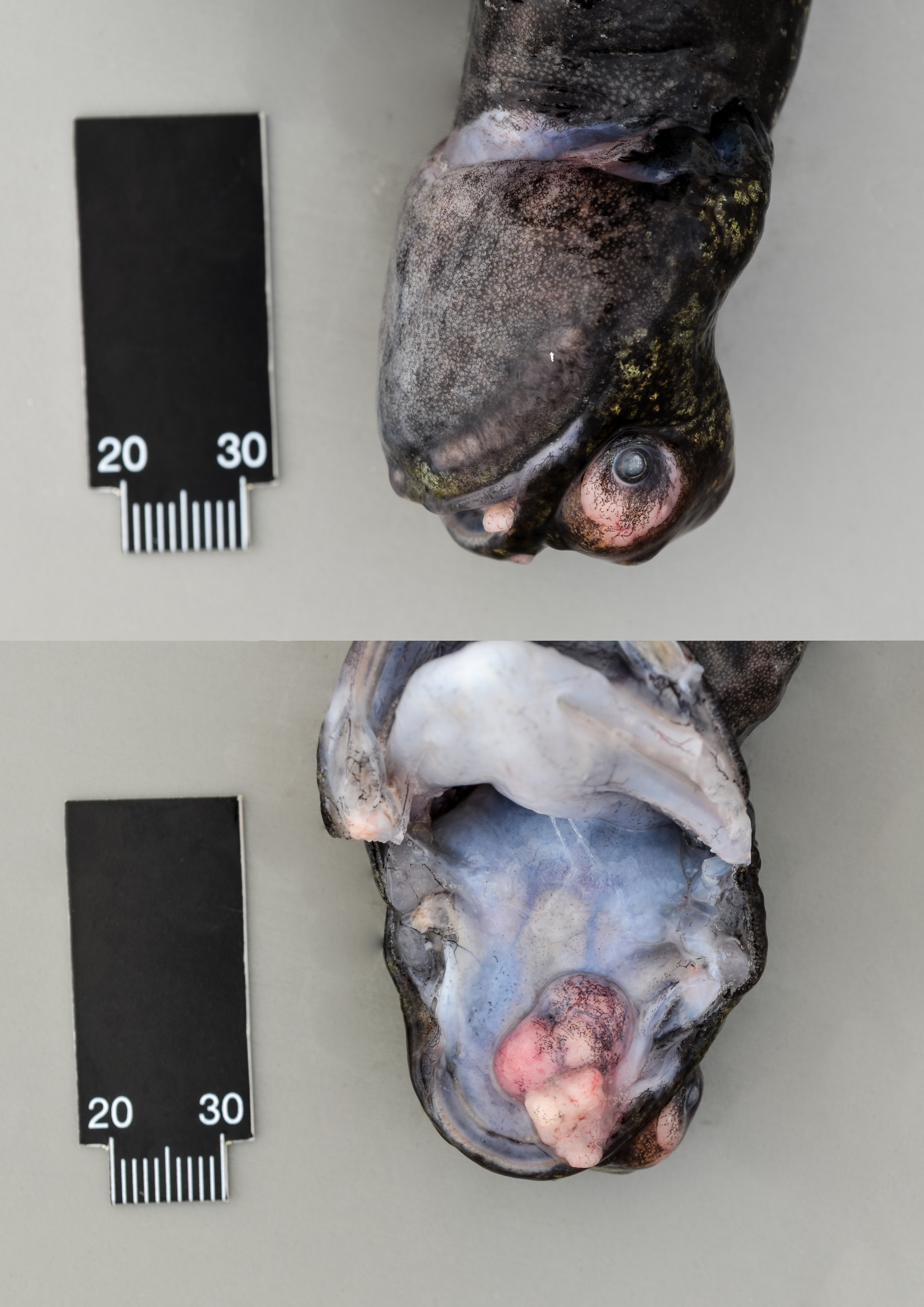
History: The animal was euthanized because of exophthalmia and poor condition due to a mass in the head which extends to the oral cavity.
Gross Pathology: On the head, between the eyes is a firm mass of about 2 x 2 x 1 cm in diameter, which reaches the oral cavity and has a multinodular appearance. The cut surface of the mass is firm, well-delineated and white. The right eye is exophthalmic.
Laboratory results:
None submitted.
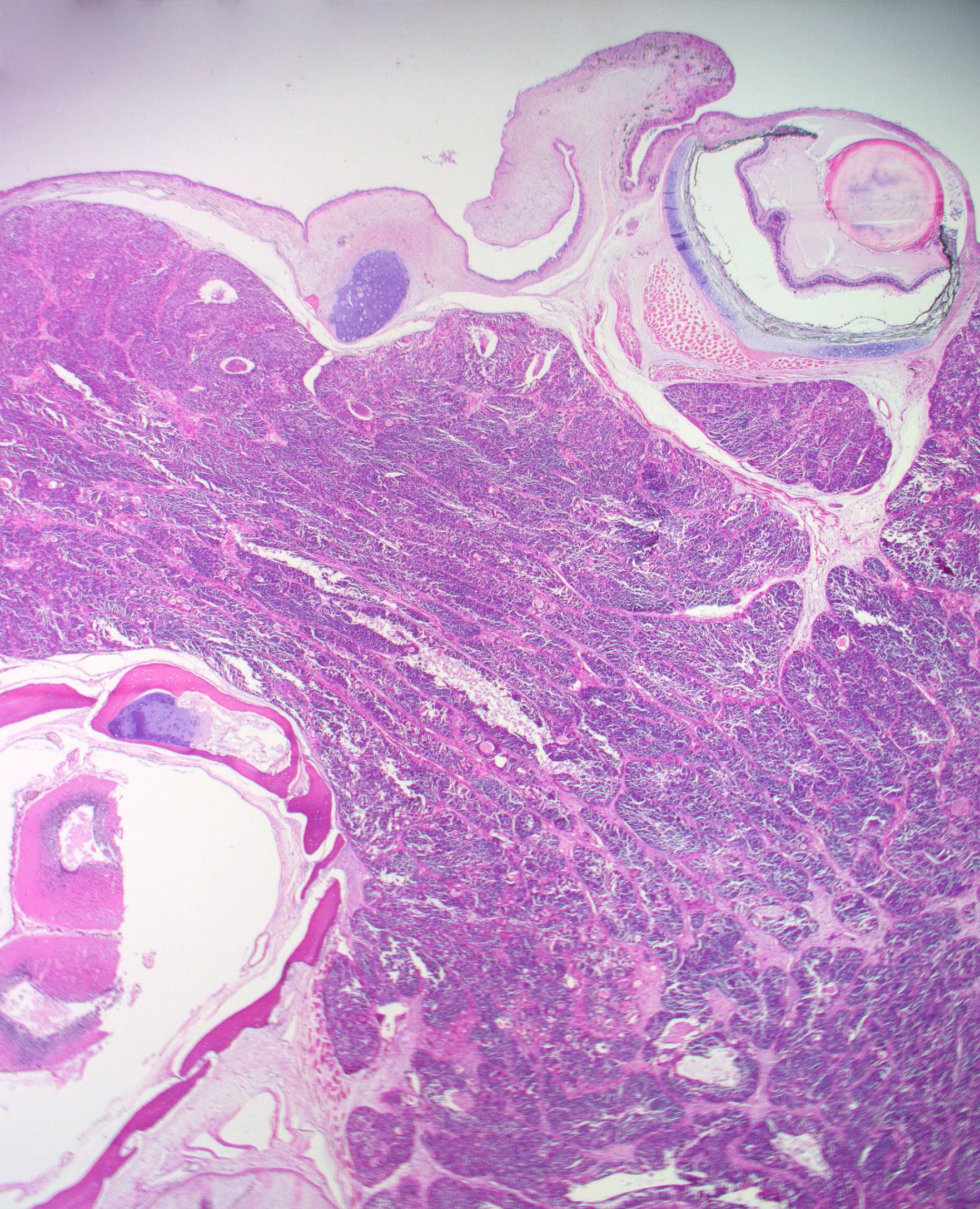
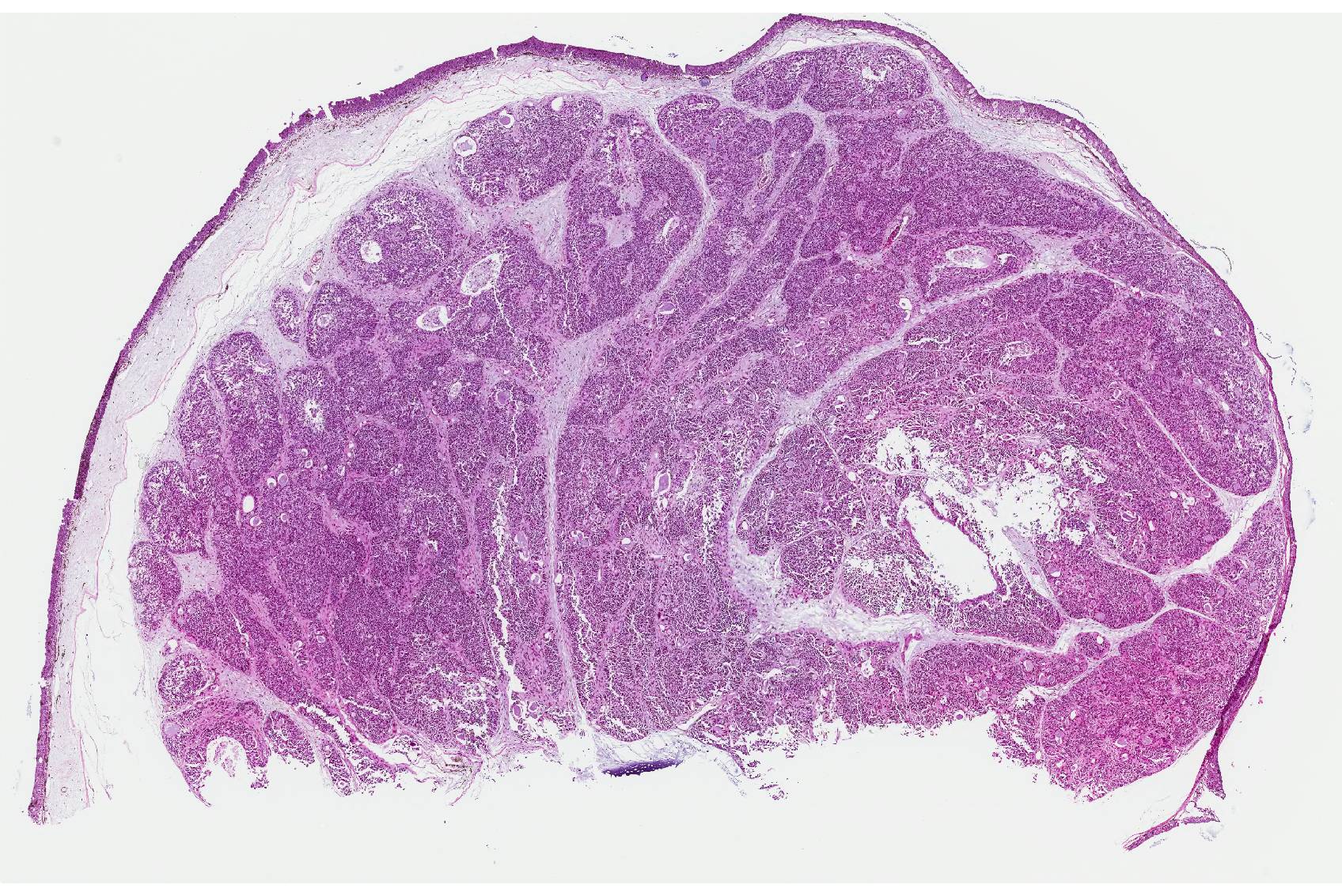
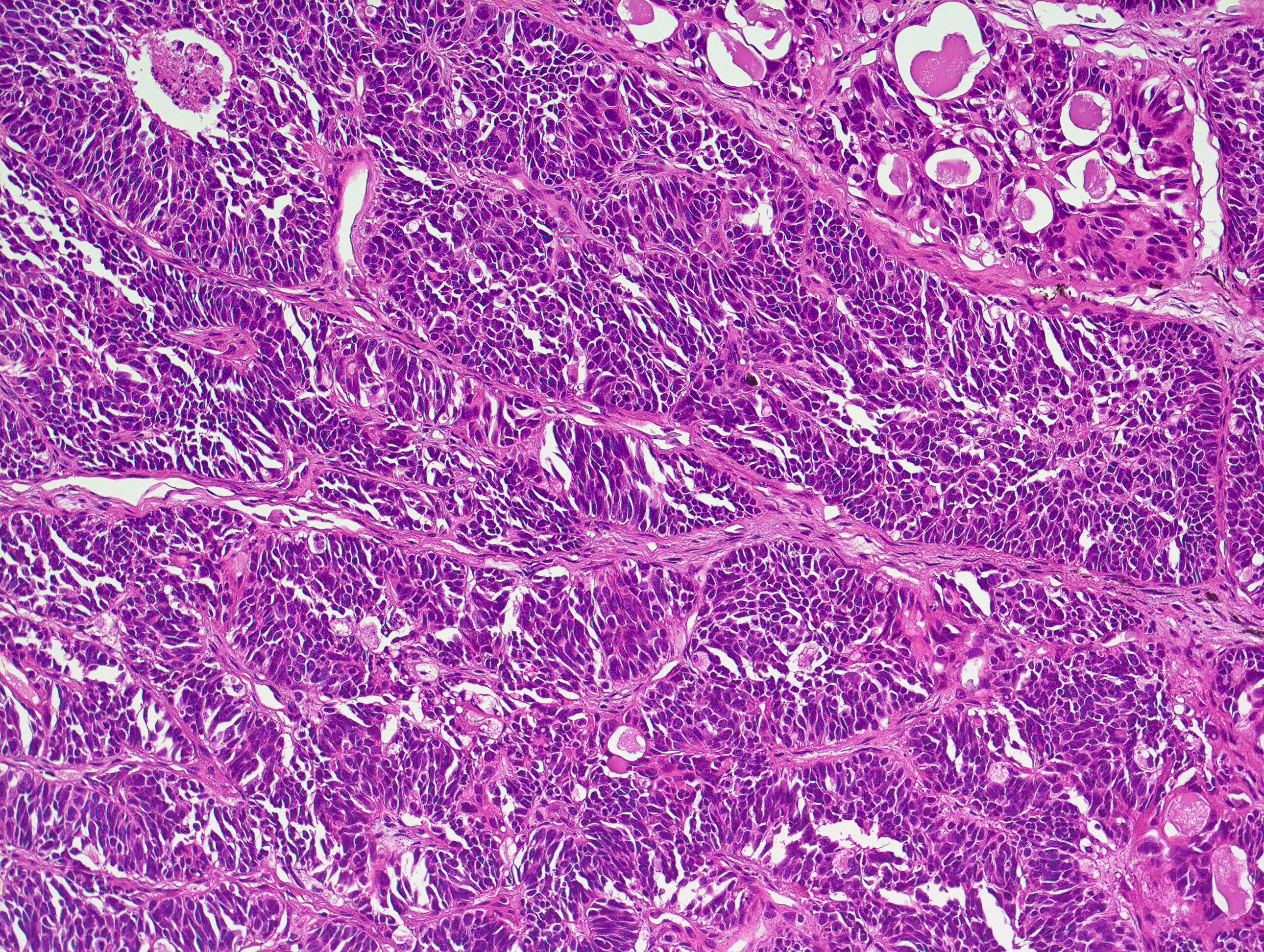
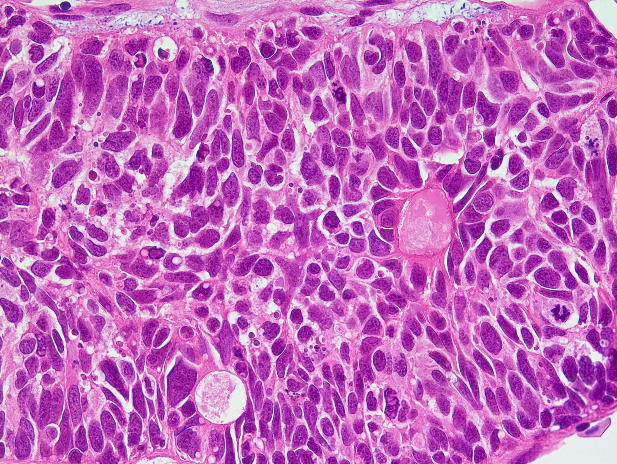
Microscopic Description: From the superficial dermis, extending to the cut borders, there is a poorly demarcated, nonencapsulated, infiltratively growing, cell rich neoplastic mass. The cells are polygonal to elongated, arranged in rosettes (Flexner-Wintersteiner-like rosettes), nests and packets and are supported by moderate amount of delicate septa of fibrovascular stroma. The neoplastic cells are 10-15 µm in diameter, have variably less distinct cell borders, moderate amount of eosinophilic, granular cytoplasm and a round to elongated nucleus with finely stippled chromatin and up to 4 nucleoli. The anisocytosis and anisokaryosis are moderate and there are 3-4 mitoses per 400X high power fields. Multifocal in the neoplasia necrosis is present. The surrounding dermis is edematous.
Contributor’s Morphologic Diagnosis:
Head: Neuromastoma (Neuroepithelioma).
Contributor’s Comment: Neoplastic disorders in amphibians are rare and limited to specific species [orders: Anura (frogs and toads), Urodela (salamander), Gymnophiona (caecilians)].7 The axolotl belongs to the Urodela and has become popular in cancer research, regenerative biology and immunology.6 Spontaneous tumors described in axolotl are melanophoroma, epithelioma, neuroepithelioma, lymphangiosarcoma, mast cell tumor, fibropapilloma, sertoli cell tumor and teratoma.4,6,7
In our case, the axolotl had a mass on the head between the eyes reaching to the oral cavity. Histologically, the neoplastic cells formed rosettes which lead to the suspicion of a neuroendocrine tumor. In a previous report from an axolotl, a mass with the same distribution and similar histological pattern was described and referred to as a neuromastoma.4 Neuromastoma (neuroepithelioma) is a neoplasia originating from neuromast cells.4 These cells are distributed on the head and body and are the end organs of the lateral line system, which is a sensory system in all fishes and permanently aquatic amphibians.2 On the head of axolotl, tumors like fibropapilloma, mast cell tumor and olfactory neuroblastoma can occur.4,6,7 These are macroscopical differential diagnoses in our case. Fibropapillomas are common in urodeles6, but in the histologic examination, fibropapilloma was excluded due to lack of the distinctive histological pattern of proliferative neoplastic fibroblasts. Mast cell tumors are common neoplasias on the head of A. mexicanum, often with ulceration of the overlying epidermis.4 In our histological examination, there was no indication of mast cells in the toluidine blue staining. Olfactory neuroblastoma and neuromastoma may have similar neuroectodermal origin, share similar histological and immunohistochemical features and are not easily distinguished from each other.4,6 Considering the similar location and histological features of the previous report of neuromastoma4, we diagnosed the tumor in our case was diagnosed as a neuromastoma.
Only few cases of neuromastoma have been reported and only in Laurenti`s alpine newt and axolotl.4 It has a prolonged course and the tumor localization impacts the health status of the animal.7
Amphibians share the same cell types as other animals, and any cell type can develop neoplastic changes.7 The knowledge about neoplasia in wild amphibians is incomplete and inconsistent due to insufficient diagnoses and misdiagnoses and difficulty to interpret the neoplasias.7 The best known amphibian neoplasm is the Lucké renal adenocarcinoma caused by ranid herpesvirus-1 (Lucké`s herpesvirus) and is endemic in the northern leopard frog (Rana pipiens) population.4,7 Interestingly, some anuran species possess anticancer secretory products and cytoprotective capabilities which make them relatively resistant to carcinogens, and the regenerative capacity of urodels is hypothesized to explain the low tumor rate in these amphibians.7
Table 1. Spontaneous neoplasias in amphibians: from Stacy et. al 2004
|
Organ system |
Tumor type |
Species |
|
Integument and soft tissues |
Epidermal papilloma
Squamous cell carcinoma
Dermal gland tumors
Melanophoroma
Fibropapilloma, Fibrosarcoma
Neuroepithelial tumors
Mast cell tumor |
Urodele species
Northern leopard frog (R. pipiens)
Grass frogs (R. temporaria) Pond frogs (R. ridibunda)
Urodele and anura species
Western tiger salamander (A. mavortium)
Axolotl (A. mexicanum) Alpine newt (T. alpestris)
Urodele species |
|
Hemolymphatic system |
Lymphoma
Granulocytic leukemia |
African clawed frog (X. laevis) Axolotl (A. mexicanum)
Toads (Bufo sp.) |
|
Hepatobiliary system |
Hepatocellular adenoma |
Barking tree frog (H. gratiosa) |
|
Alimentary system |
Gastric adenocarcinoma
Intestinal adenocarcinoma |
African clawed frog (X. laevis)
Marine toad (B. marinus) |
|
Urogenital system |
Lucké renal adenocarcinoma
|
Northern leopard frog (R. pipiens)
Urodele and anura species
Urodele and anura species
Urodele and anura species
Northern leopard frog (R. pipiens) Ornate horn frog (C. ornata)
Northern leopard frog (R. pipiens) |
|
Nephroblastoma
|
||
|
Renal carcinoma
|
||
|
Sertoli cell tumor
|
||
|
Cystadenocarcinoma |
||
|
Granulosa cell tumor
|
||
|
Ovarian teratoma |
||
|
Endocrine system |
Pancreatic carcinoma |
African clawed frog (X. laevis) Rana species |
JPC Diagnosis: Oral mucosa: Neuroepithelioma (neuromastoma), Ambystoma mexicanum, axolotl.
Conference Comment: Amystomatidae, or mole salamanders, is a family of North American amphibians that contain approximately 30 species. The axolotl, Ambystoma mexicanum, is the most common aquatic species kept in captivity and has been used for many years in biomedical research. Axolotls exhibit a distinguishing feature termed “neoteny” which is the retention of juvenile characteristics. The Anderson’s axolotl (Amystoma andersoni) are also neotenic and are common zoo residents. The Mexican axolotl is considered endangered due to habitat loss, pollution, and the introduction of fish into their habitats that predate larval axolotls4.
Neuromastomata, aptly described above, have historically been transplanted successfully, and induced experimentally by numerous carcinogens.3,4 Sensory cells of the lateral line system are found on the head, body, and tail of larval and adult aquatic amphibians. Specific cells in ampullary organs of the head in A. mexicanum contain similar cells that are called electroreceptors. It is not currently possible to distinguish olfactory, neuromastic, or electroreceptor cells from one another and neoplasms are often diagnosed as neuroepitheliomas. The lateral line and pit organs function as mechanoreceptors, while the ampullary organs are electroreceptors. The pit and ampullary organs are located only on head and the lateral line usually extends from head to tail on each side of the body. In the axolotl, specifically, there are three pairs of lateral lines (similar to the northern leopard frog, Rana pipens) and only the middle pair extend all the way to the tail.4 Oral neuroepitheliomas (neuromastomas) were first described in axolotls by Drs. Brunst and Roque in 1967 and there have been few reports since1.
Contributing Institution:
Vetsuisse Faculty, University of Bern
Institute of Animal Pathology
Laenggassstrasse 122
PF 8466
3001 Bern Switzerland
http://www.itpa.vetsuisse.unibe.ch/content/index_eng.html
References:
- Brunst VV, Roque AL. Tumors in amphibians histology of a neuroepithelioma in Siredon mexicanum. J Natl Cancer Inst. 1967;38(2):193-204.
- Coombs S, Braun CB, Donovan B. The orienting response of Lake Michigan mottled sculpin is mediated by canal neuromasts. J Exp Biol. 2001;204:337-348.
- Darquenne J. Cancerologie experimentale: actions de substances cancerigenes sur le regenerat de la queue de Triturus alpestris Comptes Rendus de l’Academie des Sciences, Paris 273D:1460-1462.
- Green DE, Harshbarger JC. Spontaneous neoplasia in Amphibia. In: Wright KM, Whitaker BR, eds. Amphibian Medicine and Captive Husbandry. Malabar, FL: Krieger Publishing Company; 2001:7, 335-400, 469.
- Matz G. Tumeurs spontanees et experimentales observes chez Triturus alpestris (Laurent) (Salamandridae). Proceedings of 1st International Colloquium on Pathology of Reptiles and Amphibians. 1982:129-133.
- Shioda C, Uchida K, Nakayama H. Pathological features of olfactory neuroblastoma in an axolotl (Ambystoma mexicanum). J Vet Med Sci. 2011;73:1109-1111.
- Stacy BA, Parker JM. Amphibian oncology. Vet Clin North Am Exot Anim Pract. 2004;7:673-695.