Joint Pathology Center
Veterinary Pathology Services
Wednesday Slide Conference
2018-2019
Conference 24
10 April, 2019
CASE II: P/2014-95 (JPC 4066219)
Signalment: 7-week-old, female Cob-X, horse, Equus caballus.
History: The filly was presented with lethargy and unwillingness to suckle over the last few days. On clinical examination she exhibited tachycardia, tachypnea, fever, increased capillary refill time, pink mucous membranes, loud and harsh respiratory sounds, and had a pasty fecal consistency. She was treated with antibiotics and nonsteroidal anti-inflammatory drugs (NSAIDs).
The following day, the foal was much the same, yet drinking milk from bucket. Additionally, she exhibited a stiff gait, which was more obvious on the hindlimbs when compared to the forelimbs. Her treatment with antibiotics and NSAIDs was continued and supplemented by intravenous fluid therapy.
The next day the foal became recumbent and died.
In the previous weeks, a number of foals, five of which died, had deteriorated with similar symptoms at the yard down the road, which belonged to the same owner. During the days following submission of the filly for post mortem examination, also other foals at the same yard started to show symptoms.
Gross Pathology: Upon gross examination, the cause of death in this foal was not apparent. The adipose tissue in this foal diffusely appeared yellow and, where associated with the mesentery, slightly nodular. Additionally, there was a mild to moderate catarrhal enteritis and very mild ascites.
Laboratory results: NA
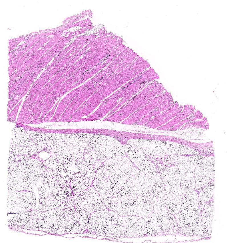
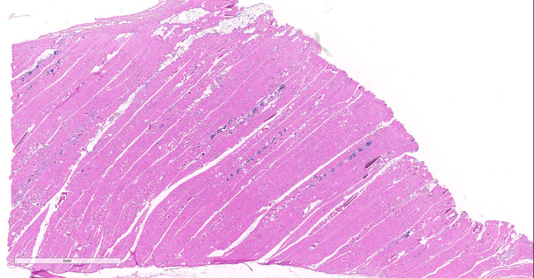
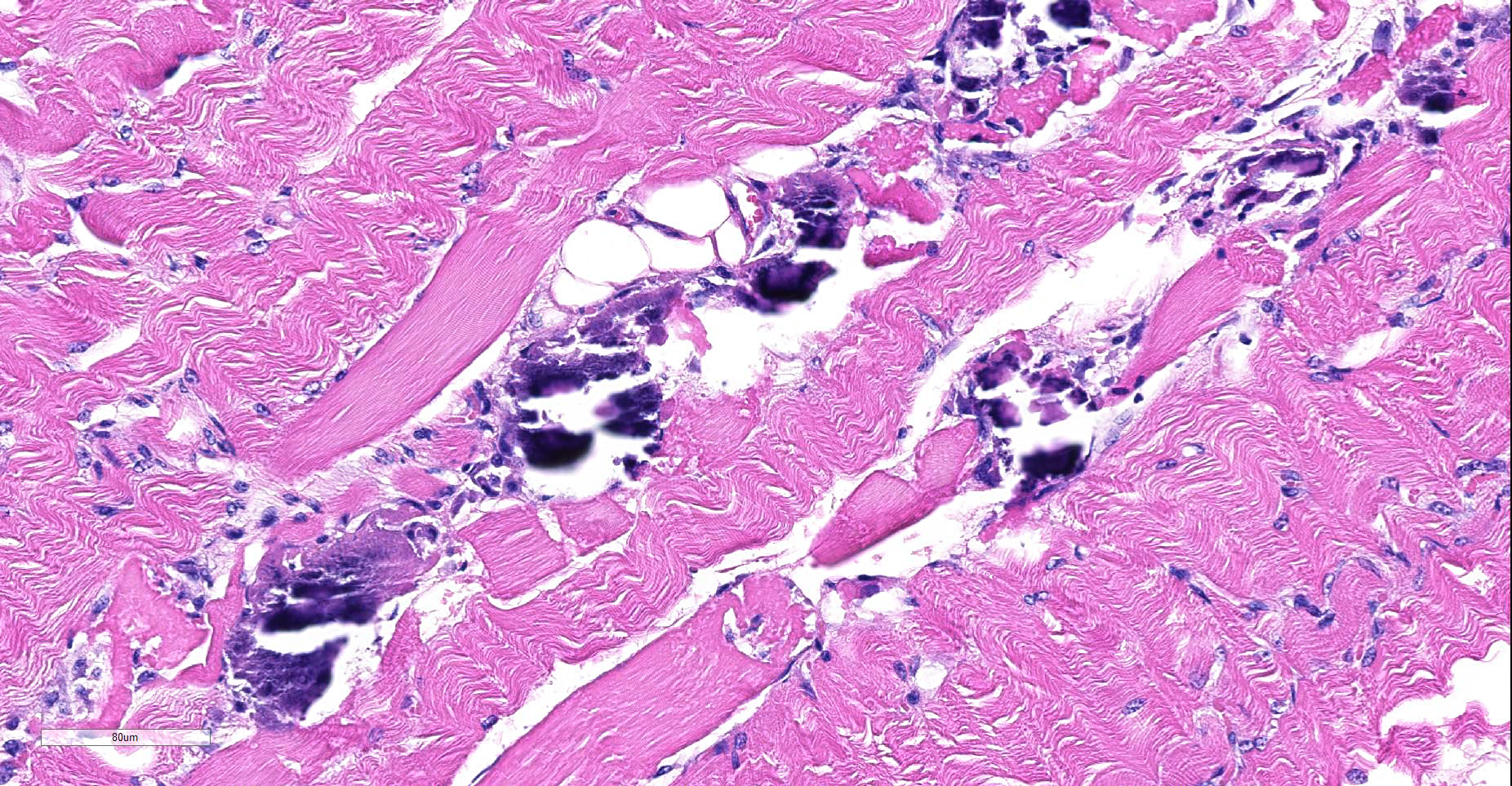
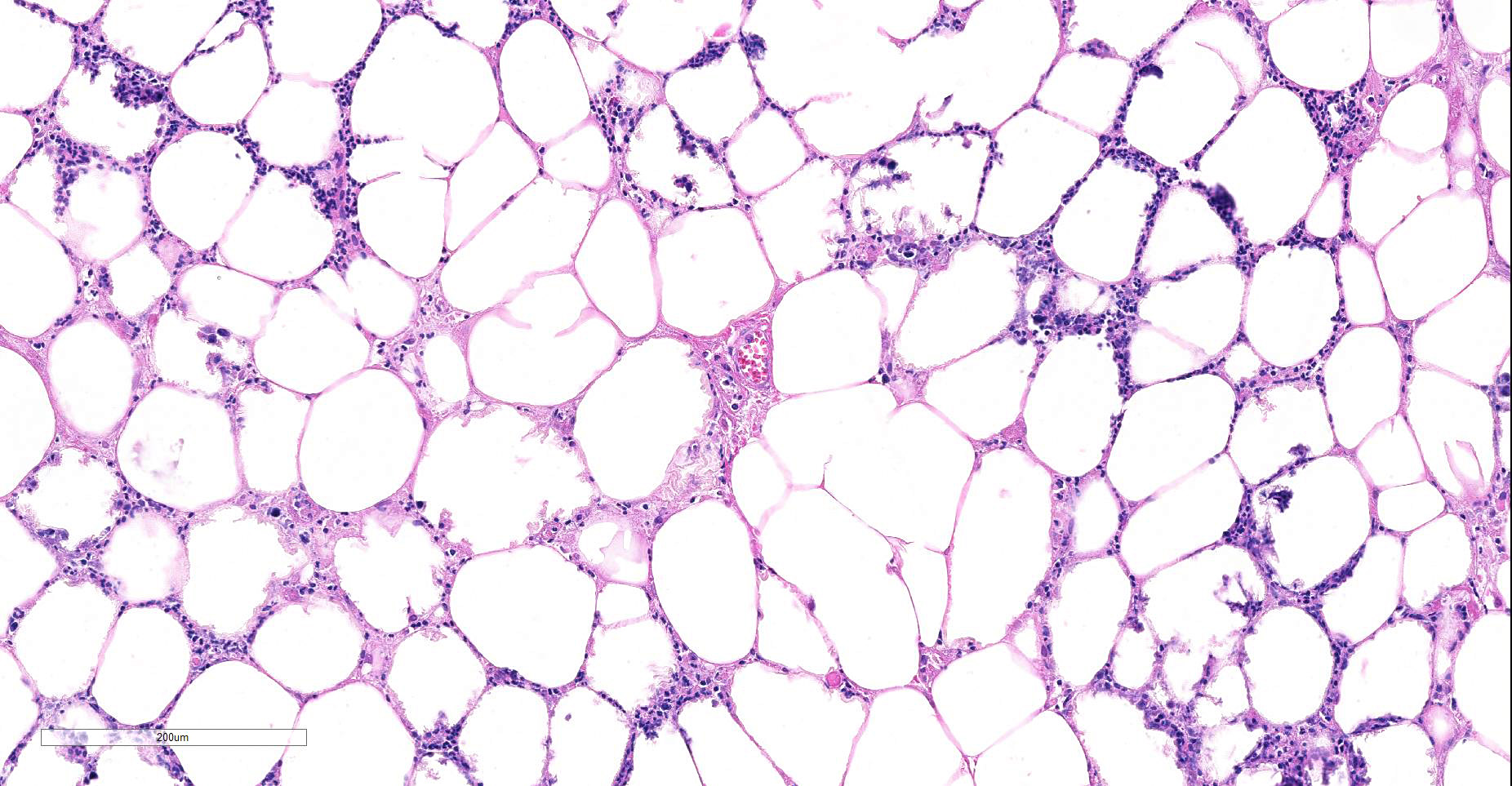
Microscopic Description:
In a multifocal distribution, the striated (skeletal) myofibers are homogenously eosinophilic, devoid of striations and frequently fragmented, exhibiting irregular contours (hyaline degeneration). Some of these fibers stain strongly eosinophilic (Zenker’s degeneration), whilst others exhibit a punctate intracytoplasmic basophilic stain (mineralization). Occasional degenerative myofibers are being infiltrated by macrophages.
In a multifocal to coalescing distribution, the interstitium of the overlying adipose tissue contains moderate numbers of neutrophils admixed with lesser numbers of macrophages and small to moderate amounts of granular basophilic material (mineralization). Numerous adipocytes, mainly in close association with the inflammatory infiltrates exhibit a blurring of their cytoplasmic vacuoles with pale eosinophilic or basophilic staining (necrosis), whilst in others small clusters of neutrophils are present within lipid vacuoles. Multifocally, the stromal tissue supporting the sheets of mature adipocytes is mildly to moderately expanded by small numbers of plump spindle cells (activated fibroblasts).
Contributor’s Morphologic Diagnoses:
Myofiber degeneration, segmental, acute to subacute, multifocal to coalescing, moderate to marked; abdominal wall with
- a) Myofiber mineralization, multifocal, moderate; and
- b) Myositis, histiocytic, subacute, multifocal, mild.
- Adipocyte necrosis, acute to subacute, multifocal to coalescing, moderate; abdominal wall with
- a) Steatitis, neutrophilic and histiocytic, subacute, multifocal to coalescing, moderate to marked; and
- Mineralization, multifocal, mild to moderate; and
- Fibrosis, subacute, multifocal, mild.
Contributor’s Comment: The combination of muscle degeneration and fat necrosis with prominent inflammatory changes in the fat (also termed “yellow fat disease”) is a disorder seen in equine and donkey foals ranging from one day to 12 weeks 2,7 and young equids up 3 years 1,5, and is thought to be the results of a deficiency of vitamin E and/or selenium, or a diet high in unsaturated fatty acids 1. However, possibly also a more complex aetiology may need to be considered, since vitamin E/selenium supplementation in these patients often is only of very little or no curative value and these foals may also exhibit normal tissue selenium or gluthathione peroxidase concentrations 6.
Whilst the metabolism of vitamin E and selenium is incompletely understood, the pathogenesis for the muscular changes observed in these foals is considered to be based on lipid peroxidation and damage to proteins of mitochondria, endoplasmic reticulum and the cytosol by radicals due to a relative lack of radical-quenching systems. Both vitamin E and selenium play a role in radical scavenging via the gluthatione peroxidase/gluthathione reductase system.7
Following injury to the cellular membranes by free radicals, these membranes are unable to upkeep the homeostasis. Mitochondria take up the surplus of calcium following the intracellular influx of this, which negatively affects their ability to produce energy. Energy is required for the recapture of calcium-ions following muscle contracture, with the increase in calcium further resulting in a hypercontractile state which in turn results in coagulative destruction of the myofilaments.7
Similarly, the changes to the adipose tissue are considered to be the result of progressive peroxidation 1. The relative distribution of necrosis and mineralization, however, in comparison to the extent of steatitis may vary between the different sites examined.6
Affected foals are considered to likely be born to selenium-deficient mares or possibly also vitamin E-deficient mares.1,6 Interestingly “yellow fat disease” has a relatively high incidence in Shetland ponies and cold-blooded horses indicating a possibly hereditary predisposition of these regarding for example transplacental passage and liver storage of selenium or colostral vitamin E, or that these horse groups may be relatively over-represented amongst suboptimally fed equids.1
Clinically, affected foals and horses exhibit depression, fever, tachycardia and variable tachypnea. 1,2,5. Additionally, mild to moderate abdominal discomfort and tenderness, painful swelling of the nuchal ligament, groin and axillary regions, stiffness and muscular weakness are present, which may affect their ability to suckle.3,5,6,7 Their feces are of abnormal consistency.1,2
In biochemistry, lactate dehydrogenase, creatine kinase and aspartate aminotransferase are increased,1,2,3 whilst serum vitamin E or selenium is decreased.1,3,5
Upon gross examination, affected foals/horses exhibit hardened, multinodular and focally hemorrhagic adipose tissues of the nuchal ligament, the coronary sulci, the subcutis and throughout the abdominal cavity with or without yellow-brown discoloration and which may be chalky on cross-section.1,2,3,5 Subcutaneous oedema, and discoloration and striation of the skeletal muscles may also be seen.1,3,6 Some horses exhibit evidence of a chronic enteritis and bronchopneumonia.1
The histological changes comprise necrosis of fat cells combined with a steatitis and mineralisation, and degeneration and mineralisation of muscle fibres combined with inflammatory infiltrates.1,5,7 Severely affected foals exhibit a monophasic myopathy, whilst subacute cases exhibit polyphasic changes.5
Interestingly, the myocardial changes are variable and range from no changes to loss of striations, mineralization and small foci of myocardial necrosis.1,5 Degenerative changes to the adrenal cortex with necrosis, calcification and replacement fibrosis of the zona fasciculata have also been reported.4
The moderator reviewed features of fat necrosis in histologic section (in the absence of saponification, which was minimal in the adipose tissue on this slide., to include loss of adipocyte nuclei, color change from basophilic to eosinophilic cell membranes, and extension of inflammation into adipocyte cytoplasm.
Contributing Institution:
Veterinary Pathology
Department of Veterinary Medicine
University of Cambridge, UK
JPC Diagnosis:
- Skeletal muscle: Degeneration and necrosis, polyphasic, multifocal to coalescing, with mineralization.
- Fat: Steatitis, necrotizing, multifocal to coalescing, marked.
JPC Comment: The contributor has done an outstanding job of reviewing the syndrome of Vitamin E/selenium deficiency in the foal, and this section is an outstanding example of the damage that may be seen in multiple tissues.
Vitamin E/selenium imbalance may have one of the most diverse repertoire of damage in many animal species, with some animals (such as pigs) manifesting in a multitude of syndromes. Most species will demonstrated muscle necrosis, often both skeletal and cardiac, in the face of Vitamin E deficiency. Decreased reproductive efficiency is also a common finding across species. Panniculitis may be seen as a result of dietary Vit E deficiency in cats, mink, foals, swine as a result of feeding high-fish diets, but is also seen in certain waterbirds, such as herons. In dogs, a vitamin E-deficient diet may result in ceroidosis of intestinal smooth muscle, so-called “brown dog gut”.
Vascular damage may be seen in animals with Vitamin E deficiency, to include cerebellar hemorrhage and necrosis in turkey poults, as well as exudative diathesis in poultry and swine, in which fluid exudes from blood vessels yielding a soft edematous overlying tissue. Vitamin E dieficiency may also result in hemolytic anemia in a number of species, including humans and non-human primates. Vitamin E deficiency in guinea pigs may result leukoencephalomalacia in feti. Cutaneous manifestations of Vitamin E deficiency include alopecia and seborrhea dermatitis in goats, as well as alopecia in calves on Vitamin E-deficient milk replacer.4
Growing pigs may suffer a number of manifestations of Vit E/Selenium imbalance, including massive hepatic necrosis (hepatosis dietetica), arteriolar degeneration and thrombosis (mulberry heart disease), exudative diathesis, and white muscle disease. These syndromes rarely occur concurrently in the same animal.
Vitamin E was discovered in 1922 by Drs. Herbert Evans and Katharine Bishop, who identified it as a dietary fertility factor for laboratory rats. In fact, the name “tocopherol” is derived from the Greek meaning “to carry a pregnancy”. It is obviously a necessary dietary factor for all animal species. As a widely used mendicant on the human market, it has enjoyed great popularity as a dietary supplement and ingredient in topical products for the skin in humans, largely without documented benefits. The benefits of megadosing on Vitamin E, begun in the 1930’s and 1940’s by the Shupe brothers (who extolled its virtues in treating heart disease) have yet to be proven beneficial in treating cardiovascular disease or a wide range of other diseases for which it has been purposed.
Participants remarked on the lack of saponification in the areas of necrotic fat on this slide. In the absence of obvious saponification, a lack of vital staining, loss of adipocytes nuclei, and extension of inflammatory cells within the cytoplasmic border of affected adipocytes are all clues to the potential viability of damaged adipocytes.
References:
- De Bruin CM, Veldhuis Kroeze EJB, Sloet van Oldruitenborgh-Oosterbaan MM. Yellow fat disease in equids. Equine Vet Educ 2006;18(1):38-44.
- Dixon RJ, Nuttal WO, Carthew DA. A case of steatitis and myonecrosis in a donkey foal. New Zeal Vet J 1983;31:62.
- Foreman JH, Potter KA, Bayly WM et al. Generalised steatitis associated with selenium deficiency and normal vitamin E status in a foal. JAVMA 1986;189(1);83-86.
- Mauldin EA, Peters-Kennedy J. Integumentary System. In: Maxie, GM, ed. Jubb Kennedy and Palmer’s Pathology of Domestic Animals, 6th ed. St. Louis, Elsevier, 2017. Vol 1, p 583.
- Platt H, Whitwell KE. Clinical and pathological observations on generalized steatitis in foals. J Comp Path 1971;81:499-508.
- Valentine BA, McGavin MD. Skeletal Muscle. In: Zachary JF, McGavin MD, eds Pathologic Basis of Veterinary Disease. 5th ed. St Louis, MO; Elsevier Mosby;2012.900-901.
- Van Vleet F, Valentine BA. Muscle and Tendon. In: Maxie MG, ed. Jubb, Kennedy and Palmer’s Pathology of Domestic animals. 5th ed. Vol. 1. Philadelphia, PA: Elsevier Saunders; 2007. 242.