Signalment:
Gross Description:
Histopathologic Description:
Morphologic Diagnosis:
Lab Results:
Condition:
Contributor Comment:
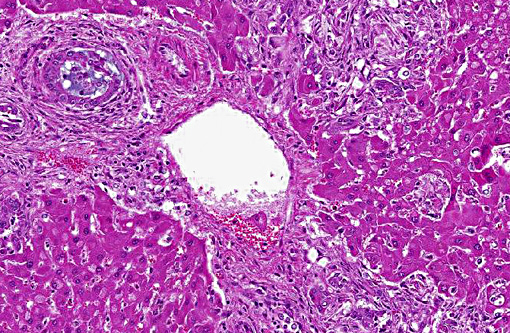
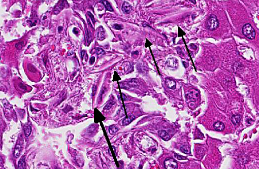
The crystals are composed principally of calcium salts of the steroidal saponins that are metabolised in the rumen and liver to form episapogenin glucuronides. In the presence of calcium they can precipitate and form crystals.(5) Crystals can obstruct bile ducts and lead to icterus and secondary photosensitization.(2) It is still unclear whether the biliary crystals alone cause liver damage or whether other toxins in the plants play a role.(1,5)
Levels of saponins vary greatly with age of plant, location and size and the plants are thus not always toxic. Outbreaks commonly occur in summer when young plants become wilted, especially if rain is followed by hot dry weather.(4)
Plants that are known to cause crystal-associated cholangiohepatopathy include:
| Scientific name | Distribution | Common name |
| Tribulus terrestris | South Africa, Australia, North America | Puncture Vine |
| Narthecium ossifragum | Norway, UK | Bog asphodel |
| Agave lecheguilla | North America | Lecheguilla |
| Nolina texana | North America | Texan bear grass |
| Brachiaria decumbens | Brazil, Australia, Malaysia | Signal grass |
| Panicum coloratum | Australia, North America, Africa | Kleingrass |
| Phytolacca octandra | NZ, South America | Inkweed |
Many plants contain steroidal saponins, but have not yet been documented as causing biliary crystals. Solanales, Primulales, Ranunculales, Fabales, Sapindales, Poales and Liliales are all orders of plants that contain steroidal saponins(1), so should not be ignored when investigating the etiology of crystal assosicated- cholangiohepatopathy.
In this case the significance of the E.coli is unclear. It may have been a post mortem contaminant, or a retrograde opportunist making use of the disrupted biliary epithelium.
JPC Diagnosis:
Conference Comment:
Conference participants contrasted this entity with that of sporidesmin, the mycotoxin that causes biliary epithelial necrosis. Biliary hyperplasia with minimal inflammation is present in both entities and photosensitization is a common sequela, albeit through slightly different mechanisms. Saponins lead to biliary obstruction rather than necrosis, but both result in accumulation of phylloerythrin due to cholestasis inciting the characteristic skin manifestation of facial eczema when the affected animal is exposed to sunlight. (2)
The contributor outlined the most well-known steroidal saponin-producing plants, of which Tribulus terrestris may be most readily recognized since it has caused enormous loss of sheep in South Africa, where it is known as yellow bighead due to icterus and marked facial edema.(5)
References:
1. Collett MG, Thompson KG & Christie RJ. Photosensitisation, crystal-associated cholangiohepatopathy, and acute tubular necrosis in calves following ingesion of Phytolacca octandra (inkweed). New Zealand Vet J 2011;59:3
2. Cullen J, Brown D. Hepatobiliary System and Exocrine pancreas. In Zachary JF, McGavin MD, ed Pathologic basics of Veterinary Disease 5th ed. Missouri, Elsevier Mosby pp440
3. Galey FD. Disorders caused by toxicants. In: Smith BP, ed. Large Animal Internal Medicine. 4th ed. St. Louis, MO: Mosby Elsevier; 2009:1697-1698.
4. McDonough SP, Woodbury AH, et al. Hepatogenous photosensitization of sheep in California associated with ingestion of Tribulus terrestris (puncture vine) J Vet Diagn Invest 1994 6:392
5. Stalker MJ, Hayes MA. Liver and biliary system. In Maxie MG, ed. Jubb, Kennedy and Palmers Pathology of Domestic Animals. 5th ed., vol 2, Philadelphia, PA; Elsevier Saunders 2007: pp 376-378