Signalment:
Histopathologic Description:
Morphologic Diagnosis:
Condition:
Contributor Comment:
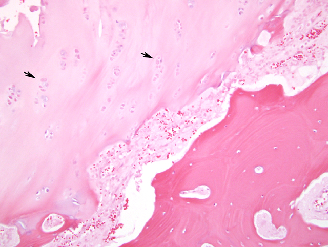
The classification of this lesion as a dysplasia in cats, appears to be appropriate but the disorganization and hypocellarity seem more likely to be secondary to failure to properly close than a primary chondrodysplasia of the growth plate. The conclusion that the growth plates WERE normal during growth is supported by the fact that the cats appear to have reached normal skeletal growth with normal appearing skeletons within normal time. Since the signals for longitudinal growth have apparently appropriately ceased in these cats but the signals for closure have either not been recognized or sent, it is understandable that the chondrocytes remaining in the non-functional growth plate would not have their normal arrangement and density. Likely the persistence of the plate and not its abnormal arrangement and density of chondrocytes is predisposing it to slip with minimal trauma in these heavy fully grown cats.
JPC Diagnosis:
Conference Comment:
Although the underlying cause of feline physeal dysplasia is not known, it has been associated with various factors including genetics, nutrition, obesity, endocrine imbalances, and other factors.1 Due to causing a delay in physeal closure, neutering has been previously considered associated with feline physeal dysplasia6, although in more recent literature this observation has been challenged.8 It is not known if the association with obesity is due to the increased stresses placed on the physis from the additional weight, causing failure under conditions of minimal trauma, or if there is an underlying endocrine abnormality that results in both obesity as well as a weakened physis.2
Epiphysiolysis in pigs is a manifestation of osteochondrosis, which has many clinical manifestations. The growth plate in affected pigs is usually characterized by a focal failure of endochondral ossification in which retained cartilage extends into the metaphysis.2,12 The chondrocytes of the cartilage core usually maintain their normal alignment. This differs from feline physeal dysplasia where the entire physis is usually affected, and the physis consists of irregular clusters of chondrocytes that have lost their normal alignment.2
The Salter-Harris classification system has been used to classify fractures of the growth plate in animals. In this system, fractures are divided into five types based on their location.Â
Salter-Harris classification system13
| Type 1 | Fracture through the physis without involvement of the epiphysis or metaphysis |
| Type 2 | Fracture involving the metaphysis and extending into the physis |
| Type 3 | Fracture involving the epiphysis and extending into the physis |
| Type 4 | Fracture involving the epiphysis and metaphysis going through the physis |
| Type 5 | Compressive fracture of the physis, crushing the growth plate |
References:
2. Craig LE: Physeal displasia with slipped capital femoral epiphysis in 13 cats. Vet Pathol 38:92-97, 2001
3. Culvenor JA, Black AP, Lorkin KF, Bradley WA: Repair of femoral capital physeal injuries in cats 14 cases. Vet Comp Orthop Traumatol 9:182-185, 1996
4. Dewey CE: Disease of the nervous and locomotor systems. In: Diseases of Swine, eds. Straw BE, Zimmerman JJ, DAllaire S, Taylor DJ, 9th ed., pp. 92-95. Blackwell Publishing, Ames, IA, 2006
5. Fischer HR, Norton J, Kobluk CN, Reed AL, Rooks RL, Borostyankoi F: Surgical reduction and stabilization for repair of femoral capital physeal fractures in cats: 13 cases (1998-2002). J Am Vet Med Assoc 224:1478-1482, 2004
6. McNicholas Jr. WT, Wilkens BE, Blevins WE, Snyder PW, McCabe GP, Applewhite AA, Laverty PH, Breur GJ: Spontaeous femoral capital physeal fractures in adult cats: 26 cases (1996-2001). J Am Vet Med Assoc 221:1731-1736, 2002
7. Moores AP, Owen MR, Fews D, Coe RJ, Brown PJ, Butterworth SJ: Slipped capital femoral epiphysis in dogs. J Small Anim Pract 45:602-608, 2004
8. Newton AL, Craig LE: Multicentric physeal dysplasia in two cats. Vet Pathol 43:388-390, 2006
9. Per+�-�z-Aparicio FJ, Fjeld TO: Femoral neck fractures and capital epiphyseal separations in cats. JSmall Anim Pract 34:445-449, 1993
10. Smith RN: Fusion of ossification centers in the cat. J Small Anim Pract 10:523-530, 1969
11. Stubbs WP, Bloomberg MS, Scruggs SL, Shille VM, Lane TJ: Effects of prepubertal gonadectomy on physical and behavioral development in cats. J Am Vet Med Assoc 209:1864-1871, 1996
12. Thompson K: Bones and joints. In: Jubb, Kennedy, and Palmers Pathology of Domestic Animals, ed. Maxie MG, 5th ed., vol. 1, pp. 136-145. Elsevier Limited, St. Louis, MO, 2007
13. Bailli+�-�res Comprehensive Veterinary Dictionary, Bailli+�-�re Tindall, Philadelphia, PA, 1988