CASE 4: T2303/14 (4068544-00)
Signalment:
11 years old, female, Lowchen dog, Canis familiaris, canine
History:
The animal suffered from pyometra and during ovariohysterectomy one ovary appeared "quite abnormal". Both ovaries were formalin-fixed and submitted for histopathological examination.
Gross Pathology:
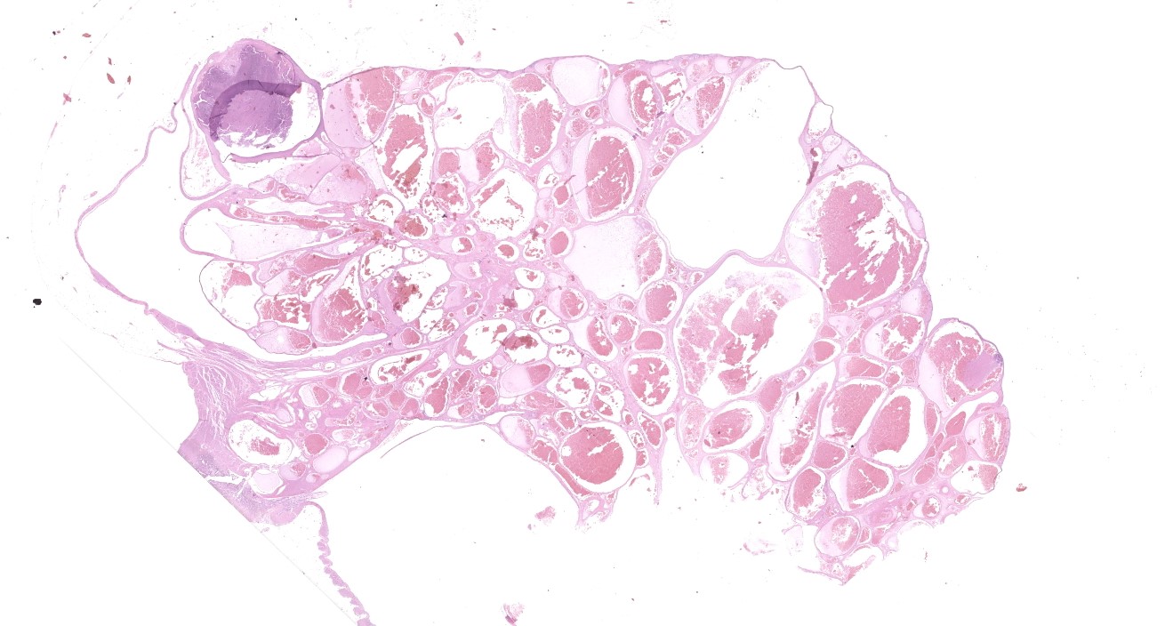
One ovary was 1.5 x 1.5 x 2 cm, with multiple small nodules and small cysts (histologically unremarkable with follicles and Corpora lutea in different stages, not submitted). The other ovary was 3 x 3 x 2 cm, with a bramble-like appearance. On cut surface multiple cyst-like structures filled with greasy brownish material were obvious.
Laboratory results:
Immunohistochemically endothelium was positive for von Willebrand factor and vimentin (not submitted).
Microscopic description:
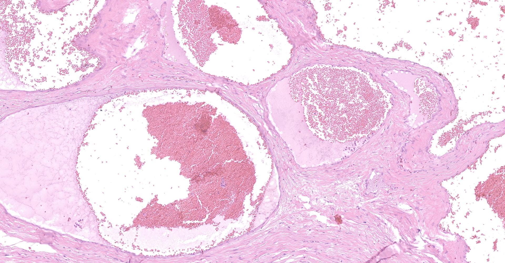
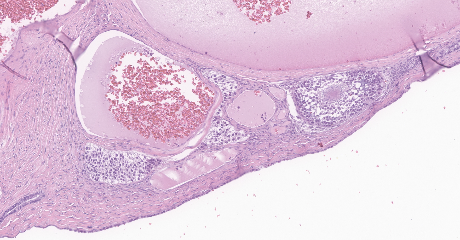
Ovary: Compressing and replacing normal ovarian tissue is a 3 x 2 cm, moderately cellular, well-demarcated, nodular, unencapsulated neoplasm composed of large blood-filled vascular spaces measuring up to 500 mm, separated by variably sized bands of fibrous connective tissue. Neoplastic cells lining the blood-filled spaces are flattened and spindeloid, with indistinct cell borders, scant amounts of fibrillar eosinophilic cytoplasm, elongate nuclei with finely stippled chromatin and indistinct nucleoli. Mitoses are less than 1 per high power field. Few vascular spaces contain thrombi measuring up to 0.5 mm, characterized by concentrically arranged fibrin with entrapped erythrocytes, completely or partially occluding the lumen. Multifocally fibrin and stromal collagen is replaced by granular basophilic material (mineralization).
Contributor's morphologic diagnosis:
Ovary, hemangioma, cavernous
Contributor's comment:
Ovarian neoplasia is a regularly found condition in dogs, but epidemiological data are rare. In a statistical analysis of cases of a private diagnostic laboratory in Germany the incidence of ovarian tumors was low (0.225 %) in a period of 10 years.10 In the recently published Swiss Canine Cancer Registry (1955-2008) tumors of female reproductive organs account only for about 0.89 %.1
Ovarian neoplasia occurs more often in older animals. In dogs often owners note behavioral changes. Additionally, lactation, vaginal discharge or pyometra can be diagnosed. The most relevant tumors of the ovarian tissue are divided into three broad categories: deriving from the surface coelomic epithelium, from the gonadal stroma or from germ cells. Nongonadal stromal tumors (e.g. vascular, fibroblastic, smooth-muscle) are very uncommon in all species.3,5,9
Ovarian hemangioma is rare in domestic animals with the exception of mature and aged sows, in which hemangioma is the most common ovarian neoplasia. Tumors are described as globular, well circumscribed and tan to red-brown. They are located within the ovarian cortex and may occur bilaterally. The tumor is composed of well differentiated endothelium lining vascular spaces and clefts. There are no signs of malignancy.2,6,8 Vascular hamartomas may also occur in the ovaries (observed in cows and sows) and a distinction between hemangioma and vascular hamartoma can be challenging.9
Hemangiomas are more common in dogs as benign mesenchymal skin tumors with distinct histological subtypes (e.g. cavernous and capillary).10
Contributing Institution:
Institut fuer Veterinaer-Pathologie, Justus-Liebig-Universitaet Giessen
Frankfurter Str. 96, 35392 Giessen, Germany
http://www.uni-giessen.de/cms/fbz/fb10/institute_klinikum/institute/pathologie
JPC diagnosis:
Ovary: Vascular hamartoma.
JPC comment:
There was spirited debate about how best to classify this unusual lesion. The robust, fibrous nature of the separating connective tissue, and the few retained follicles within the affected tissue pushed the moderator and conference participants to diagnose vascular hamartoma over hemangioma. Unfortunately, additional unstained slides were not available for this case. However, a short discussion about ovarian hemangioma is provided.
Ovarian hemangiomas are rare occurrences, and while most often are discovered as an incidental lesion in humans, they may present in association with other ovarian neoplasms, or as their own primary lesion resulting in a mass, pain, ascites, and rarely widespread abdominal malignancy. Approximately 2/3 of ovarian hemangiomas are of the cavernous type, but capillary types are found either as the primary type or as part of a cavernous type.6
Human ovarian hemangiomas are most often found in the medulla or hilus but must also be differentiated from the numerous vessels of the ovarian medulla of older animals. Some ovarian hemangiomas have been associated with stromal luteinization, which would need to be differentiated from an ovarian steroid producing tumor with pseudo-vascular degenerative change. Other uncommon vascular tumors reported in the ovary include lymphangioma, infantile hemangioendothelioma, hemangiopericytoma, and glomus tumor.4
References:
- Grüntzig K, Graf R, Hässig M, Welle M, Meier D, Lott G, Erni D, Schenker NS, Guscetti F, Boo G, Axhausen K, Fabrikant S, Folkers G, Pospischil A. The Swiss Canine Cancer Registry: a retrospective study on the occurrence of tumours in dogs in Switzerland from 1955 to 2008. J Comp Pathol. 2015;152:161-71.
- Hsu FS. Ovarian hemangioma in swine. Vet Pathol. 1983;20:401-409.
- Kennedy PC, Cullen JM, Edwards JF, Goldschmidt MH, Larsen S, Munson L, Nielsen. Histological classification of tumors of the genital system of domestic animals. WHO, Washington, DC: Armed Forces Institute of Pathology; 1998.
- Lerwill MF, Clement PB, Young RH. Miscellaneous primary tumors, secondary tumors, and nonneoplastic lesions of the ovary. In: Mills SE, ed. Sternberg's Diagnostic Surgical Pathology, 6th Ed. Philadelphia, PA: Wolters Kluwer Health. 2015:7294-7393.
- MacLachlan NJ, Kennedy PC: Tumors of the genial system. In Meuten DJ: Tumors in domestic animals. 4th ed. Ames, IO: Iowa State Press;2002:547-573.
- McEntee M: Reproductive Pathology of Domestic Mammals. San Diego, CA: Academic Press; 1990: 87-88.
- Muronda M, et al. Ovarian haemangiomas with peripheral hilus cell proliferation. Pathology. 2017; http://dx.doi.org/ 10.1016/j.pathol.2017.01.012.
- Sheikh-Omar AR, Jaafar M. Ovarian haemangioma in sows. Vet Rec. 1985;117:110.
- Schlafer DH, Miller RB: Femal genital system. In: Maxie MG, ed., Jubb, Kennedy, and Palmer's Pathology of Domestic Animals. 5th ed. Philadelphia, PA: Elsevier Limited; 2007, vol 3: 450-456
- v. Bomhard D: Epidemiologie. In Nolte I, Nolte M: Praxis der Onkologie bei Hund und Katze. Stuttgart: Enke Verlag; 2001.