CASE I: NO21-0000475 (JPC 4170014)
Signalment:
10-year-old male African Elephant (Loxodonta africana)
History:
This 10-year-old male African elephant had been diagnosed and treated over the last week for elephant endotheliotropic herpesvirus (EEHV) infection. It had chronic anemia for several years. After several days of rapidly progressing debilitation, it went down in the elephant restraint device and died, despite antiviral treatment.
Gross Pathology:
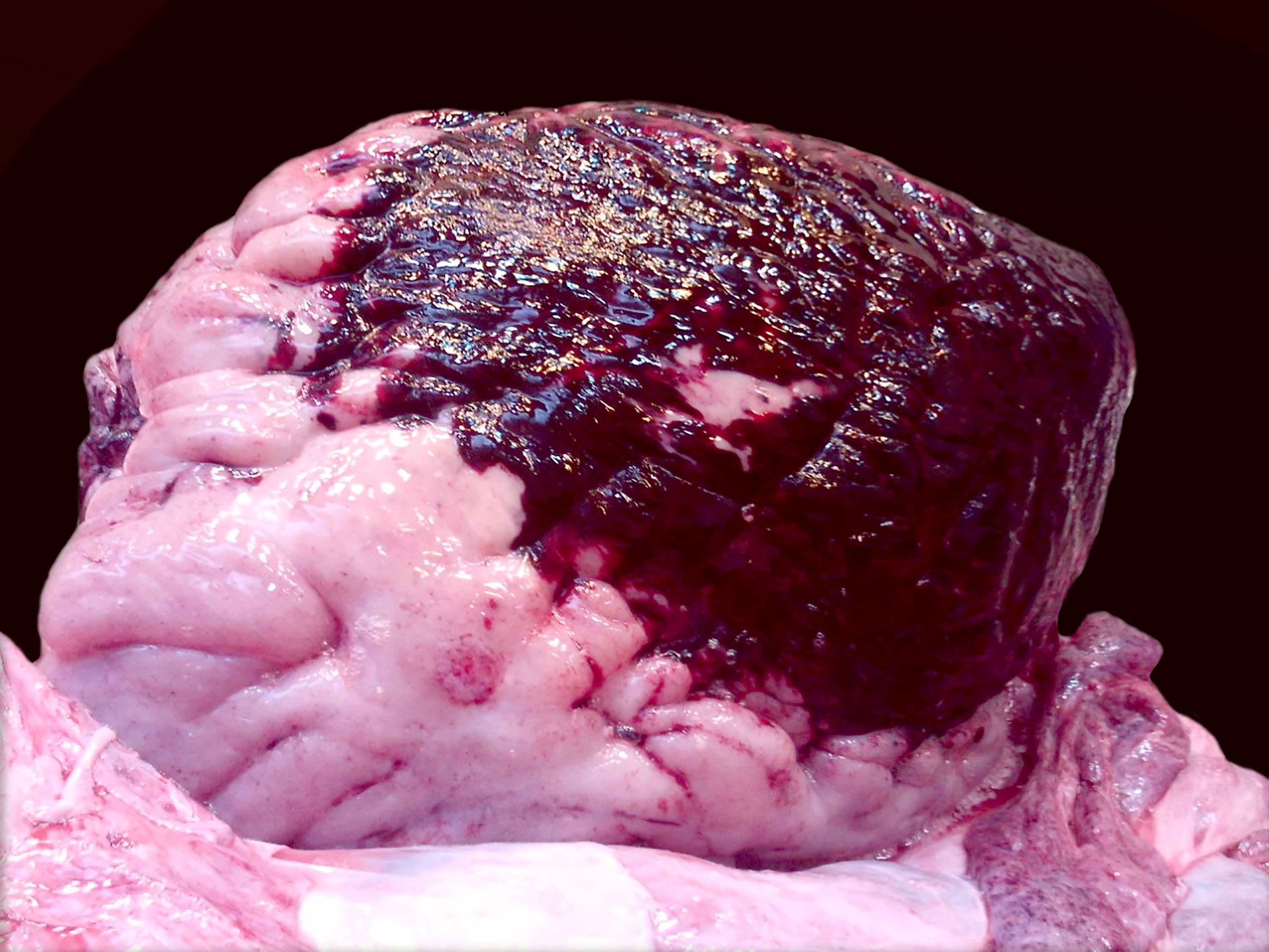
The epicardium, myocardium, and endocardium were diffusely dark red to black and there was a 5 x 5 cm region of hemorrhage at the base of the pulmonary artery. Petechial hemorrhages were noted on the gastrointestinal serosa, mesentery, lymph nodes, and there was extensive mural edema multifocally in the intestine. The bone marrow was pale.
Laboratory Results:
Perimortem, elevated EEHV-2 levels were noted with plasma levels at 435,000 vge/ml.
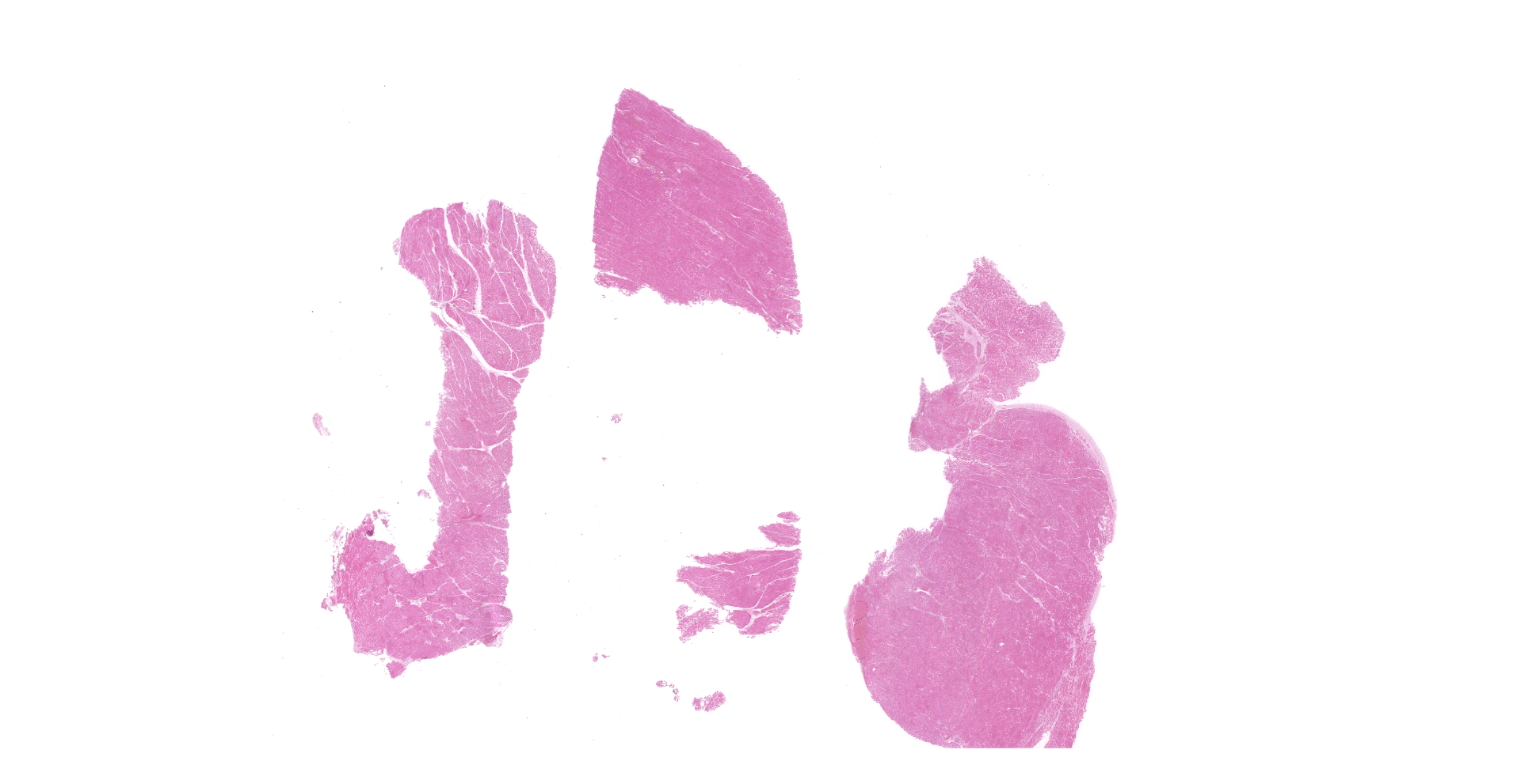
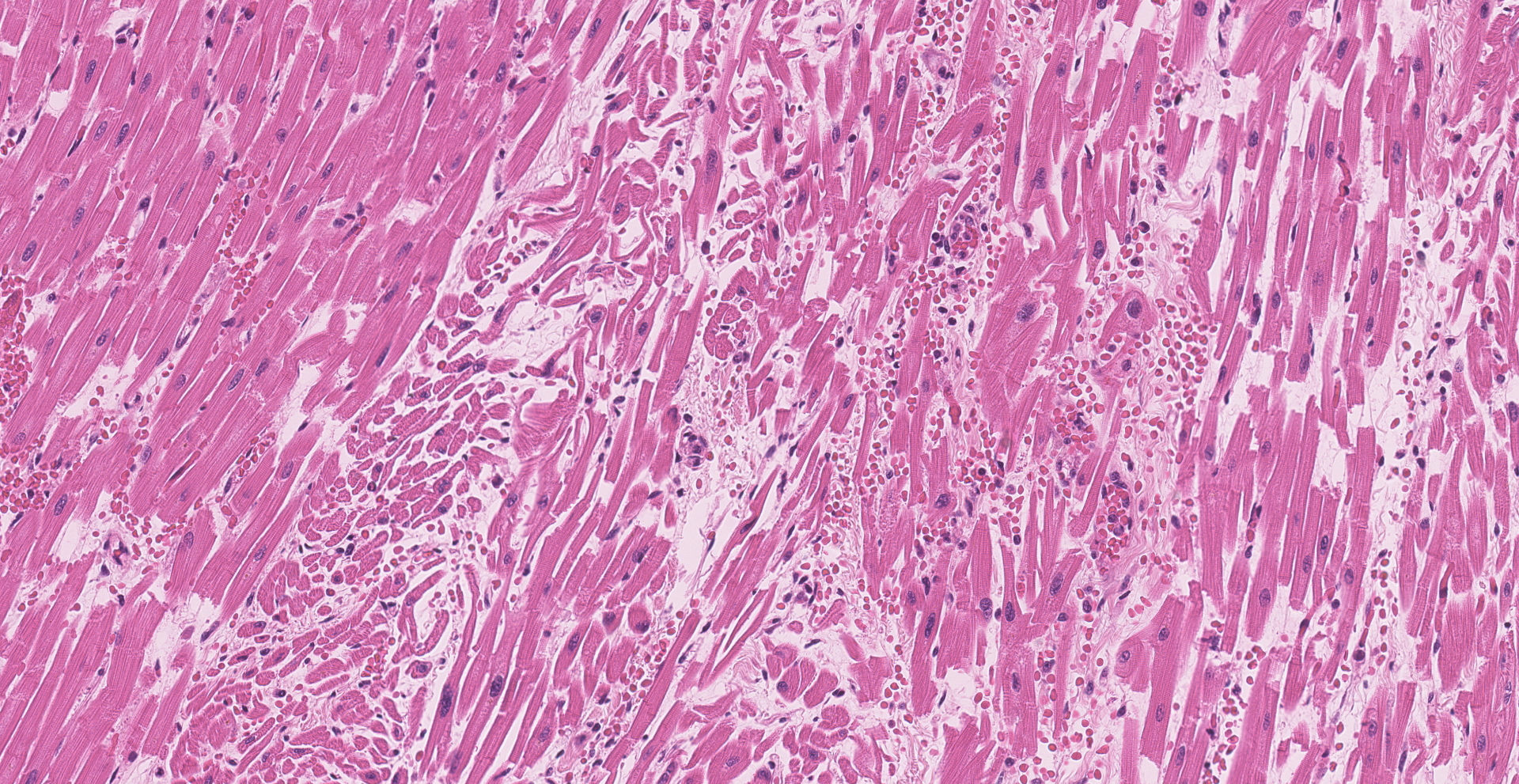
Microscopic Description:
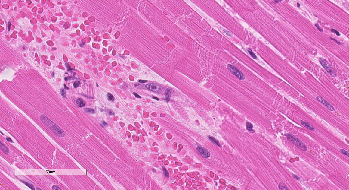
Within the heart, capillary vessels are frequently disrupted and infiltrated by neutrophils. Edema and hemorrhage are extensive in these areas, disrupting the adjacent cardiomyocytes which are shrunken, fragmented, and hypereosinophilic with moderate to marked myofiber disarray. Endothelial cells rarely contain intranuclear inclusions.
Contributor?s Morphologic Diagnoses:
Heart: Severe myocardial necrosis, degeneration, and hemorrhage with intranuclear endothelial inclusion bodies.
Contributor?s Comment:
The histologic findings are consistent with elephant endotheliotropic herpesvirus (EEHV) infection. In this case, EEHV-2 was isolated. Elephant endotheliotropic herpesvirus (EEHV) is a double stranded DNA, beta herpesvirus that causes an acute hemorrhagic syndrome (EEHV-HD) that mostly affects young Asian elephants. Studies of Asian and African elephants have shown that most Asian elephants carry strains EEHV1b, EEHV4 and EEHV5, while African elephants carry EEHV2, EEHV6 and EEHV7. Despite its widespread presence in African elephants, there have been only 13 confirmed cases of EEHV in African elephants with an approximately 50% mortality rate EEHV is known to infect endothelial cells, causing edema, hemorrhage, and coagulopathies, with capillary endothelial cells being the most affected. The heart is also often severely affected. Nonclinical African elephants may have cutaneous papillomas along the trunk and lymphoid hyperplasia in the urogenital mucosa and lungs. Currently, it is not known why some EEHV-infected elephants develop EEHV-HD, while others do not. The cause of chronic anemia in this elephant was not determined, but there was a marked decrease in the bone marrow in the samples examined.
The described virus: dsDNA, betaherpesvirus
Target à primarily young elephants
Cause: acute hemorrhagic syndrome (EEHV-HD)
Asian Elephants EEHV1b, EEHV4, and EEHV5
· only 13 cases; 50% mortality; targets endothelial cells; coagulopathy
· non-clinical à papillomas along trunk; lymphoid hyperplasia in urogenital mucosa and lungs
· Unknown why some develop disease and not
· Unknown why anemia in this particular elephant, though marked bone marrow decrease
African Elephants: EEHV2, EEHV6, and EEHV7
Contributing Institution:
National Institutes of Health
Bethesda, MD 20892
https://www.ors.od.nih.gov/sr/dvr
JPC Diagnosis:
Heart: Vasculitis, necrotizing, multifocal, moderate, with myocardial hemorrhage and edema, and rare endothelial intranuclear viral inclusions.
JPC Comment:
The contributor provides a concise review of elephant endotheliotropic herpesvirus (EEHV), a significant cause of mortality in predominately juvenile Asian and African elephants.
In 1988, a young Indian elephant in a Swiss circus died from an unknown acute hemorrhagic disease, which was followed by nearly 20 similar cases in North America and Europe during the 1990s. In 1995, a veterinary pathology resident, Dr. Laura Richman, identified herpesvirus-like particles using electron microscopy while investigating endothelial cell nuclear inclusion bodies in heart and liver samples from an Asian elephant from the Smithsonian National Zoo. Four years later, Richman and her colleagues published a report describing the detection of DNA from a novel herpesvirus called ?elephant endotheliotropic herpesvirus?. Since its discovery, EEHV has been identified in both captive and wild elephants and is the most common cause of mortality in juvenile Asian Elephants (Elephas maximus) in both North America and Europe.5 As a testament to the virus' significance, EEHV was responsible for the deaths of 29.6% of Asian elephants born in captivity in the United Kingdom and Ireland between 1995 and 2013.3
One retrospective review6 of 27 cases of EEHV in Indian elephants found the most common gross lesion to be myocardial hemorrhage, followed by multisystemic hemorrhage, blue or purple tongue discoloration, pericardial effusion, edema, and ascites. Common areas of non-cardiac hemorrhage included the gastrointestinal mucosa and serosa, liver, pancreas, and lungs. Histologically, all cases demonstrated hemorrhage within the heart, spleen, and thymus, with the most severe hemorrhage noted in the heart and spleen. Cardiac hemorrhage was most severe in the sub-endocardial and sub-epicardial regions. Microthrombi were present in 63% of cases, most commonly in the lungs in addition to other organs such as the kidneys and liver. 100% of lung sections demonstrated endothelial cell damage characterized by separation, sloughing, denudation, or loss, followed by edematous expansion of the tunica intima (95%) and migrating leukocytes (67%). Similar lesions were present within the hepatic blood vessels, predominantly within the sinusoids and portal capillaries. The most consistent organs demonstrating endothelial viral nuclear inclusions were the heart and liver.6
The same study concluded disseminated intravascular coagulation (DIC) is likely associated with end stage EEHV, with key histologic features including the presence of microthrombi and widespread hemorrhage in multiple tissues. DIC is an acquired coagulopathy that commonly precedes multiorgan failure, cardiovascular failure, and death. The most likely pathogenesis associated with EEHV stems from endothelial cell damage with secondary exposure of subendothelial tissue factor. Exposure of tissue factor results in both platelet activation and initiation of the extrinsic coagulation pathway. Extensive endothelial damage, as is likely in cases of EEHV, can overwhelm localized homeostasis control mechanisms, resulting in generalized activation of the coagulation cascade. If the underlying cause is unable to be resolved, both platelets and coagulation factors become depleted and hemostasis can no longer be maintained, resulting in multifocal areas of hemorrhage.6
Asian elephants with EEHV have successfully been treated with famciclovir, an antiviral medication, in addition to supportive therapy such as rectal and intravenous fluids, conspecific plasma, diuretics for edema, antibiotics, and oxygen. However, administration of antivirals and supportive care is most likely to be successful when initiated prior to onset of clinical signs. Subclinical viremia has been detected in Asian elephants up to 28 days prior to onset of clinical signs. Therefore, the optimal method suggested for identification of subclinical cases is regular PCR analysis of blood samples of young Asian elephants.3 Additional methods of sample collection for surveillance may include trunk washes, conjunctival swabs, and saliva swabs as viral shedding typically commences 10 days after onset of viremia.1 A recently published study demonstrated the ability to detect EEHV in elephant feces using qPCR, although sensitivity was significantly lower compared to saliva swabs. However, a similar method may one day provide a suitable non-invasive method of early detection of EEHV in captive elephants while also facilitating surveillance of shedding in wild herds.1
A notable feature of EEHV is severe and lethal disease is practically non-existent in animals less than one year of age, suggesting maternal antibodies likely provide protection against disease, if not infection.5
During the conference, the moderator discussed encephalomyocarditis virus (ECMV) as a differential diagnosis in this case. In comparison to EEHV, ECMV is typically associated with more severe myocardial degeneration and necrosis, less hemorrhage, and endothelial intranuclear viral inclusions are not associated with this entity. However, based on the moderator's experience, viral inclusions are infrequently present cases of EEHV and additional diagnostics (e.g. PCR) are frequently required for definitive diagnosis.
References:
1. Common SM, Yun Y, Silva-Fletcher A, et al. Developing a non-invasive method of detecting elephant endotheliotropic herpesvirus infections using faecal samples. Vet Rec. 2022;190(2):e833.
2. Fayette MA, Brenner EE, Garner MM, Bowman MR, Latimer E, Proudfoot JS. Acute hemorrhagic disease due to elephant endotheliotropic herpesvirus 3A infection in five African elephants (loxodonta africana) at one North American zoological institution. J Zoo Wildl Med. 2021;52(1):357-365.
3. Kendall R, Howard L, Masters N, Grant R. The impact of elephant endotheliotropic herpesvirus on the captive Asian elephant (Elephas maximus) population in the United Kingdom and Ireland (1995-2013). J Zoo Wildl Med. 2016;47(2):405-418. doi:10.1638/2015-0217.1
4. Landolfi JA, Terrell SP. Proboscidae. In: Terio KA, McAloose D, Leger JS, eds. Pathology of Wildlife and Zoo Animals. San Diego, CA: Elsevier, Inc; 2018:420-423.
5. Long SY, Latimer EM, Hayward GS. Review of Elephant Endotheliotropic Herpesviruses and Acute Hemorrhagic Disease. ILAR J. 2016;56(3):283-296.
6. Perrin KL, Kristensen AT, Bertelsen MF, Denk D. Retrospective review of 27 European cases of fatal elephant endotheliotropic herpesvirus-haemorrhagic disease reveals evidence of disseminated intravascular coagulation. Sci Rep. 2021;11(1):14173. Published 2021 Jul 8.