CASE III:18-0933b Case 1 (JPC 4125007)
Signalment:
Red kangaroo (Osphranter rufus)
History:
Culled female free-ranging red kangaroo examined as part of investigations into mass mortalities.
Gross Pathology:
No significant external findings
Laboratory results:
No laboratory findings reported.
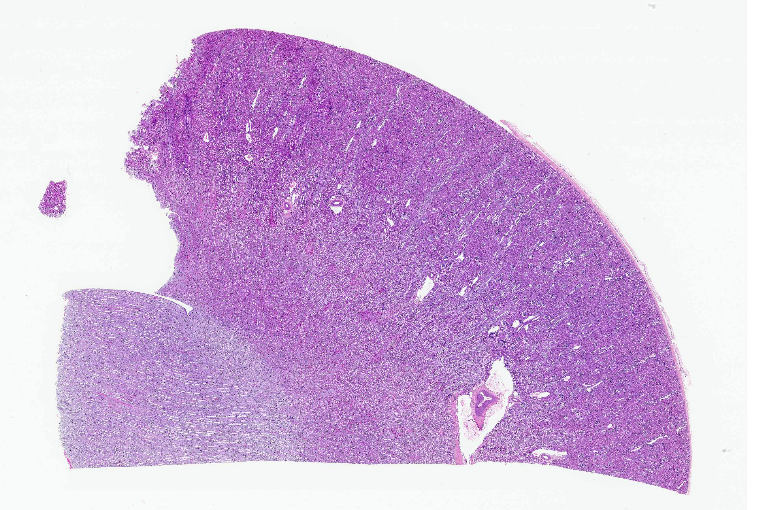
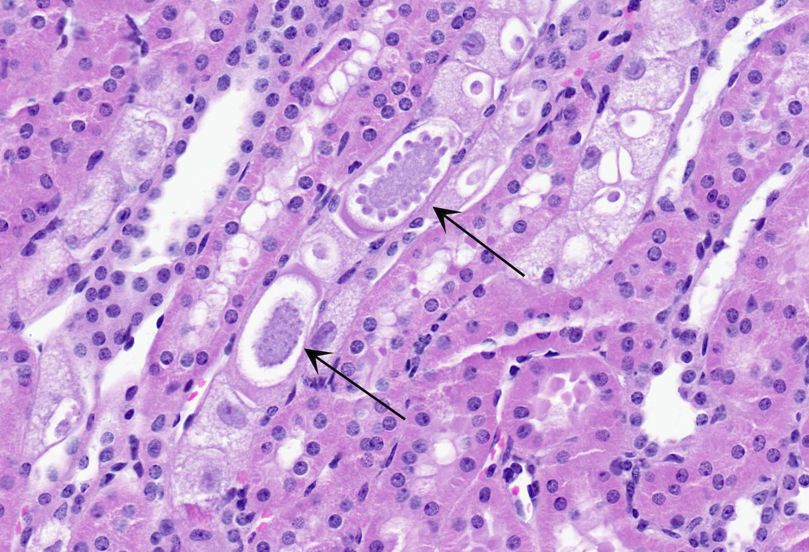
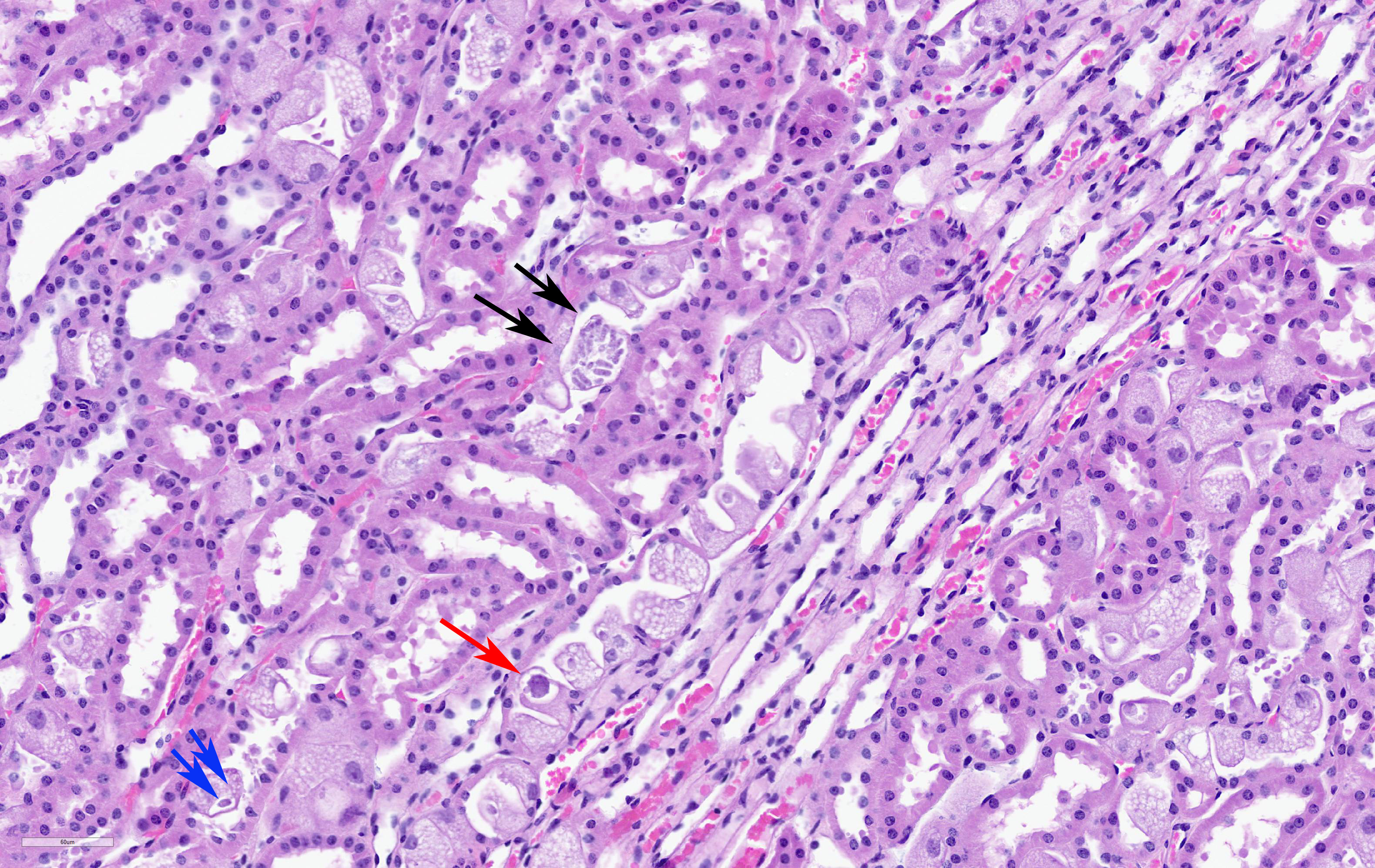
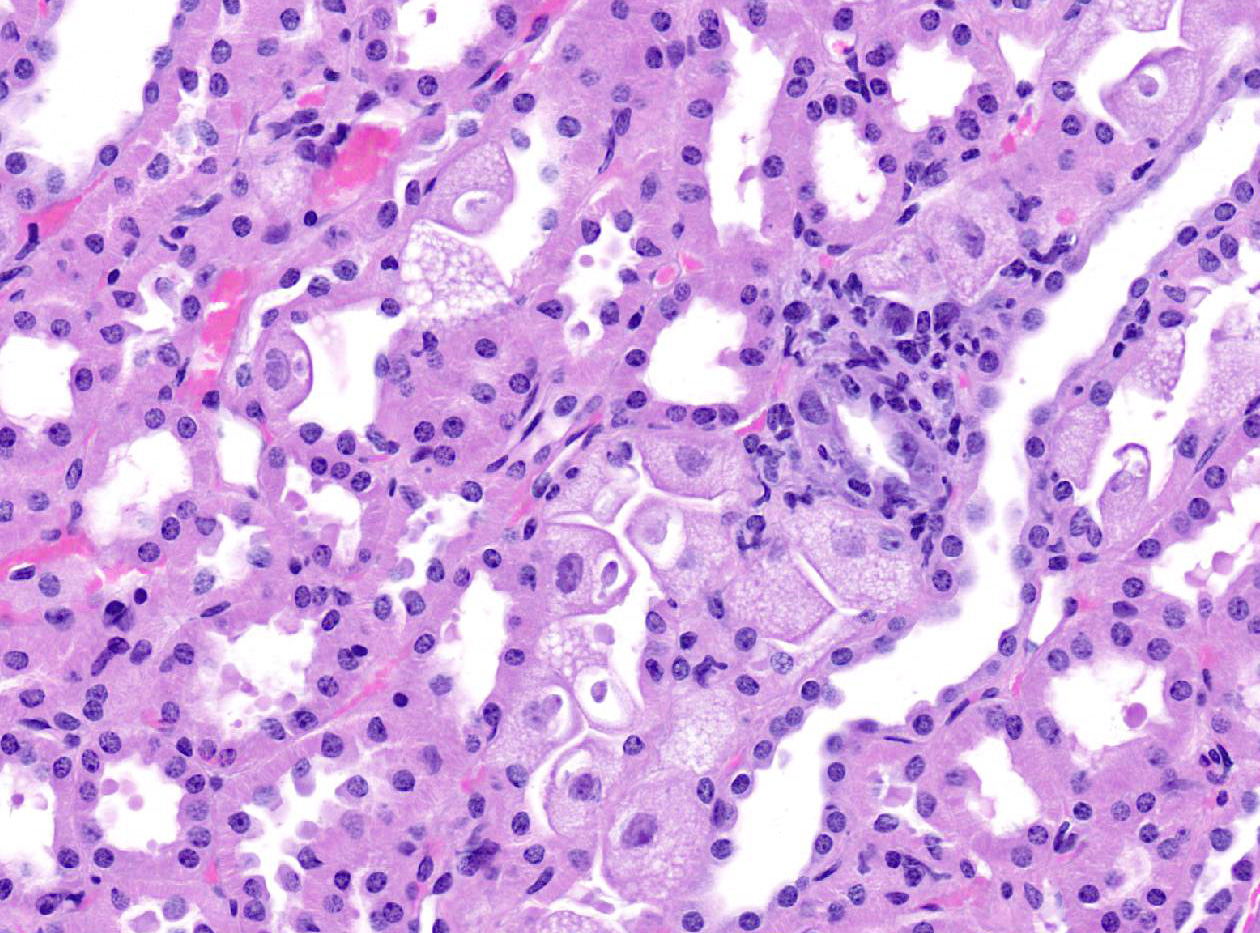
Microscopic Description:
Kidney: Renal tubular epithelial cells, concentrated in the corticomedullary region and medullary rays are frequently hypertrophied, characterised by karyomegaly and cytomegaly and large cytoplasmic vacuoles. Vacuoles often contain protozoal macrogametes, sporonts, and sporozoites (parasitopherous vacuoles). Macrogametes, 6- 8 um in diameter, are most frequently observed within hypertrophied cells that bulge into the tubular lumina. Intraepithelial sporoblasts and basophilic crenated sporocysts containing closely packed sporozoites are also common. Sporonts (with circumferential nuclei) are rarely observed, intraepithelial shizonts are not observed. Epithelial cells of parasitized tubules are sometimes degenerate or attenuated, but minimal appreciable inflammation is observed.
Contributor's Morphologic Diagnoses:
Kidney:
renal coccidiosis (Klossiella sp)
Contributor's Comment:
Protozoal parasites within the renal tubules of this kangaroo are morphologically consistent with Klossiella sp. This parasite is widely spread in both wild and captive Australian marsupials and is thought to be largely non-pathogenic in otherwise healthy individuals.1-5,8
Klossiella spp. are apicomplexan coccidians with a global distribution, known to infect the kidneys of mammals. Usually infecting the renal tubular epithelium, it may also be found within glomerular endothelial cells in some cases. There are 19 recognized species in this genus and additional reports without species characterization.9 The life cycle is assumed to be direct with schizogony and sporogony occurring in the host's renal tissue, and infection likely acquired by the oral route after contamination of food and drinking water with urine containing sporocysts.9 Species identification is based on the morphology of the different life-cycle forms and the parasitized tissues; although not definitively determined in this case, the host and parasite morphology suggest this species is most likely K. rufi.3
Contributing Institution:
The University of Adelaide, Veterinary Diagnostic Laboratory
JPC
Diagnosis:
Kidney, tubular epithelium: Hypertrophy, multifocal, severe, with numerous intraepithelial apicomplexan sporonts and gametes, etiology consistent with Klossiella sp.
JPC Comment:
Apicomplexans of the genus Klossiella have been reported in multiple species, including mice (K. muris), guinea pig (K. cobayae), horse, zebra, jackass, burro, donkey (K. equi), South American opossum and mouse opossum (K. tejarai), Australian water rat (K. hydromyos), African bats (K. killicki), African muridae (K. mabokensis), snakes (K. boyae), birds, and six different species in Australian marsupials.5
Klossiella sp. are intracellular apicomplexan protists that infect renal epithelium. Following ingestion of an infective sporocyst, sporozoites are released and move to the kidney, where they enter tubular epithelial cells and become trophozoites. Trophozoites form schizonts and merozoites, and from these schizonts, gametes are thought to form. Fertilized gametes are believed to develop into sporonts, which bud to form sporoblasts. Each sporoblast undergoes successive divisions to form sporocysts that contain sporozoites. Mature infective sporocysts are surrounded by a thick wall and pass from the body into the urine to be later ingested by another host, completing the cycle.7
Most coccidians infect the gastrointestinal tract in their natural hosts; however, genus Klossiella's renal tropism is not unique. In contrast to Klossiella, renal coccidiosis due to Eimeria truncata is known to be a highly fatal disease of geese, sometimes wiping out entire flocks within a few days. Symptomatic geese are typically extremely weak and emaciated. Grossly, kidneys of severely affected birds are greatly enlarged and discolored, corresponding with the microscopic features of enormous numbers of coccidia within tubules, which are sometimes extensively destroyed.6 Multiple species of the genus Eimeria infect the avian renal tubular epithelium, including E. somateriae in long tailed ducks, E. boschadis in mallard ducks, and a novel species in great-horned owls.7,9
References:
1. Ardiaca M, Bennett MD, Montesinos A, Juan-Salles C, Soriano-Navarro M: Klossiella Dulcis N. Sp. (Apicomplexa: Klossiellidae) in the Kidneys of Petaurus Breviceps (Marsupialia: Petauridae). J Zoo Wildl Med 2016:47(2):622-627.
2. Barker IK, Munday BL, Harrigan KE: Klossiella spp. in the kidneys of peramelid, petaurid, and macropodid marsupials. Z Parasitenkd 1975:46(1):35-41.
3. Barker IK, Munday BL, Hartley WJ: Klossiella (Apicomplexa, Klossiellidae) in petaurid and macropodid marsupials in Australia. J Protozool 1985:32(3):520-522.
4. Bennett MD, Woolford L, O'Hara AJ, Nicholls PK, Warren KS, Friend JA, et al.: Klossiella quimrensis (Apicomplexa: Klossiellidae) causes renal coccidiosis in western barred bandicoots Perameles bougainville (Marsupialia: Peramelidae) in Western Australia. J Parasitol 2007:93(1):89-92.
5. Duncan, M. Perissodactyls. In: Terio KA, McAloose D, St. Leger J, eds. Pathology of Wildlife and Zoo Animals. San Diego, CA; Elsevier Inc., 2018: 450-451.
6. Farr MM, Wehr EE. Eimeria truncata associated with morbidity and death of domestic goslings. Cornell Vet. 1952;42(2):185-187.
7. Gardiner CH, Fayer R, Dubey JP, eds. An Atlas of Protozoan Parasites in Animal Tissues, 2nd ed. Armed Forces Institute of Pathology, Washington, DC, 1998:61-62.
8. Jankovsky JM, Brand M, Gerhold RW. Identification of a Novel Renal Coccidian (Apicomplexa: Eimeriidae) from the Great-Horned Owl ( Bubo virginianus ), USA. J Wildl Dis. 2017;53(2):368-371.
9. Taylor JL, Wagner JE, Kusewitt DF, Mann PC: Klossiella Parasites of Animals - Literature-Review. Veterinary Parasitology 1979:5(2-3):137-144.
10. Wobeser G. Renal coccidiosis in mallard and pintail ducks. J Wildl Dis. 1974;10(3):249-255.