Signalment:
Gross Description:
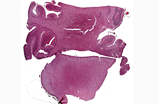
Histopathologic Description:
Kluver and Barerra stains showed the material was bright blue (consistent with myelin). No PAS positive material was observed.
Morphologic Diagnosis:
Lab Results:
Condition:
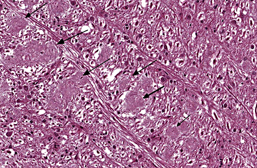
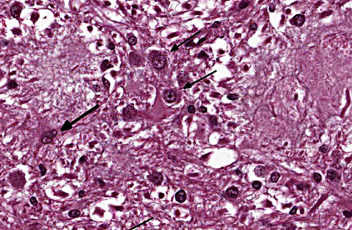
Contributor Comment:
Ultrastructurally, the disease is characterized by hypertrophic and hyperplastic oligodendrocytes emitting numerous and disorganized small processes around the node of Ranvier, leading to myelin sheaths malformation in the white matter. The histological examination displays haphazard, multifocal, pale, eosinophilic, granular to fibrillar, 20-100 μm plaques which are pathognomonic for this disease, along with mild oligodendroglial hyperplasia, discrete astrocytosis, lymphocytic infiltration and cuffing.(2,4,5,6)
JPC Diagnosis:
Conference Comment:
This condition is reported in both sexes of Charolais cattle, and has also been reported in three-quarter crossbred as well as purebred animals. Gross lesions are absent, but the microscopic lesions are unique and distinctive. Axons are found to traverse the plaques with mostly normal myelin and no sign of degradation or phagocytosis of the myelin. Evidence of axonal degeneration, when found, is mild. As mentioned above, ultrastructural examination suggests the abnormality lies within the paranodal region of axons. Younger plaques are characterized by axons wrapped with hypertrophied oligodendrocyte processes, in a thin myelin sheath, near the nodes of Ranvier. In older plaques the axons may be demyelinated and surrounded by more abundant oligoden-drocyte processes. The defect lies in the inability of oligodendrocytes to maintain the paranodal myelin structure with resultant oligodendrocyte hypertrophy.(2)
There are two types of oligodendrocytes within the CNS, interfascicular oligodendrocytes and satellite cells. The interfascicular oligodendrocytes are respon-sible for myelination of axons and normally they appear as small cells with hyperchromatic nuclei; special staining techniques, such as myelin basic protein, are normally required to visualize their processes. The nuclei of oligodendrocytes are often seen aligned in rows parallel to myelinated axons, where they maintain the internodal segments of the myelin sheath.(7)
References:
1. Duchesne A, Eggen A. Radiation hybrid mapping of genes and newly identified microsatellites in candidate regions for bovine arthrogryposis-palatoschisis and progressive ataxia based on comparative data from man, mouse and rat. J Anim Breed Genet. 2005;122(Suppl.1):28-35.
2. Maxie MG, Youssef S. The nervous system. In: Maxie MG ed. Jubb, Kennedy and Palmers Pathology of Domestic Animals. 5th ed. Vol 1. Edinburgh, UK: Saunders Elsevier; 2007:381-385.
3. Morrison JP, Schatzberg SJ, De Lahunta A, Ross JT, Bookbinder P, Summers BA. Oligodendroglial dysplasia in two Bullmastiff dogs. Vet Pathol. 2006 ;43:29-35.
4. Parodi AL. Progressive ataxia in cattle study of histologic lesions. Recueil de Medecine Veterinaire de l'Ecole d'Alfort. 1981 ; 339-345. (in French)
5. Patton CS. Progressive ataxia in Charolais cattle. Vet Pathol. 1977;14:535-537.
6. Vandevelde M, Higgins R, Oevermann A . Veterinary Neuropathology: Essentials of Theory and Practice. Chichester, UK: Wiley-Blackwell; 2012:176-177.
7. Zachary JF. Nervous System. In: McGavin MD, Zachary JF, eds. Pathologic Basis of Veterinary Disease. 5th ed. St. Louis, MO: Mosby Elsevier; 2012:777-778.