Signalment:
10-year-old Holstein-Friesian cow from a dairy herd (
Bos Taurus). This cow presented to the large animal teaching hospital with a history of being off feed and having decreased milk production. A swelling in the subcutis of her left ventral abdomen was noted. This cow had a history of clinical mastitis in the left front quarter, and was being treated with intravenous antibiotics including sulfadimethoxine and oxytetracycline. Heart sounds were muffled, and multiple comet tails were noted on ultrasound examination of the thorax.
Gross Description:
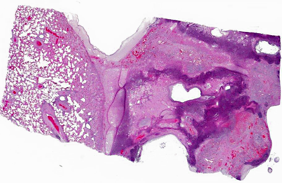
There is a moderately increased amount of blood-flecked foam in the distal 1/2 of the trachea and bronchial tree. The lungs contain approximately 100, 0.5 cm to 1 cm, firm, round, red-brown nodules with caseous or purulent centers; these nodules are evenly distributed through all lung lobes. Additionally, there are smaller numbers of subpleural and randomly distributed foci comprising solitary or multiple closely apposed or coalescing, 1 to 2 cm diameter, air-filled bullae surrounded by firm white tissue and lined by green-black, tenacious necrotic material and scattered, variably sized, velvety, white to brown plaques.
Histopathologic Description:
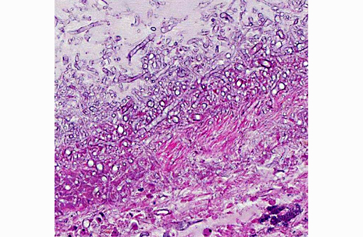
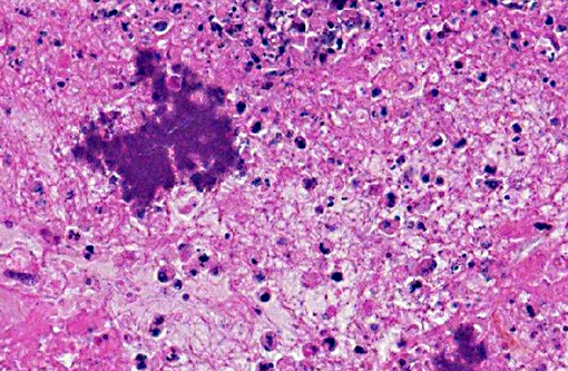
Obliterating over 50% of the lung parenchyma in the examined section is a focally extensive, empty space bounded by a dense band of fungal hyphae embedded in eosinophilic, amorphous to granular necrotic material and fibrin. Fungal hyphae stain relatively poorly with PAS and GMS, and are characterized by nonparallel walls, a lack of septa, irregular, non-dichotomous branching at right angles, and occasional bulbous dilatations. The adjacent pulmonary parenchyma is characterized by lytic or coagulative necrosis, multifocal hemorrhage, granulation tissue, and florid infiltrates of viable and degenerate neutrophils with fewer macrophages, eosinophils, lymphocytes and plasma cells. Within this surrounding tissue are many large colonies of gram positive short rods. Bronchiolar epithelium is multifocally attenuated or eroded, and airway lumina rarely contain proteinaceous material admixed with small numbers of degenerate inflammatory cells. In less affected areas of the lung, alveolar spaces are filled by edema, fibrin, and foamy macrophages and bronchioles are spared.
Morphologic Diagnosis:
Pneumonia, necrosuppurative, chronic-active, multifocal, severe, with intralesional fungal hyphae consistent with zygomycetes and gram positive rods. (
Arcanobacterium pyogenes and
Rhizomucor spp. [not further classified] were cultured from the lung).
Condition:
Zygomycotic pneumonia
Contributor Comment:
The class Zygomycetes is divided into the orders
Mucorales and
Entomophthorales. Members of the order
Mucorales tend to cause disseminated disease characterized by angioinvasion, while members of the
Entomophthorales tend to cause localized subcutaneous granulomas.(2) Mucormycosis generally manifests as subcutaneous, systemic or rhinocerebral infections.(2) The main portal of entry in bovine mucormycosis is reported to be the gastrointestinal tract; however, a recent report of 194 slaughtered feedlot steers with granulomatous lymphadenitis and intralesional fungal hyphae noted that no evidence of gastrointestinal disease or systemic spread was present in any of the animals, and that other portals of entry could not be completely ruled out.(1) Zygomycetes are cosmopolitan and ubiquitous, and at least one member of the genus
Rhizopus, R. pusillis, is thermophilic.(1) The presence of broad, infrequently septate hyphae with irregular branching is characteristic of these fungi and can be seen in routine H & E stains, but visualization is greatly augmented by the periodic acid-Schiff reaction or Gomoris methanamine silver stain. In the present case, because
Arcanobacterium pyogenes (but not fungi) were cultured from the mammary gland, it is likely that the bacterial mastitis was the primary source of an embolic pneumonia that was further complicated by aerogenous zygomycete infection. There was no gross or histological evidence of fungal infection in any other tissue, including mammary gland and gastrointestinal tract.
JPC Diagnosis:
Lung: Pleuropneumonia, necrosuppurative, chronic, focally extensive, severe, with vasculitis, numerous colonies of bacilli and fungal hyphae.
Conference Comment:
The contributor provides a good summary of the fungal aspect of this mixed infection. Conference participants debated on the bacterial agent; based on the combination of lytic and coagulative necrosis observed, some participants considered
Mannheimia or
Mycoplasma. These entities were ruled out, however, as neither of them would be present in large colonies as seen in this slide. Recently
Arcanobacterium pyogenes (formerly
Actinomyces pyogenes and
Corynebacterium pyogenes) has undergone another taxonomic reclassification and subsequent name change and is now known as
Trueperella pyogenes. Based on phylogenetic and chemotaxonomic differences, the genus
Arcanobacterium was divided into two genera--
Arcanobacterium and a novel genus,
Trueperella. T. pyogenes is the type species for this new genus.(4) Both genera are in the family
Actinomycetaceae, a diverse group of gram positive bacteria that also includes the genera
Actinomyces and
Actinobaculum.(3)
T. pyogenes is an opportunistic pathogen that clinically causes abscesses, mastitis, suppurative pneumonia, endometritis, pyometra, arthritis and umbilical infections in cattle, sheep and pigs. Its pathogenicity is due to several virulence factors, primarily pyolysin, a hemolytic exotoxin that lyses neutrophils, macrophages and other cell types. Additionally, they produce neuraminidases, adhesins, extracellular matrix-binding proteins and fimbriae.Â
T. pyogenes produces proteases as well, but their role in pathogenicity is yet to be determined. Other members of veterinary importance in the family
Actinomycetaceae include: Actinomyces bovis (lumpy jaw in cattle), Actinomyces species (pyogranulomatous mastitis in pigs and poll evil and fistulous withers in horses),
Actinomyces viscosus (cutaneous pyogranulomas in dogs and horses, proliferative pyogranulomatous pleuritis in dogs, abortion in cattle),
Actinobaculum suis (cystitis and pyelonephritis in pigs), and
Actinomyces hordeovulneris (cutaneous and visceral abscessation, pleuritis, peritonitis, and arthritis in dogs).(3)
References:
1. Ortega J, et al. Zygomycotic Lymphadenitis in Slaughtered Feedlot Cattle.Â
Vet Pathol. 2010;47(1):108-115.
2. Ginn P, Mansell JEKL, Rakich PM. In: Maxie MG, ed.
Jubb, Kennedy and Palmers Pathology of Domestic Animals. Vol. 1. 5th ed. Philadelphia, PA; Saunders Elsevier: 2007;707-8.
3. Quinn PJ, et al. Actinobacteria. In:
Veterinary Microbology and Microbial Disease. 2nd ed. Ames, Iowa: Wiley Blackwell; 2011, Kindle edition, location 6945 of 35051.
4. Yassin AF, Hupfer H, Siering C, Schumann O. Comparative chemotaxonomic and phylogenetic studies on the genus
Arcanobacterium Collins et al. 1982 emend. Lehnen et al. 2006: proposal for
Trueperella gen. nov. and emended description of the genus
Arcanobacterium. Int J Syst Evol Microbiol. 2011;61:1265-74.