Signalment:
Gross Description:
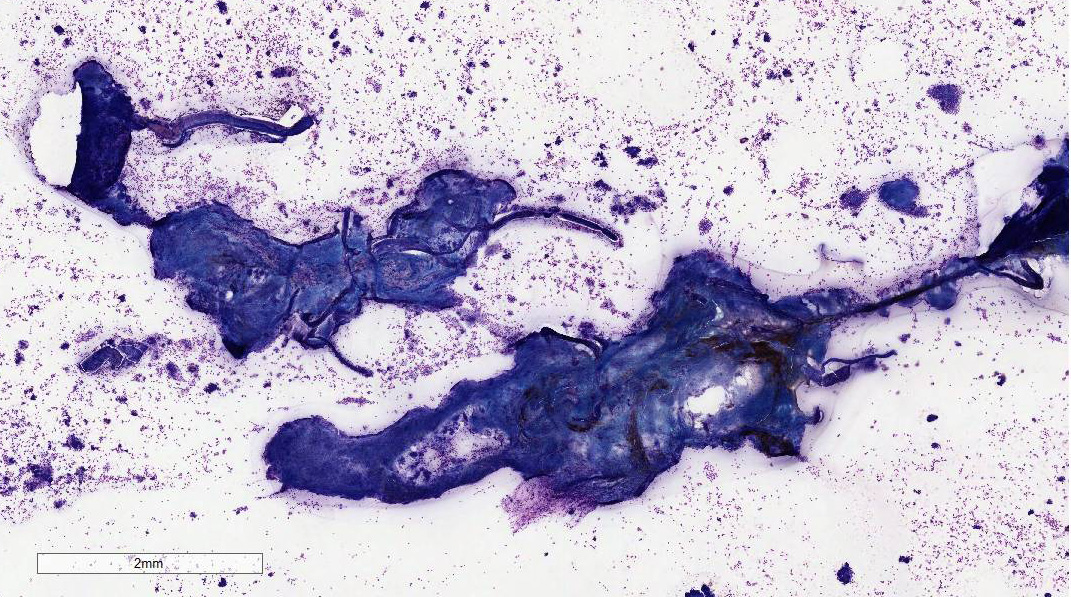
Histopathologic Description:
showing the same
morphology as those within the dense clusters. There are areas of monolayer
adjacent to the dense clusters with a moderate number of neutrophils and
eosinophils. Eosinophils, neutrophils, and small lymphocytes are found
throughout the background. Occasional small lymphocytes contain a few
eosinophilic to azurophilic granules.
Table 1:
Capillarid Species Name Location Host Eucoleus
bouhmi frontal sinus fox, dog Eucoleus
aerophilus bronchi dog, cat, fox Aonchotheca
putorii stomach,
intestine bear,
hedgehog, raccoon, swine, bobcats, mustelids, cat Aonchotheca spp intestine ruminants Calodium
hepaticum liver rodents, many
occasional hosts including humans. Pearsonema
plica urinary
bladder dog, fox, wolf Pearsonema
feliscati urinary
bladder cat Table 2:
Urinary Nematodes Name Host Dioctophyme
renale dog, mink Pearsonema
plica,
P. feliscati dog, fox, wolf,
cat Stephanurus
dentatus swine Trichosomoides
crassicauda rats Crassicauda
boopis whales embedded
parasite. The lifecycle for P. feliscati is not determined.2,6
It is assumed to be similar to P. plica which is thought to be through
the earthworm as an intermediate host or a transport host such as a bird.
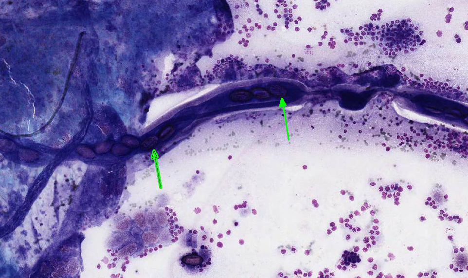
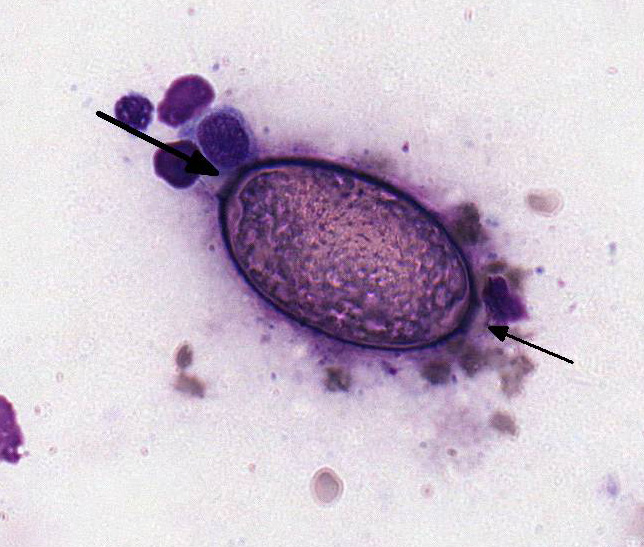
Adult worms are 2.5 to 5 cm in length. The ova are elongate and approximately
60 microns in length and 27 microns in width. They have bipolar plugs
(bioperculate), a thick shell with globular ridges enclosing a single cell.10
The literature does not provide morphologic or biological characteristics other
than the host to distinguish P. plica and P.
feliscati. The cat is included in host species for both P. feliscati
and P. plica in Georgis Parasitology for Veterinarians with no
mention of differential characteristics.2 The capillarid in this
case is given as P. feliscati based on host. JPC Cytologic
Interpretation: Presence
of Peasonema spp nematode fragments and ova, mild transitional cell
dysplasia, and low grade eosinophilic and neutrophilic inflammation, Siamese
cross, Felis catus.
embedded
parasite. The lifecycle for P. feliscati is not determined.2,6
It is assumed to be similar to P. plica which is thought to be through
the earthworm as an intermediate host or a transport host such as a bird.
Adult worms are 2.5 to 5 cm in length. The ova are elongate and approximately
60 microns in length and 27 microns in width. They have bipolar plugs
(bioperculate), a thick shell with globular ridges enclosing a single cell.10
The literature does not provide morphologic or biological characteristics other
than the host to distinguish P. plica
and P.
feliscati. The cat is included in host species for both P. feliscati
and P. plica in Georgis Parasitology for Veterinarians with no
mention of differential characteristics.2 The capillarid in this
case is given as P. feliscati based on host.
Morphologic Diagnosis:
Lab Results:
Condition:
Contributor Comment:
In the cat, the
disease is rarely reported in the United States;6 however, in
Australia, an incidence of greater than 30% was found in one survey study.8
No evidence of clinical cystitis was seen in the infected cats. On histological
examination of the urinary bladder in the infected cats, the nematodes were
superficially embedded in the mucosa with no breaching of the basal layer or
basement membrane. A moderate inflammatory infiltrate that included eosino-phils
was seen in association with the
JPC Diagnosis:
Conference Comment:
In
both dogs and cats, Peasonema plica has been reported in the urinary
bladder and ureter submucosa and is typically associated with mild subclinical
inflammation and edema.1,7 There is a higher
prevalence of the parasite in wild red foxes (Vulpes vulpes) in many
European countries and it is associated with an increased pathogenicity. This
can result in severe cystitis, pollakiuria, dysuria and hematuria in this
species.1,3 In this case, both Pearsonema plica and Pearsonema
feliscati have been implicated in urinary bladder infection in cats,
conference participants could not distinguish between two cytologically. Conference participants readily identified
numerous bioperculate eggs free within the sample and inside the reproductive
tract of adult nematode fragments. Pearsonema spp are a
subclassification of aphasmid nematodes of the family Trichuridae.
Histologically, aphasmid nematodes are characterized by thin eosinophilic
cuticle, reduced polymyarian-coelomyarian musculature, two hypodermal bacillary bands stichosome esophagus,
a spiny sheath and oval
bioperculate eggs.4 Cytologically, specific features of the adult
nematode can be more difficult to appreciate than on histologic tissue
section. However, the presence of the highly characteristic ova
confirms the presence of a urinary capillarid nematode.
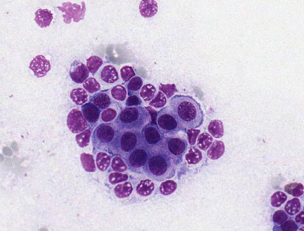
Participants
also noted vacuolation, binucleation, anisokaryosis, and prominent nucleoli
within the reactive sloughed urothelium. Transitional epithelial cells are
among the most pleomorphic cells in the body and can demonstrate marked
reactive change in response to a variety of insults. For this reason, the
conference moderator reminded participants that care must be taken prior to
over-interpreting reactive urothelium as malignancy.
References: