Signalment:
Gross Description:
Histopathologic Description:
In lesions not shown, this animal also had an exocrine pancreatic adenocarcinoma, with surrounding inflammation. Vacuolar change was discovered in the liver, along with bile stasis. Pyogranulomas were found in the spleen and were immunohistochemically positive for FIP antigens.
Morphologic Diagnosis:
Lab Results:
Condition:
Contributor Comment:
PCR is the test capable of identifying the greatest number of cats (34/36), versus culture (in pouch 24/36 or Diamonds media (5/36).(2) Other tests can include wet mount smears of loop samples from the rectum and chromogenic in situ hybridization.(4) A confounding factor is the presence of Pentatrichomonas hominis.(3)
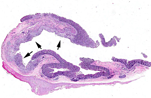
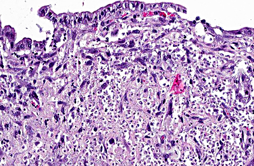
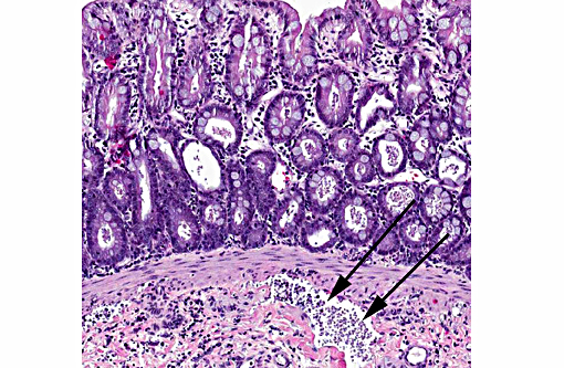
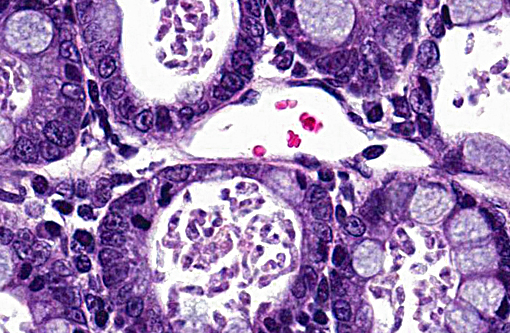
In sections from spontaneously infected cats, lesions included an increase in lymphocytes and plasma cells in the lamina propria as measured across the width of the villi, and a few infiltrating neutrophils. Trichomonads were tear-shaped to crescentic organisms; flagella were not visible. Organisms were on the mucosa and less frequently in crypts. Organisms were visible only in 56% of sections in colon and 6 sections were needed to have 95% confidence of seeing them. Some cats, such as this one, had ulcers containing trichomonads as well.(10)
Initial infection of na+â-»ve cats with a feline isolate of T. foetus produced persistent infection in all cats and resulted in diarrhea that resolved after 7 weeks.(7) Experimental infection of cats with 2 bovine isolates resulted in colonization of the intestine and lesions that varied in severity with the isolate.(6) Conversely heifers inoculated with a feline organism developed similar endometritis to that caused by a bovine isolate and organisms were recovered over an 11 week period.(3) Some authors believe the bovine and feline pathogens are separate species.(5,8)
JPC Diagnosis:
Conference Comment:
Two trichomonads have been identified as inhabiting the intestinal tract of cats, Tri-trichomonas foetus and Pentatrichomonas hominis, with T. foetus being considered the cause of large bowel diarrhea and P. hominis being considered a commensal.(4) Although most commonly considered an infection of kittens, older cats may be asymptomatically infected with T. foetus as well.(9) Generally thought to be the same organism which causes diarrhea in cats as well as venereal disease in cattle, recent studies have suggested the organisms may indeed be different species. A new species, Tritrichomonas blagburni n.sp., has been suggested as the cause of intestinal tritrichomonas in cats.(8)
Giardia sp. trophozoites can be difficult to distinguish from those of T. foetus. Differentiating features of T. foetus includes a distinct undulating membrane, lack of cyst formation and being refractory to treatment with common antiprotozoals used for treatment of giardia. Pathogenicity of T. foetus may be related to alterations in normal host flora, epithelial adherence and elaboration of cytotoxins. An important consideration in obtaining biopsy samples for evaluation is sampling from multiple locations, as literature indicates organisms may not be present in all samples.(10)
References:
1. Gookin JL, Levy MG, Law JM, et al. Experimental infection of cats with Tritrichomonas foetus. Am J Vet Res. 2001;62:1690-1897.
2. Gookin JL, Stebbins ME, Hunt E, et al. Prevalence of and risk factors for feline Tritrichomonas foetus and Giardia infection. J Clin Microbiol. 2004;42:2707-2710.
3. Levy MG, Gookin JL, Poore M, et al. Tritrichomonas foetus and not Pentatrichomonas hominis is the etiologic agent if feline trichomal diarrhea. J Parasitol. 2003; 89:99-104.
4. Mostegl MM, Wetscher A, Richter B, et al. Detection of Tritrichomonas foetus and Pentatrichomonas hominis in intestinal tissue specimens of cats by chromogenic in situ hybridization. Vet Parastitol. 2012;183:209-214.
5. +à-lapeta J, Muller N, Stack CM, et al. Comparative analysis of Tritrichomonas foetus (Riedmuller 1928) cat genotype, T. foetus (Riedmuller 1928) cattle genotype and Tritrichomonas suis (Davaine, 1875) at 10 DNA loci. Int J Parasitol. 2012;42:1143-1149.
6. Stockdale HD, Dillon AR, Newton JC, et al. Experimental infections of cats (Felis cattus) with Tritrichomonas foetus isolated from cattle. Vet Parasititol. 2008;154:156-161.
7. Stockdale HD, Rodning S, Givens M, et al. Experimental infection of cattle with a feline isolate of Tritrichomonas foetus. J Parasitol. 2007;93:1429-1434.
8. Walden HS, Dykstra C, Dillon A, et al. A new species Tritrichomonas (Sarcomastigophora: Trichomonida) from the domestic cat (Felis catus). Parasitol Res. 2013;112: 2227-2235.
9. Xenoulis PG, Lopinski DJ, Read SA, et al. Intestinal Tritrichomonas foetus infection in cats: a retrospective study of 104 cases. J Feline Med Surg 2013; 15: 1098-1103.
10. Yaeger MJ, Gookin JL. Histologic features associated with Tritrichomonas foetus-induced colitis in domestic cats. Vet Pathol 2005;42: 797-804.