Signalment:
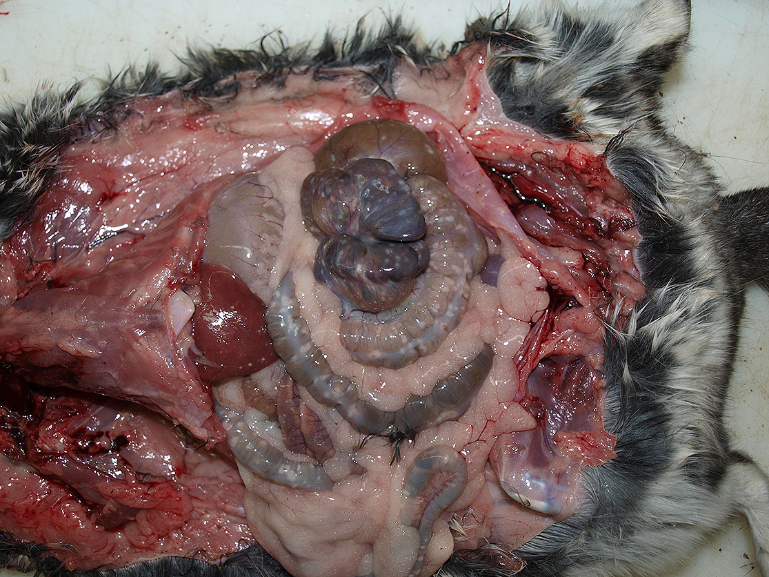
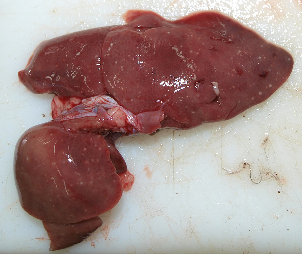
Gross Description:
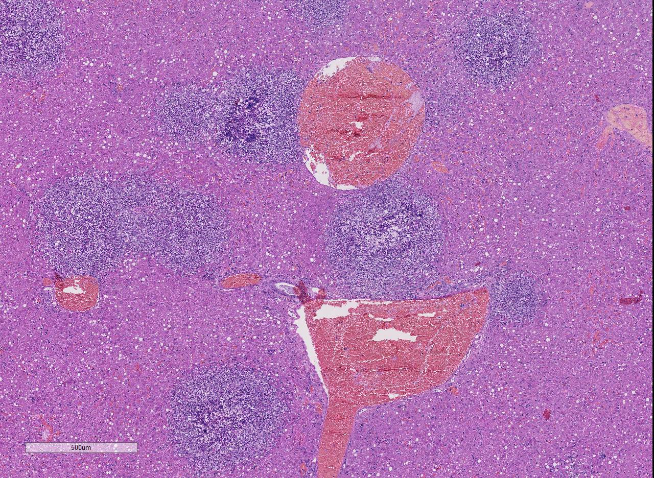
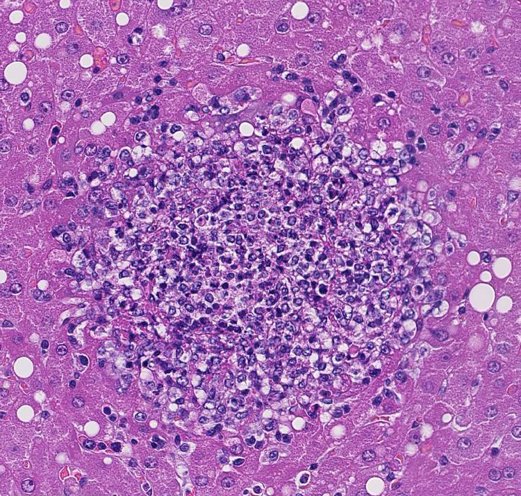
Histopathologic Description:
Morphologic Diagnosis:
Lab Results:
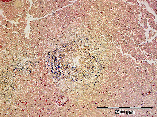
Tissue gram stain: in necrotic foci, there are numerous intra- and extracellular road-shaped bacteria consistent with Listeria monocytogenes.
Condition:
Contributor Comment:
Listeria monocytogenes is a facultative intracellular pathogen that invades host macrophages, neutrophils, and epithelial cells. Important virulence factors include the surface protein internalin, which interacts with host cell E-cadherin on the host cells, allowing the bacterium to cross the intestine, placenta, and blood-brain barrier. Once inside the cell, the organism also utilizes cholesterol-binding hemolysin to lyse pha-golysosomes and escape into the cytoplasm. The organism proliferates in the host cell cytoplasm and migrates against the cell membrane to form protrusions that can then be taken up by adjacent cells.1
Listeria monocytogenes behaves as three separate rarely overlapping diseases or syndromes: infection of the gravid uterus with abortion; septicemia with miliary visceral abscesses or necrosis; and encephalitis.1 Septicemic (systemic) listeriosis occurs in aborted fetuses and neonatal lambs, calves, foals up to one week of age, and young rabbits; it is characterized by multisystemic bacterial colonization and multifocal multisystemic areas of coagulative necrosis or microabscesses formation.1, 7 Necrotic foci are numerous in the liver, but much less numerous in the heart and other viscera. Neonates generally become infected in utero.1
Chinchillas are particularly susceptible to infection with Listeria monocytogenes and most reports mention massive outbreaks involving a significant number of animals on chinchilla farms.3, 8A single case report of listeriosis in a chinchilla caused by Listeria ivanovii has been described, and lesions were also characterized with necrotic and suppurative hepatitis.2 Currently, chinchillas are becoming more common as pets, and less are kept for fur production. In the presented case, the source of infection remains unknown, but it may be speculated that infection occurred due to contamination of water or food during the time when animals were kept in the garden.
JPC Diagnosis:
Conference Comment:
After ingestion of the bacteria and translocation from the intestinal tract, the main target organ is the liver.1,4,6 The organism has a tropism for hepatocytes and has the ability to penetrate the host cell using the surface protein internalin, men-tioned by the contributor.1 The bacterium then replicates within the host cell cytoplasm. Listeria monocytogenes also has the ability to co-opt the host cell actin filaments using the bacterial actin assembly-inducing protein (ActA) to migrate to the host cell membrane and induce pseudopod-like protrusions that can be transferred to another host cell.4 Subsequent recruitment of neutrophils leads to lysis of hepatocytes, release of the organism, and ensuing bacteremia.6 Dissemination of the organism to multiple tissues such as the heart, brain, spleen, lymph nodes, and intestinal tract causes the classic lesion of multiple random miliary white foci of necrosis in the chin-chilla.6 Conference participants discussed the differential diagnosis for the macroscopic finding of miliary white foci in the liver of rodents, which includes: Yersinia pseudotuberculosis, Yersinia pestis, Francisella tularensis, Escherichia coli, and group b Streptococcus spp, among others. Conference participants also noted multifocal moderate macrovesicles within hepatocytes that often peripheralize the nuclei, interpreted as lipid vacuolar de-generation. This animal had a reported clinical history of inappetence for four days prior to death. Gross photographs document abundant mesenteric and subcutaneous fat stores. Anorexia in this animal likely caused excessive delivery of free fatty acids from abundant adipose tissue. Excessive delivery of fatty acids, in conjunction with hepa-tocyte damage from the bacterial infection, likely impaired synthesis and secretion of lipoproteins leading to excessive accumulation of triglycerides in hepatocytes.9 Listeria monocytogenes was first described in 1926 by microbiologist Dr. Everitt G. Murray based on six cases of sudden death in young rabbits. He described it as a disease that causes an infiltration of large mono-nuclear leukocytes and named it Bacterium monocytogenes.5 The bacterium was re-named in 1940 to Listeria monocytogenes in honor of famed surgeon and pioneer of antiseptic technique, Dr. Joseph Lister. Interestingly, Listeria monocytogenes was not identified as a foodborne pathogen until 1981.4
References:
2. Kimpe A, Decostere A, Hermans K, Baele M, Haesebrouck F. Isolation of Listeria ivanovii from a septicaemic chinchilla (Chinchilla lanigera). Veterinary Record. 2004; 154: 791-792.
3. KirinusI JK, KrewerI C, ZeniI D, MonegoI F, da SilvaII MC, KommersII GD, de VargasI AC. Outbreak of systemic listeriosis in chinchillas. Ciência Rural, Santa Maria 2010; (40): 686-689.
4. McAdam A, Milner D, Sharpe A. Infectious Diseases. In: Kumar V, Abbas A, Aster J, eds. Robbins and Cotran Pathologic Basis of Disease. 9th ed. Philadelphia, PA; Saunders Elsevier; 2015: 366.
5. Murray E, Webb R, Swann M. A disease of rabbits characterized by a large mononuclear leucocytosis, caused by a hitherto undescribed bacillus Bacterium monocytogenes. J Pathol Bacteriol. 1926; 29:407439.
6. Norton J, Reynolds R. Diseases and Veterinary Care. In: The Laboratory Rabbit, Guinea Pig, Hamster, and other Rodents. Oxford, UK: St. Louis, MO: Elsevier, 2012: 995.
7. Percy DH, Barthold SW. Guinea Pig. In: Percy DH, Barthold SW, eds. Pathology of Laboratory Rodents and Rabbits. 4th ed. Ames, IA: Blackwell; 2016: 226-227.
8. Wilkerson MJ, Melendy A, Stauber E. An outbreak of listeriosis in a breeding colony of chinchillas. J Vet Diagn Invest. 1997; (9): 320-323.
9. Zachary JF. Mechanisms of Micro-bial Infections. In: Zachary JF, McGavin MD, eds. Pathologic Basis of Veterinary Disease. 5th ed. St. Louis, MO: Elsevier Mosby; 2012: 192-195