Signalment:
Four-month-old male Labrador retriever, (
Canis familiaris).A 3-month-old Labrador retriever pup developed progressively
worsening tetraparesis with a spastic swimming-puppy-like position of the
thoracic limbs and a flattened chest. One month later mild vestibular signs and
myoclonic jerks in the head and cervical region became obvious. General
clinical examination was within normal limits. Neurological examination
revealed absent patellar reflexes, weakness on the 4 limbs with an abnormal
spasticity of the thoracic limbs and mild generalized muscle atrophy. During the
second visit a vestibular strabismus in the right eye, a mild right-sided head
tilt and regular myoclonic jerks at the head and thoracic limbs were noticed.
Electrophysiological
examination was normal. RX of the thorax only confirmed the dorsoventral
flattening of the thorax. Due to the worsening neurological signs further
examinations were declined by the owner and the pup was euthanized at the age
of 4.5 months.
Gross Description:
Except for the dorso-ventral flattening of the thorax, no
gross abnormalities were noted at necropsy.
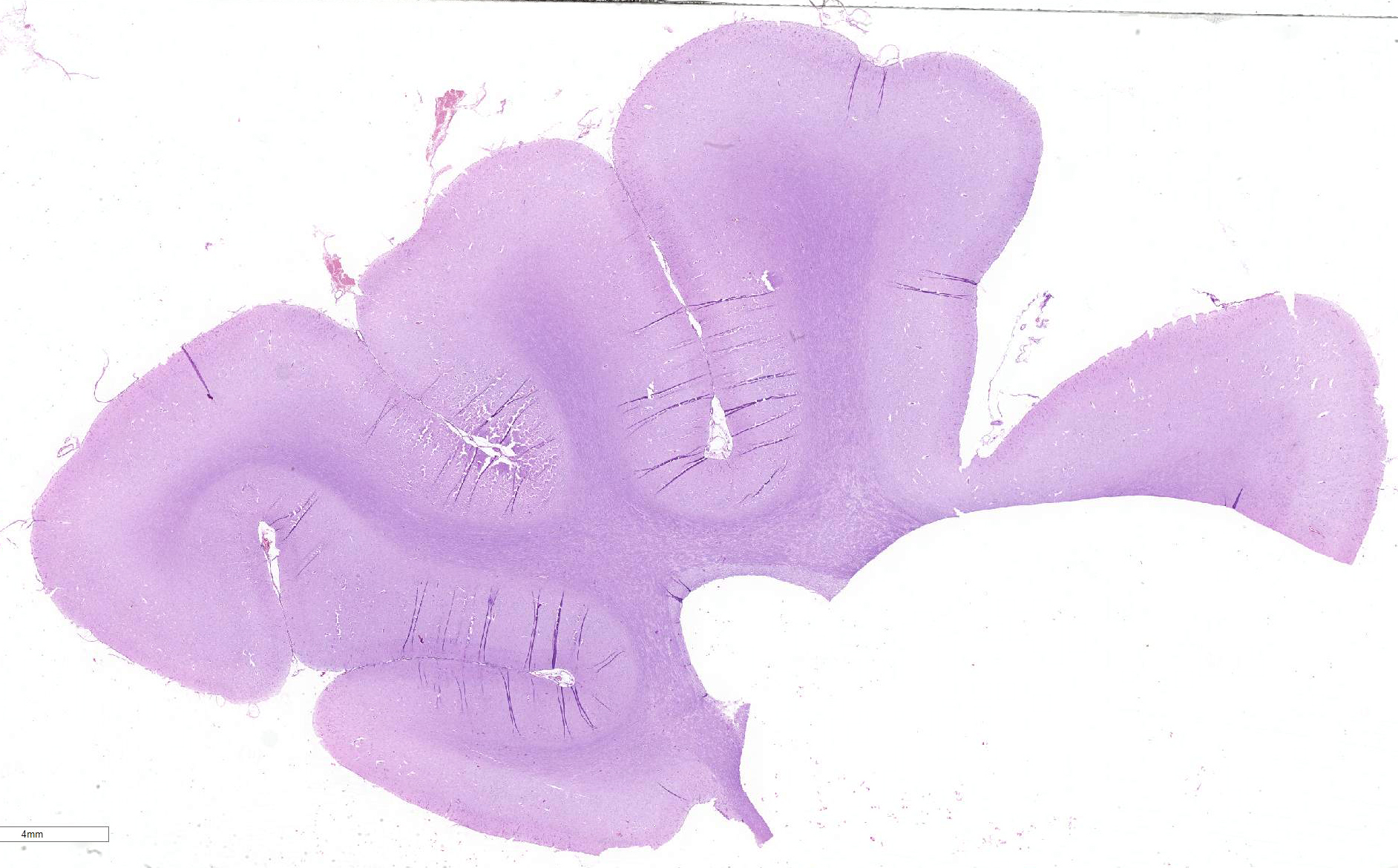
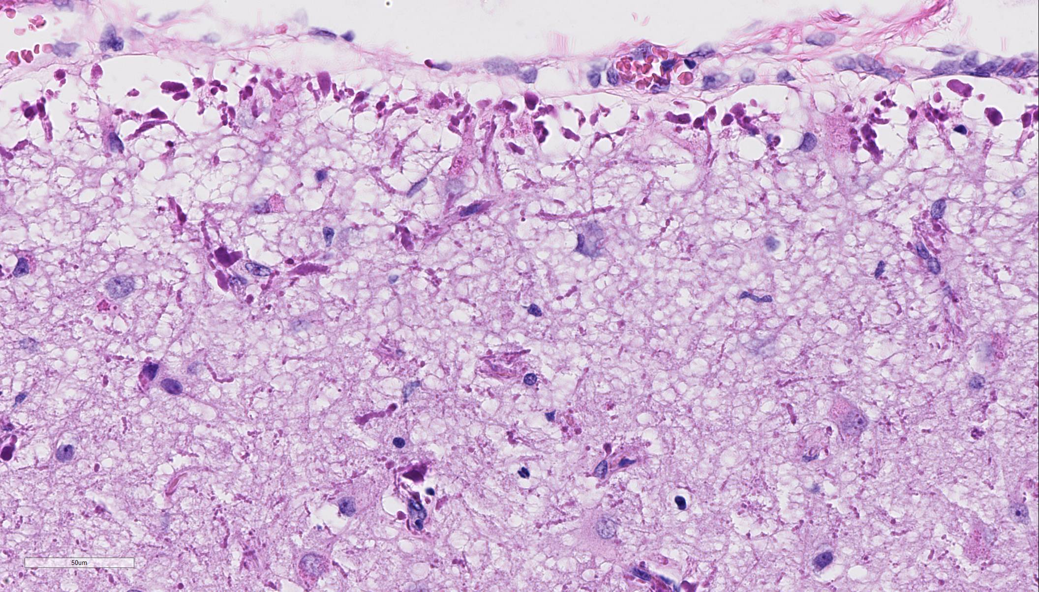
Histopathologic Description:
Cerebrum: Within the white and gray matter of the brain, some blood vessels
are surrounded by numerous short, perpendicularly oriented, hypereosinophilic,
amorphous intraastro-cytic accumulations, varying in diameter from 4 to 20 µm
(Rosenthal fibers). These Rosenthal fibers are also found in the astrocytic
endfeet in the subpial tissue and to a lesser extend throughout the parenchyma.
Mainly in the white matter, there is proliferation of abnormal astrocytes with
large nuclei, prominent nucleoli and glassy eosinophilic to pale cytoplasm.
Occasionally, there are binucleated astrocytes. GFAP-staining:
All Rosenthal fibers are strongly immunopositive for GFAP.
Morphologic Diagnosis:
Cerebrum, gray and white matter,
encephalopathy, multifocal, chronic, moderate, perivascular and subpial
accumulation of Rosenthal fibers, astro-cytosis and astrocytic hypertrophy.
Lab Results:
Blood examination and cerebrospinal fluid analysis were within
normal limits.
Condition:
Astroglial dystrophy with Rosenthal fibers
Contributor Comment:
In
humans, Rosenthal fibers are found in Alexander disease (AxD), and, albeit in greatly
reduced numbers in chronic reactive astrocytosis and low-grade astrocytomas.
4,5
They are seldom encountered in animal neuropathology. Alexander disease, or
fibrinoid leuko-dystrophy, is a rare neurodegenerative disorder of astrocyte
dysfunction in human. In veterinary medicine, Alexander disease is very rare
and has been reported in a few dogs (two Labrador Retrievers, one Scottish
Terrier dog, one Miniature Poodle, three Bernese Mountain dogs, one Bernese
Mountain cross breed, one French bulldog, and one Chihuahua) and four sheep
(one white Alpine sheep and three Merino sheep).
1-7
There
are no specific gross lesions of AxD. The classic histological lesions are
Rosen-thal fibers. These fibers are deeply eosinophilic, irregularly shaped,
elongated, round to oval intra-astrocytic aggregates. Rosenthal fibers have
been shown to be ubiquinated aggregates of GFAP, αβ-crystalin and
HSP27.
3,4
In
humans AxD is classified based on the age of onset as infantile, juvenile and
adult. Recently, Prust
et al. proposed a reclassification in two
age-dependent clinical subtypes: type I, characterized by an early age of
onset, seizures, macrocephaly, encephalopathy, developmental delay, paroxysmal
deterioration, failure to thrive and typical MRI features, and type II,
characterized by a later age of onset, autonomic dysfunction, bulbar symptoms,
ocular movement abnormalities and atypical MRI features.
5 The characteristic
pathological feature of both types of AxD are widespread and abundant Rosenthal
fibers. All
known genetic causes of AxD are attributed to GFAP mutations (explaining more
than 95% of the cases), mostly
de novo dominant missense mutations with
hotspots at R79 and R239, the latter one inducing the most aggressive form.
1
JPC Diagnosis:
Cerebrum: Astroglial dystrophy, diffuse, severe, with marked
subpial, subependymal, and perivascular Rosenthal fiber formation, Labrador
retriever,
Canis familiaris.
Conference Comment:
The contributor provides a concise review of Alexander disease
(AxD), a rare neurodegenerative disorder previously reported in dogs, sheep,
and humans.
1-7 Conference participants identified the large brightly
eosinophilic and irregularly shaped Rosenthal fibers (RF) with astrocytes
scattered throughout the white matter and aggregated in the subpial,
subependymal, and perivascular spaces. This is the classic histologic lesion
distribution associated with previously reported cases of AxD in all reported
species.
1,7 As mentioned by the contributor, these accumulated
fibers consist of large aggregates of glial fibrillary acidic protein (GFAP),
αB-crystallin, heat shock protein (hsp-27) and ubiquitin, within markedly
expanded astrocytic processes distributed throughout the central nervous
system.
1,4,7 Prior to the conference, the Joint Pathology Center ran
a GFAP immunohistochemical stain which demonstrated intense immuno-staining of
the RF surrounding vessels and in the subpial and subependymal areas. RF have
also been reported to be immuno-positive for ubiquitin.
The presence of RF is not pathognomonic for AxD and has been reported in
glial scars and multiple sclerosis in humans; however, the distribution of the
RF in the subpial, subependymal, and perivascular areas
in this and other reported cases is unique to AxD. In
addition to Rs, other common histologic lesions of AxD include white matter
demyelination and astrogliosis.
1,7
As mentioned by the contributor, it is thought
that a mutation in GFAP leads to glial intermediate filament disorganization,
decreased solubility, and defective degradation of the protein.
1,3,7
This results in accumulation of the aberrant and misfolded protein, leading to
cellular stress and the unfolded protein response (UPR) in the endoplasmic
reticulum. Cellular stress and the resulting UPR are postulated to be the
initiating factor for the production of ubiquitin and heat shock proteins
(αB-crystallin, hsp-27) accumulating with GFAP and forming the RFs seen
histologically.
1,4,7 Accumulation of these insoluble
fibers is
likely progressively toxic to astrocytes and degrades oligodendrocyte function, affecting myelin formation in the white matter.
As a result, animals affected with AxD typically
present as juveniles with rapidly progressive depression, ataxia,
paresis, generalized tremors, decreased spinal reflexes, and seizures.
1
References:
1. Aleman
N, Marcaccini A, Espino L, et al. Rosenthal fiber encephalopathy in a dog
resembling Alexander disease in humans.
Vet. Pathol. 2006; 43:
1025-1028.
2. Gruber A, Pakodzy A, Leschnik M, et al. Morbus
Alexander: 4 Fälle bei Hunden in Österreich. Wien. Tierärztl.
Mschr.
Vet Med Austria. 2010; 97:1-4.
3. Ito
T, Uchida K, Nakamura M, et al. Fibrinoid leukodystrophy (Alex-ander's
disease-like disorder) in a young adult French bulldog.
J Vet Med Sci.
2010; 72:1387-1390.
4. Kessell
A, Finnie J, Manavis J, et al. A Rosenthal fiber encephalo-myelopathy
resembling Alexander's disease in three sheep.
Vet Pathol. 2012; 49: 248-254.
5. Prust M, Wang J, Morizono H, et al. GFAP mutations, age
at onset, and clinical subtypes in Alexander disease.
Neurology. 2011; 77:
1287-1294.
6. Richardson
J, Tang K, Burns D. Myeloencephalopathy with Rosen-thal fiber formation in a
Miniature Poodle.
Vet Pathol. 1991; 28: 536-538.
7. Wrzosek M, Giza E, Płonek M et al. Alexander
disease in a dog: Case presentation of electrodiagnostic, magnetic resonance
imaging and histopathologic findings with review of literature.
BMC Vet Res.
2015; 11:115.