Signalment:
10-year-old spayed female Welsh corgi dog,
Canis familiars.The dog presented in 2011 for a 6-week history of hematuria. Urine culture was negative and no signs of uroliths were seen on radiographs. Ultrasound revealed two, 1.7 mm, mineralized foci in the left renal pelvis, but the kidneys were normal in shape and size. No further workup was performed at that time. The dog presented again in July 2013 with continued hematuria. On ultrasound, the left renal cortex had a 2.2 cm, round, heterogeneous mass with multiple, internal, anechoic regions.
Gross Description:
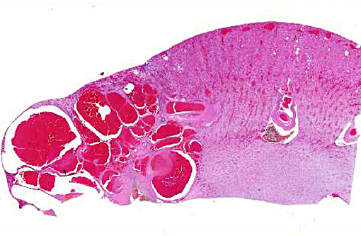
The renal cortex was extensively disrupted by coalescing, dark red, blood-filled nodules ranging from 0.7 x 0.5 x 0.2 cm to 2.5 x 2 x 2 cm.
Histopathologic Description:
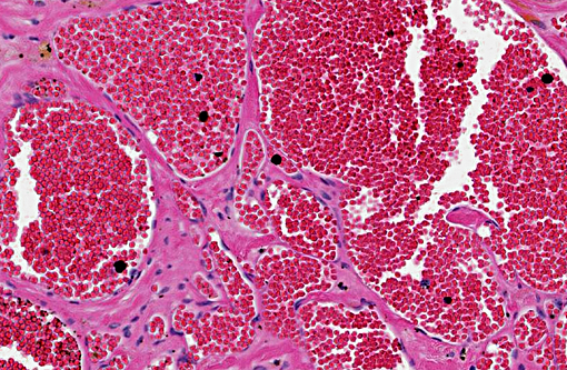
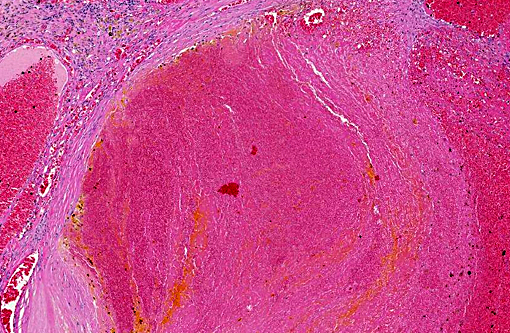
Kidney: The renal interstitium is markedly expanded by many, severely dilated, blood-filled vascular spaces lined by mature endothelial cells surrounded by abundant fibrous tissue. Some vascular spaces contain thrombi with fibrin arranged as lines of Zahn separated by red and white blood cells that are occasionally attached to the vascular wall by fibrous tissue. The intervening and adjacent renal parenchyma is markedly atrophic with replacement of many nephrons by fibrosis, many lymphocytes and plasma cells, and variable hemorrhage. Remaining tubules and glomeruli in these areas are often atrophic or sclerotic, respectively. Some tubules contain proteinaceous fluid. The renal pelvis contains degenerating erythrocytes and brown, granular pigment. The more distant renal parenchyma is unremarkable apart from congestion.
Morphologic Diagnosis:
Kidney: Renal telangiectasia.
Condition:
Renal telangiectasia
Contributor Comment:
Renal telangiectasia is a rare, non-neoplastic proliferation of blood vessels.(3) It has been described in the Welsh corgi as a familial disease, and similar lesions have been described in cattle, dogs, cats, mink and ferrets.(2) Affected corgis typically present for hematuria and may have bilateral lesions or additional lesions in other tissues, such as the liver, duodenum, brain, vertebrae, subcutaneous tissue, and spleen.(3,4) In this case, no contralateral or extra-renal lesions were identified by abdominal ultrasound or at surgery. The primary clinical differential in this case was renal hemangiosarcoma. Renal telangiectasia is differentiated histologically from hemangiosarcoma by the fact that the vascular spaces are lined by a bland, mature endothelium without mitotic activity or cellular atypic.(4)
JPC Diagnosis:
Kidney: Vascular malformation with multifocal thrombosis.
Conference Comment:
Conference participants discussed three optional diagnoses for this case: telangiectasia, hemangioma or vascular hamartoma. The familial lesion of renal telangiectasia in Pembroke Welsh corgis as described in the literature characteristically arises bilaterally with frequent occurrences in other organs. The clinical history in this case suggests the lesion is isolated to one kidney; however, the lesion lacks the well-circumscribed appearance of a hemangioma. The signalment and clinical signs do not correlate well with a hamartoma, which implies a congenital proliferation. The diagnosis of vascular malformation reflects these inconsistencies and participants inability to arrive at a consensus.
The pattern of presentation in corgis interestingly resembles a familial condition in people known as hereditary hemorrhagic telangiectasia (HHT). Also known as Osler-Weber-Rendu disease, this autosomal dominant disorder occurs at a rate of 1 in 5,000 people and manifests as telangiectasia of the oral mucosa and arteriovenous malformations in the lungs, liver and less often brain. Three mutations have been identified in HHT, all with protein products influencing the TGF-β superfamily.(5)
TGF-β is a growth inhibitor of most epithelial cells but a potent stimulator of fibroblasts. Its binding to cell-surface receptors leads to phosphorylation of SMAD transcription factors, specifically Co-SMAD & R-SMAD. When phosphorylated, these factors overcome SMAD-7 inhibition, thereby promoting transcription and activation of fibroblasts. Additionally and likely pertaining to these vascular lesions, TGF-β is strongly implicated in the stabilization of nascent blood vessels, following their sprouting and migration through the finely orchestrated events coordinated by VEGF and the Notch pathway.(1)
This inheritable condition has serious consequences in people due to their predisposition for hemorrhages and thrombosis. The understanding of disease pathogenesis has also shed new light on the specific molecular interactions of angiogenesis, one of the hallmarks of cancer. The authors speculated on the value of Pembroke Welsh corgis serving as an animal model for vascular malformations over 30 years ago.(3) While knockout mice have been developed which reproduce these lesions, this naturally occurring canine model may offer further valuable insight in piecing together the complex interactions of angiogenesis and its role in vascular malformations and neoplasia.
References:
1. Kumar V, Abbas AK, Fausto N, Aster JC. Tissue renewal, regeneration and repair. In: Kumar V, Abbas AK, Fausto N, Aster JC, eds. Robbins and Cotran Pathologic Basis of Disease. 8th ed. Philadelphia, PA: Elsevier Saunders; 20010:89, 100-101.
2. Maxie MG, Newman SJ. Urinary system. In: Maxie MG, ed. Jubb, Kennedy and Palmers Pathology of Domestic Animals. 5th ed. Vol 2. Philadelphia, PA: Elsevier Saunders; 2007:495.
3. Meuten DJ. Tumors of the urinary system. In: Tumors in Domestic Animals. 4th ed. Ames, IA; Iowa State Press; 2002:524.
4. Moore FM, Thornton GW. Telangiectasia of Pembroke Welsh corgi dogs. Vet Pathol. 1983;20(2):203-8.
5. Shovlin CL. Hereditary haemorrhagic telangiectasia: pathophysiology, diagnosis and treatment. Blood Reviews. 2010;24:203-219.