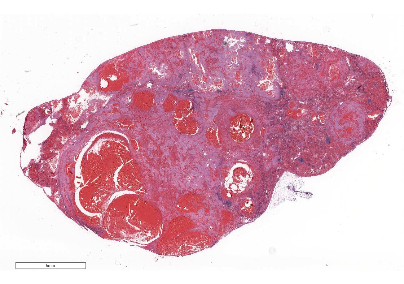
Signalment:  

Three-year-old castrated male domestic shorthair cat (Felis catus).History of inflammatory bowel disease treated with prednisolone. Presented with lethargy, anorexia, and a change in behavior (less social).


Gross Description:  

Hemoabdomen with multiple splenic masses on abdominal ultrasound and exploratory laparotomy with subsequent splenectomy. On gross evaluation of the spleen, the spleen was enlarged with diffuse nodularity and rounded edges. Expanding the right ventral aspect, there was an approximately 7 x 3.5 x 2.5 cm mass-like swelling. On cut section, the parenchyma was dark red-purple with foci of hemorrhage.


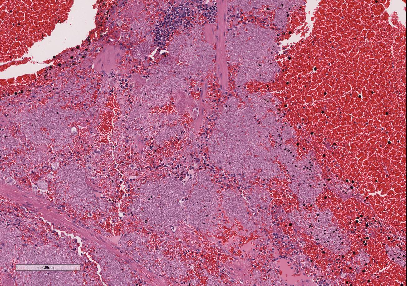
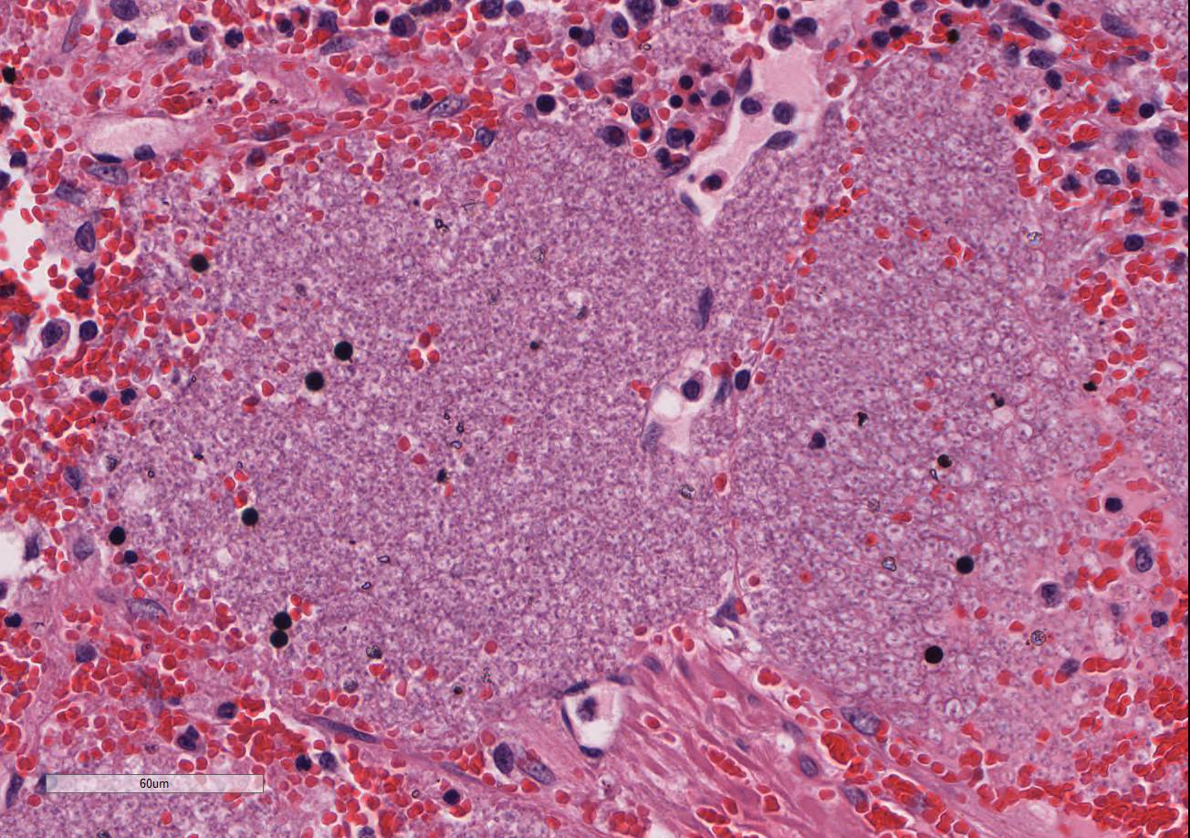
Histopathologic Description:

Multifocally throughout the splenic parenchyma, there are large areas of congestion, hemorrhage, necrosis, fibrin deposition and extensive accumulation of extracellular pale basophilic to amphophilic foamy to stippled material composed of 2-4 um diameter round structures each with a thin wall and a central basophilic granular core, consistent with cyst and trophozoite structures. There is a mild to moderate infiltrate composed of macrophages with fewer neutrophils and occasional macrophages contain similar round structures in the cytoplasm. In the intervening congested red pulp, there are numerous hematopoietic precursors including predominantly erythroid and megakaryocytic cells. Special stain: The cell wall of the round structures is multifocally faintly positive for GMS (Gomori methenamine silver) stain and acid-fast negative.


Morphologic Diagnosis:  

Spleen: Severe multifocal to coalescing congestion, hemorrhage, and necrosis with myriad extracellular and intrahistiocytic cysts and trophozoites, consistent with Pneumocystis spp.

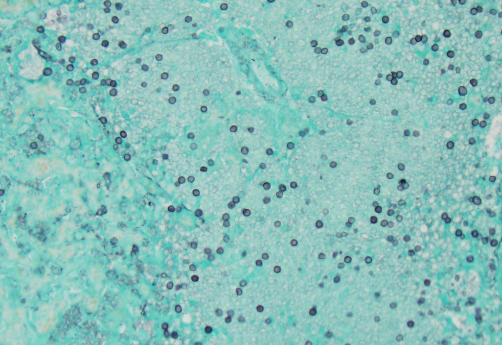
Pneumocystis spp. confirmed with immuno-histochemistry, electron microscopy, and PCR testing. Sequencing suggests a novel species/strain.


Lab Results:  

Moderate regenerative anemia (Hct 18.7%, reticulocyte count 171,000/uL), WBC within reference (8,900/uL), and moderate thrombocytopenia (90,000/uL).


Condition:  

Pneumocystis carinii


Contributor Comment:  

Given the lack of clinical signs referable to the respiratory tract and the extensive splenic involvement by the infection, the findings are consistent with extrapulmonary pneumocystosis in this case. A small splenic choristoma in the pancreas was also affected and a minor intravascular population of these organisms was identified in the liver at the time of biopsy.

Pneumocystis is a saprophytic organism that has somewhat uncertain taxonomy. Pneumocystis has been classified as a unicellular protozoan of the phylum Sarcomastigophora, subphylum Sarcodina. The reproductive behavior is similar to the ascospore formation of yeast cells and the staining properties resemble that of pathogenic fungi. Based on 16S-like rRNA, Pneumocystis is phylogenetically most closely related to the fungi of the class Ascomycetes. Multiple species have been designated, although there is some controversy in this area and several strains likely exist. The most widely referenced species are Pneumocystis carinii in dogs and P. jiroveci in humans, with several species indicated in the laboratory animal literature. The life cycle of Pneumocystis involves a tropho-zoite and a cyst form, both of which occur in the infected tissue.5

Pneumocystis is best known as an opportunistic organism that most commonly causes pneumonia, in particular in immuno-compromised people and animals. Clinical pneumonia occurs in many cases, but subclinical or latent infections are also common in many animal species, including cats. Clinical disease may occur via new infection or reactivation of a latent infection under conditions of stress, immuno-suppressive therapy, or with other underlying infection, such as canine distemper virus in dogs.5 In dogs, most reported cases of pneumocystic pneumonia occur in animals with underlying immuno-deficiency, such as miniature Dachshund, Pomeranian and Cavalier King Charles spaniel dogs with combined immunodeficiency syndrome.2,4 In cats, subclinical or latent infections are most common, and although Pneumocystis has been identified in the lungs in the cat, natural cases of clinical disease have not been reported.5 Under experimental conditions, immunosuppression with glucocorticoid administration has led to pneumocystic pneumonia in cats.5

Extrapulmonary infection with Pneumo-cystis has been reported in humans, particularly with HIV/AIDS, and involves the spleen in some cases.1,3 A case of extrapulmonary pneumocystosis has also been reported in a dog.5 In humans with extrapulmonary infections, some cases have concurrent pneumocystic pneumonia, while others do not, and those that do not are thought to either acquire a primary extra-pulmonary infection or have reactivation of a latent infection at extrapulmonary sites.1 Other than spleen, reported sites for extrapulmonary involvement in humans include lymph node, liver, bone marrow, adrenal gland, gastro-intestinal tract, kidney, thyroid gland, heart, pancreas, central nervous system, bone, eyes or ears.1,3 In a minority of cases, intravascular organisms are identified in the tissues,1 which was present in the liver biopsy from this patient.

In pneumocystic pneumonia, the classic histologic appearance is of intra-alveolar aggregates of foamy eosinophilic material with only a minor infiltrate of macrophages and without significant neutrophilic inflammation. GMS staining highlights the 4-7um diameter cyst wall and more easily demonstrates the ovoid, irregular or crescent shape of the cysts. At extrapulmonary sites, Pneumocystis has similar tissue destruction with necrosis and a similar appearance of foamy eosinophilic material.1,3 Methods of confirmation of Pneumocystis include cytologic or histologic morphologic appearance and GMS staining characteristics, PCR evaluation, immuno-histochemistry or immunofluorescence.3,5


JPC Diagnosis:  

Spleen: Splenitis, necrotizing and hemorrhagic, diffuse, severe with numerous extracellular and intra-histiocytic trophozoites, domestic shorthair, Felis catus.


Conference Comment:  

We thank the contributor for providing a thorough review of the epidemiology, pathogenesis, and comparative pathology of Pneumocystis spp. in humans and veterinary species. This outstanding case provided conference participants the opportunity to describe and identify a relatively common opportunistic pathogen in an extremely rare extra-pulmonary location and an uncommonly affected species. Despite the lack of case reports of feline splenic pneumocystosis in the veterinary literature, many conference participants included Pneumocystis sp. on their list of differential diagnoses due to the classic histomorphology, characterized by the presence of abundant extracelluar and intrahistiocytic foamy lightly eosinophilic material and absence of overwhelming inflammation. Although definitive vasculitis and thrombosis are not seen, conference participants also readily identified the numerous brilliant hemodynamic changes present in the tissue section, indicated by severe congestion, marked dilation of sinusoids, and hemorrhage. Multifocal areas of extramedullary hematopoiesis are also identified, although its association with the infectious etiology is unclear.

As mentioned above, extrapulmonary pneumocystosis is rare but has been reported in a dog and immunocompromised people, typically affected with HIV/AIDS.3,5,6 Postmortem analysis from previously reported human cases indicates that Pneumocystis sp. can disseminate throughout the body via hematogenous and/or lymphatic routes. It is thought that the vast majority of cases of extrapulmonary pneumocystosis result from hematogenous or lymphatic spread from the lung. However, in cases where the lungs are unaffected, such as in this cat, there may be reactivation of latent infection in extra-pulmonary organs due to severe immunosuppression.6


References:

1. Cohen OJ and Stoeckle MY. Extrapulmonary Pneumocystis carinii infections in the acquired immunodeficiency syndrome. Arch Intern Med. 1991; 151:1205-1214.
2. Kanemoto H, Morikawa R, Chambers JK, et al. Common variable immune deficiency in a Pomeranian with Pneumocystis carinii pneumonia. J Vet Med Sci. 2015; 77(6):715-719.
3. Karam MB and Mosadegh L. Extra-pulmonary Pneumocystis jiroveci infection: A case report. Braz J Infect Dis. 2014; 18(6):681-685.
4. Lobetti R. Common variable immunodeficiency in miniature dachshunds affected with Pneumocystis carinii pneumonia. J Vet Diagn Invest. 2000; 12:39-45.
5. Lobetti R. Pneumocystosis. In: Greene CE, ed. Infectious Diseases of the Dog and Cat. 4th ed. St. Louis, MO: Elsevier Inc; 2012:689-695.
6. O'Neal CB, Ball SC. Splenic pneumocystosis: An atypical presentation of extrapulmonary Pneumocystis infection. AIDS Reader. 2008; 8:503-508.


Click the slide to view.


4-1. Spleen, cat.


4-2. Spleen, cat.


4-3. Spleen, cat.


4-4. Spleen, cat.


4-5. Spleen, cat.


Back | VP Home | Contact Us |