Signalment:
Gross Description:
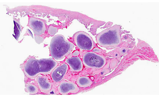
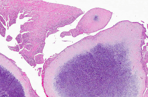
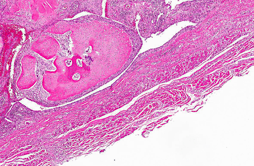
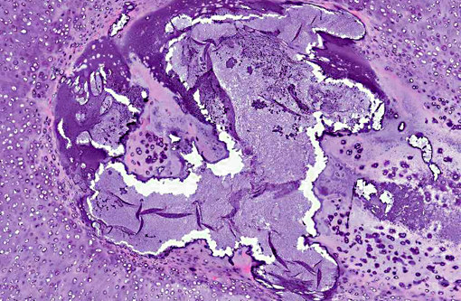
Histopathologic Description:
Morphologic Diagnosis:
1. Synovial osteochondromatosis.
2. Synovial hyperplasia with lymphocytic plasmacytic synovitis.
Condition:
Contributor Comment:
In humans, SOC is considered to be a metaplastic condition rather than a neoplastic disease.(11) There are however, at least two case reports of malignant transformation of synovial osteochondromatosis to chondrosarcoma in dogs.(1,2)
Based on the description of the condition in humans numerous subcategories have been described. These include chondromatosis where the nodules are strictly cartilaginous in nature and osteochondromatosis where some of the nodules have undergone endochondral ossification as in this case. They are further subdivided based upon the suspected origin as either primary or secondary. SOC is defined as being secondary when there is a preexisting history of joint trauma or chronic mechanical irritation, while primary SOC is an idiopathic condition.12 In this case, there was no previous history of joint injury or disease and careful examination of the articular surfaces of the joints did not reveal any sign of disease suggesting that this is a primary SOC. There are limited numbers of cases describing the treatment of this condition; those that do exist suggest that removal of the nodules and partial synovectomy may improve the clinical signs, although radiographic reoccurrence of nodules was noted in some of these cases.(4)
JPC Diagnosis:
Conference Comment:
| Primary synovial chondromatosis | Secondary synovial chondromatosis | |
| Occurrence | Rare | Fairly common |
| Joints affected | Single | Multiple |
| Degenerative joint disease | Mild | Severe |
| Number of nodules | More | Fewer |
Participants also considered chondroma and chondrosarcoma as potential diagnoses; however, most agreed that a chondroma would more likely present as a single, discrete mass rather than as multiple nodules, and chondrosarcoma would likely not appear so well-demarcated or demonstrate such a repeatable and orderly appearance within each nodule.Â
The classification of primary synovial chondromatosis is controversial, as there is evidence of both a reactive process and a metaplastic/proliferative process.(1) Recent cytogenic studies demonstrate that in human primary osteochondromatosis, a subpopulation of mesenchymal stem cells undergo clonal proliferation, suggesting a benign neoplastic process.(1)
References:
2. D+�-�az-Bertrana C, Durall I, Rial JM, Franch J, Fontecha P, Ramis A. Extra- and intra-articular synovial chondromatosis and malignant transformation to chondrosarcoma. Vet Comp Orthop Traumatol. 2010;4:277-283.
3. Edinger DT, Manley PA. Arthrodesis of the shoulder for synovial osteochondromatosis. J Small Anim Prac. 1998;39:397-400.
4. Flo GL, Stickle RL, Dunstan RW. Synovial chondrometaplasia in five dogs. J Am Vet Med Assoc. 1987;191:1417-1422.
5. Gregory SP, Pearson GR. Synoval osteochondromatosis in a Labrador retriever bitch. J Small Anim Prac. 1990;31:580-583.Â
6. Howard MO, Nieves MA, Miles KG. Synovial chondromatosis in a Great Horned Owl (Bubo virginianus). J Wildlife Dis. 1996;32:370-372.
7. Hubler M, Johnson KA, Burling RT, Francis DF, Ratcliffe RCC. Lesions resembling osteochondromatosis in two cats. J Small Anim Pract. 1986;27:181-187.
8. Kirk MD. Radiographic and histologic appearance of synovial osteochondromatosis of the femorotibial bursae in a horse: a case history report. Vet Radiol. 1982;23:167-170.
9. Smith TJ, Baltzer WE, Lohr C, Steiger-Vanegas SM. Primary synovial osteochondromatosis of the stifle in an English Mastiff. Vet Comp Orthop Traumatol. 2012;2:1-7.
10. Stone EG, Walser MW, Redig PT, Rings B, Howard DJ. Synovial chondromatosis in raptors. J Wildlife Dis. 1999;35:135-140.
11. Thompson K. Diseases of joints. In: Jubb, Kennedy and Palmers Pathology of Domestic Animals. 5th ed. 2007;130-136.
12. Villacin AB, Brigham LN, Bullough PG. Primary and secondary synovial chondrometaplasia. Hum Path. 1979;10:439-451.
13. Zimmerman W, Kircher P, Hani H. Synovial osteochondromatosis in swine: a case report. Schwiez Arch Tierheilkd. 2000;142:289-291.