Joint Pathology Center
Veterinary Pathology Services
Wednesday Slide Conference
2017-2018
Conference 2
August 30th, 2017
CASE IV: T17-15923 (JPC 4101085).
Signalment: 8-month-old, female, German Shepherd Dog (Canis familiaris).
History: A 5 x 5 x 3 cm gingival mass was observed on right mandible.
Gross Pathology: A multilobulated exophytic growth.
Laboratory results: None provided.
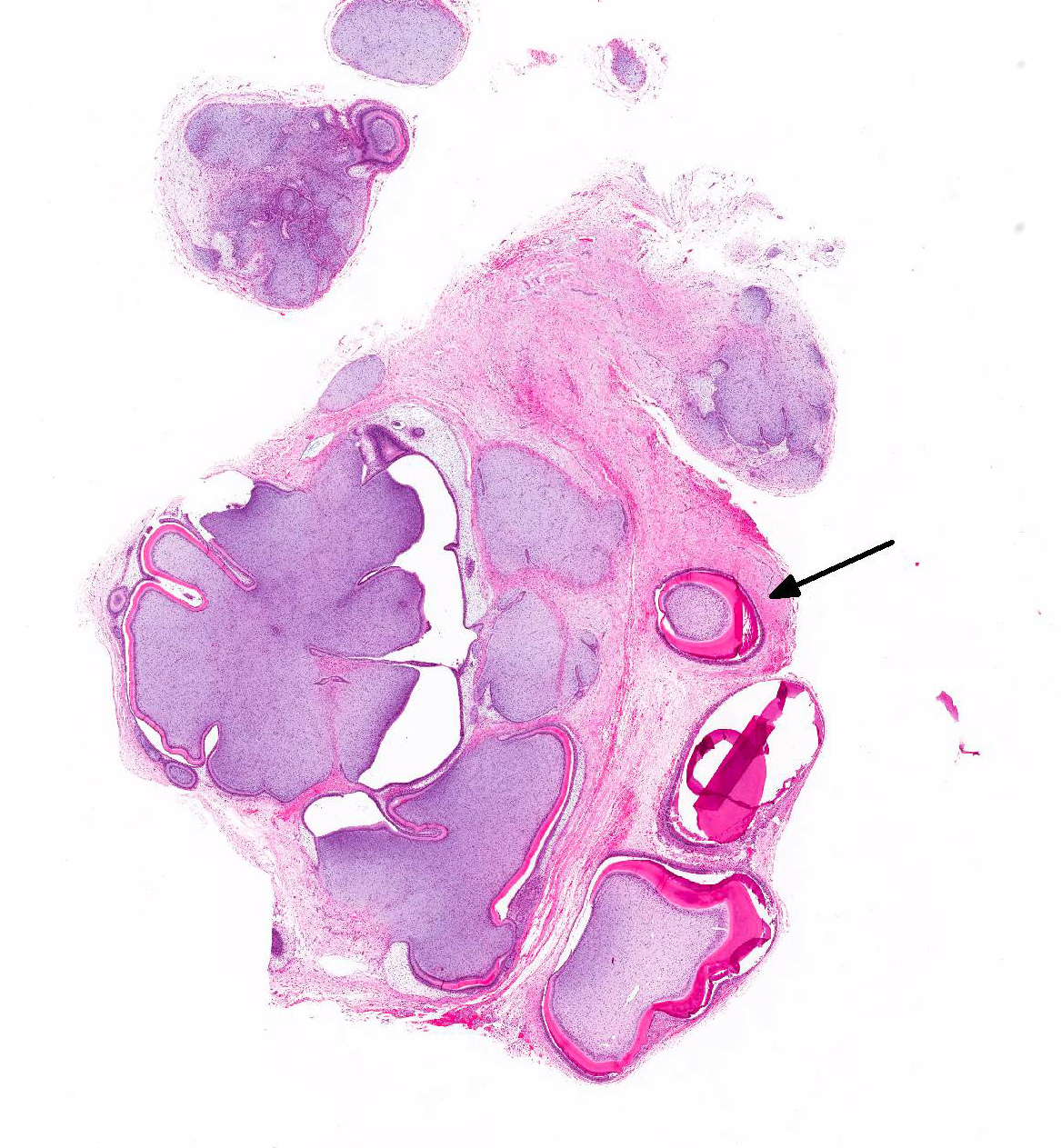
Microscopic Description: Gingiva: The mass was a non-demarcated and non-capsulated mass that contained epithelial and mesenchymal elements composed of islands of odontogenic tissue lined with a palisading columnar epithelium along a basement membrane consistent with ameloblasts supported on ample spindled to stellate mesenchymal cells (dental pulp). Multiple section consistent with a developing tooth like-structures (denticles) composed of odontoblasts, dentin and enamel were observed. Mitotic cells and malignant features were not present.
Contributors Morphologic Diagnosis: Gingiva: Odontoma, German Shepherd dog, canine (Canis familiaris).
Contributors Comment: Differential diagnoses for maxillary and mandibular swelling in immature dogs include trauma, infection, developmental disorders, and neoplastic lesions. Neoplastic oral maxillary or mandibular masses in immature, young dogs are typically of odontogenic origin.2 Odontomas are rare1 slow-growing masses that occur during odontogenesis, mainly in young dogs, resulting in dentition disruption and in prevention of eruption or displacement of normal teeth.3 The masses cause swelling and deformity of mandible and/or maxilla. The cause and pathogenesis of odontomas, either in humans or nonhuman species, is unknown.2,3 However, hereditary factors, genetic alterations during dental development, infections or trauma have been suggested to be involved in odontoma formation.3
Odontomas are odontogenic tumors with features of resembling the embryonic pattern of tooth development. Production of enamel, dentin, cementin, and sometimes small teeth are characteristic. Tumors with patterns resembling normal teeth are compound odontomas, whereas a more disorganized arrangement is a characteristic of complex odontomas. Complex odontomas are composed of well-differentiated but disorganized dental hard tissues bearing no resemblance to a tooth and surrounded by a thin fibrous capsule. Compound odontomas are composed of many separate, small tooth-like structures known as denticles, each one containing enamel, dentin, cementum, and pulp which are anatomically similar to normal teeth.3,4 Compound odontomas are localized in the mandible or maxilla. However, in most reported cases in dogs, compound odontomas are localized in the mandible3.
In dogs and other species, clinical signs associated with odontomas include facial or mandibular swelling, ocular symptoms, missing teeth, or tooth-like structures erupted into the oral cavity. Odontomas can become extremely large, even in very young dogs.2
Surgical excision is the treatment of choice for odontomas. Complete surgical excision of the mass in combination with aggressive curettage would result in a favorable outcome.2,3 For prognostic and therapeutic considerations, it is important to differentiate odontomas from ameloblastic odontoma and ameloblastoma, which develop in old dogs.4
JPC Diagnosis: Gingiva: Compound odontoma, German Shepherd Dog, canine.
Conference Comment: This case provides an excellent example of a compound odontoma in a dog. The contributors review of odontomas is excellent and mirrors much of the initial conference discussion. Conference participants described hamartomatous proliferation of odontogenic epithelium and primitive mesenchyme forming variably sized tooth-like structures in variable stages of development (denticles).
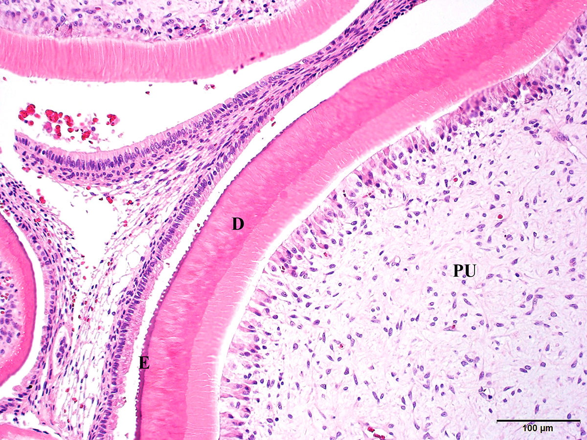
The moderator began with a review of tooth development which begins with two embryonic tissues: buccal cavity squamous epithelium (BCSE) and embryonic mesenchyme (EM). The EM forms dentin, cementum, and pulp, and the BCSE invaginates into the EM to form the dental lamina. The dental lamina grows into the adjacent tissue to form the dental bud which progresses through cap and bell stages eventually leading to crown eruption. The epithelium (BCSE) differentiates to form the ameloblastic cell layer at the outer surface of the tooth that produce enamel and the mesenchymal layer (EM) differentiates into odontoblasts, cementoblasts, and pulp (also periodontal ligament and alveolar bone) which move toward the center of the tooth and makes dentin and cementum respectively. Enamel covers the outside of the tooth and is composed of hydroxyapatite (mineral) crystal and heavily mineralized calcium salts. Dentin comprises the bulk of the tooth mass under the enamel and is slightly softer with tubules that contain odontoblast processes. Cementum covers the root of the tooth (doesnt project above gumline) and is a bone-like substance (contains basophilic reversal lines) with embedded cementocytes.5
The microscopic characteristics of epithelium of odontogenic origin (also known as ameloblastic epithelium) are: peripheral palisading, anti-basilar nuclei, basilar epithelial cytoplasmic clearing, and non-basilar epithelial cells (stellate reticulum) have intercellular bridges which separate the inner and outer layer of enamel epithelium during development.3 There are two types of teeth in mammals, brachydont (carnivores) and hypsodont (ruminants and horses). Brachydont teeth are characterized by a short crown above the gingiva, constricted neck at the gumline, and a root embedded in the jawbone. Hypsodont teeth are high-crowned teeth extending past the gum line that continue to erupt throughout life.5
The moderator compiled a very complete description of various dental tumors classified as: epithelial with mature fibrous stroma (no odontogenic mesenchyme, not inductive) and mixed epithelial and mesenchymal (odontogenic epithelium and ectomesenchyme, inductive).
The tumors and common microscopic findings are outlined in the chart below:
|
Tumor |
Odontogenic epithelium |
Stroma |
Mesenchyme |
Matrix |
Species affected |
Misc. |
|
Ameloblastoma |
Yes |
Not essential for diagnosis |
None |
None |
Dog, cat, horse |
Keratinization may occur |
|
Amyloid producing odontogenic tumor |
Yes |
Not essential for diagnosis |
None |
Amyloid |
Dog, cat, horse |
Matrix composed of enamel proteins which are still Congophilic and exhibit apple-green birefringence; IHC + for laminin |
|
Canine acanthomatous ameloblastoma |
Yes |
Stellate fibroblasts in dense collagen; regularly spaced dilated, empty blood vessels |
Periodontal ligament |
None |
Dog |
Interconnected sheets of odontogenic epithelium
|
|
Ameloblastic fibroma |
Yes |
Loose, collagen poor, resembles dental pulp |
Dental pulp |
None |
Young animals, cattle
|
Most common oral neoplasm in cattle
|
|
Ameloblastic fibro-odontoma |
Yes |
Loose, collagen poor, resembles dental pulp |
Dental pulp
|
Dentin or enamel
|
Young animals, cattle |
|
|
Complex odontoma |
Yes |
Well-differentiated dentinal tissue |
Dental pulp |
Dentin, enamel (may be mineralized) |
Dog, rodent, primates, horse |
Horse, rodents produce cementum; balls of disorganized dental hard substance |
|
Compound odontoma |
Yes |
Well-differentiated dentinal tissue; dense collagen and vascular connective tissue |
Dental pulp |
Dentin, mineralized enamel |
Young dogs |
Multiple tooth-like structures (denticles) |
Chart adapted from Munday JS, Lohr CV, Kiupel M. Tumors of the alimentary tract. In: Meuten DJ, ed. Tumors in Domestic Animals. 5th ed. Ames, IA: John Wiley & Sons, Inc.; 2017:530-542.
Tumors of mesenchyme or odontogenic ectomesenchyme with or without sparse odontogenic epithelium were briefly presented: Peripheral odontogenic fibromas (POFs) are composed of connective tissue consisting of delicate fibrillar collagen of varying density and evenly spaced stellate cells. Relatively small amounts of odontogenic epithelium are present in the connective tissue. At times, this tumor can be difficult to differentiate from fibrous gingival hyperplasia. However, POFs present as a single mass whereas gingival hyperplasia is multifocal and extend linearly along the gingiva.
Within the domestic animal species, cementomas have only been reported in the horse. They appear as radiopaque masses surrounding tooth roots of the incisors (most common). Microscopically they are composed of thick deposits of irregular cementum-like material with basophilic reversal lines that are fused to the base of the tooth. Osteomas can look similar but will be fused with adjacent bone, not with the tooth.
Finally, the various types of odontogenic cysts, both developmental and inflammatory, were discussed. Developmental or dentigerous cysts are the most common and occur usually in sheep and brachycephalic dogs. The epithelial lining of dentigerous cysts is thought to be derived from the reduced enamel epithelium. There are two types of inflammatory cysts: periapical (radicular) cysts which form around the apex of non-vital teeth and are due to periodontal or endodontal disease, and lateral periodontal cysts which are less common and occur lateral to the root of a vital tooth. In general, all types of odontogenic cysts have a true epithelial lining and usually an underlying loose fibrous wall.3
Contributing Institution:
The University of Georgia
College of Veterinary Medicine
Department of Pathology
Tifton Veterinary Diagnostic & Investigational Laboratory
Tifton, GA 31793
http://www.vet.uga.edu/dlab/tifton/index.php
References:
- Felizzola CR, Martins MT, Stopiglia A, et al. Compound odontoma in three dogs. J Vet Dent. 2003; 20 (2):79-83.
- Hoyer NK, Bannon KM, Bell CM, et al. Extensive maxillary odontomas in 2 dogs. J Vet Dent. 2016; 33(4):234-242.
- Munday JS, Lohr CV, Kiupel M. Tumors of the alimentary tract. In: Meuten DJ, ed. Tumors in Domestic Animals. 5th ed. Ames, IA: John Wiley & Sons, Inc.; 2017:530-542.
- Papadimitriou S, Papazoglou LG, Tontis D, et al. Compound maxillary odontoma in a young German shepherd dog. J Small Anim Pract. 2005; 46(3):146-50.
- Uzal FA, Plattner BL, Hostetter JM. Alimentary system. In: Maxie MG, ed. Jubb, Kennedy, and Palmers Pathology of Domestic Animals. Vol. 2. 6th ed. St. Louis, MO: Elsevier; 2016: 4-7.
- Valentine BA, Lynch MJ, May JC. Compound odontoma in a dog. J Am Vet Med Assoc. 1985; 186 (2):177-9.