Signalment:
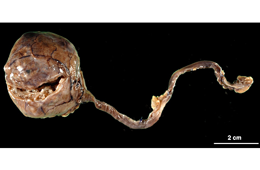
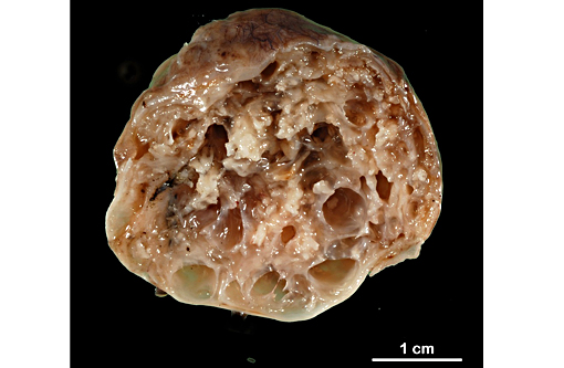
Gross Description:
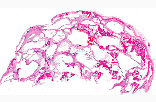
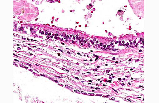
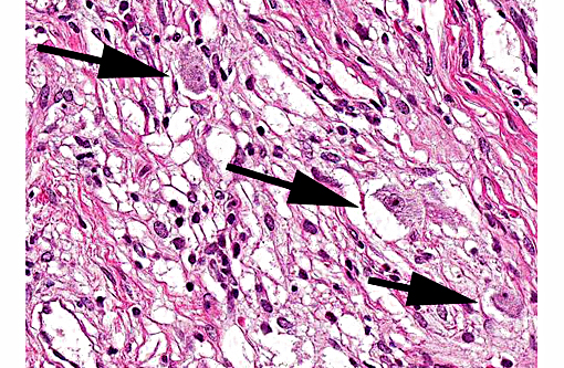
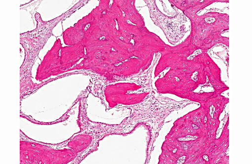
Histopathologic Description:
Morphologic Diagnosis:
Lab Results:
Condition:
Contributor Comment:
One of two ovarian teratomas reported by Willis(7) spread to peritoneal surfaces. Of 10 cases of ovarian teratoma found at necropsy of about 13,000 guinea pigs over an 8-year period,(6) none had metastasized, though some cases had resulted in abdominal hemorrhage.
Tissues from at least 2 germ layers were found in all 10 tumors; most tumors had all 3 germ layers, and nervous tissue tended to be the dominant ectodermal component. Nervous tissue also figured prominently as the ectodermal component in another case of ovarian teratoma that had spread to the peritoneal surface of the diaphragm.(1)
Granulosa cell tumor is reported far less commonly than ovarian teratoma in guinea pigs;(3) however, because of its potentially cystic nature, it should be included in the differential diagnosis for an ovarian mass along with cystic rete ovarii.(4) Though distant metastasis has not been recorded in ovarian teratomas in guinea pigs, a few cases have seeded peritoneal surfaces. In this guinea pig, no recurrence or spread of the teratoma was clinically evident at 10 weeks after ovariohysterectomy. The alopecia had resolved, and the guinea pig had gained weight.
JPC Diagnosis:
Conference Comment:
Alzarin red or Dunn-Thompson stains can be used to confirm the crystals as hemoglobin origin. Other tissue types that can be seen in teratomas which were not present in this example include ectodermal components such as hair, tooth enamel, sebaceous glands and cornified squamous epithelium. Other mesodermal elements can include adipose tissue, bone marrow, skeletal/cardiac/smooth muscle, embryonic mesenchyme and tooth structures including dentin and pulp. Other endodermal components include salivary gland epithelium, renal epithelium and thyroid gland (when thyroid tissue predominates the neoplasm is referred to as struma ovarii literally, goiter of the ovary).
Other species in which teratoma is common include 129 strain mice where the tumor most commonly occurs in the testis, but can also occur in extragonadal locations such as along the midline. Ovarian teratomas in mice are uncommon,(4) however; malignant teratoma has been documented in the ovary of transgenic mice. Teratomas are also common in cryptorchid testis of male horses. They are uncommon in other domestic/lab animal species, but have been documented in ferrets, dogs, cats, cattle, sheep and domestic poultry.(2) As a general rule, malignant teratomas are far less common in all species than their benign counterparts.
Ovarian neoplasms can be broadly divided into three groups: a) sex-cord stromal tumors which include granulosa-theca cell tumors, and thecoma/luteoma; b) tumors of the epithelial surface which include papillary adenoma/carcinoma and cystic adenoma; and c) germ cell tumors which include dysgerminoma and teratoma. Teratomas arise from totipotential germ cells that have differentiated along two or more somatic lines. Dysgerminomas, in contrast have not undergone somatic differentiation and still resemble germ cells, similar to their testicular counterpart, the seminoma. Other rare germ cell tumors include yolk sac carcinoma, choriocarcinoma and embryonal carcinoma.(5)
References:
1. Frisk CS, Wagner JE, Doyle RE. An ovarian teratoma in a guinea pig. Lab Anim Sci. 1978; 28:199-201.
2. Leylek RA, Blankenship-Paris TL, Boyle MC. Pathology in practice. Malignant teratoma of the left ovary of a mouse. J Am Vet Med Assoc. 2014; 245(2):191-193.
3. Manning PJ. Neoplastic diseases. In: Wagner JE, Manning PJ, Eds. The Biology of the Guinea Pig. New York: Academic Press; 1976:211-225.
4. Percy DH, Barthold SW. Pathology of Laboratory Rodents and Rabbits. 3rd ed. Ames, IA: Blackwell; 2007:121, 248-251.
5. Schlafer DH, Foster RA. Female genital system. In: Maxie GM, ed. Jubb, Kennedy and Palmers Pathology of Domestic Animals. Vol 3. 6th ed. St. Louis, MO: Elsevier; 2016:375-377.
6. Vink HH. Ovarian teratomas in guinea-pigs: A report of ten cases. J Pathol. 1970; 102:180-182.
7. Willis RA. Ovarian teratomas in guinea-pigs. J Pathol Bacteriol. 1962; 84:237-239.