Joint Pathology Center
Veterinary Pathology Services
Wednesday Slide Conference
2018-2019
Conference 15
16 January 2019
CASE II: NCDS-02203/1003-17 (JPC 4067571).
Signalment: 10-12 week old male Sprague-Dawley rat (Rattus norvegicus, SD:Crl)
History: This animal was a control animal in a 1 week study for an oncology agent.
Gross Pathology: Dozens of petechiae, ecchymoses and pinpoint to 1.4mm diameter, soft, yellow-tan foci and were scattered throughout the parenchyma of both lungs. Additional petechiae and ecchymoses were scattered throughout the left lateral lobe of the liver and urinary bladder. The epicardium of the right ventricle was irregularly thickened by off-white opaque material.
Gross Pathology: None.
Laboratory results: Ophthalmic examinations were not performed in this study.
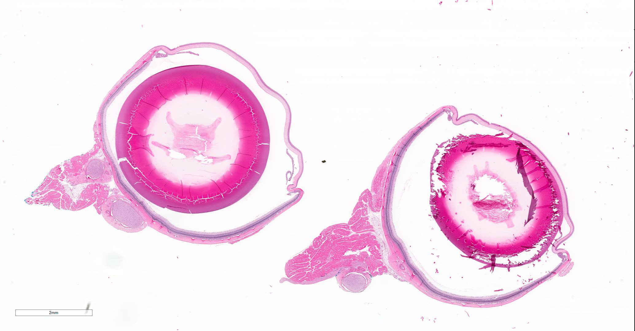
Microscopic Description:
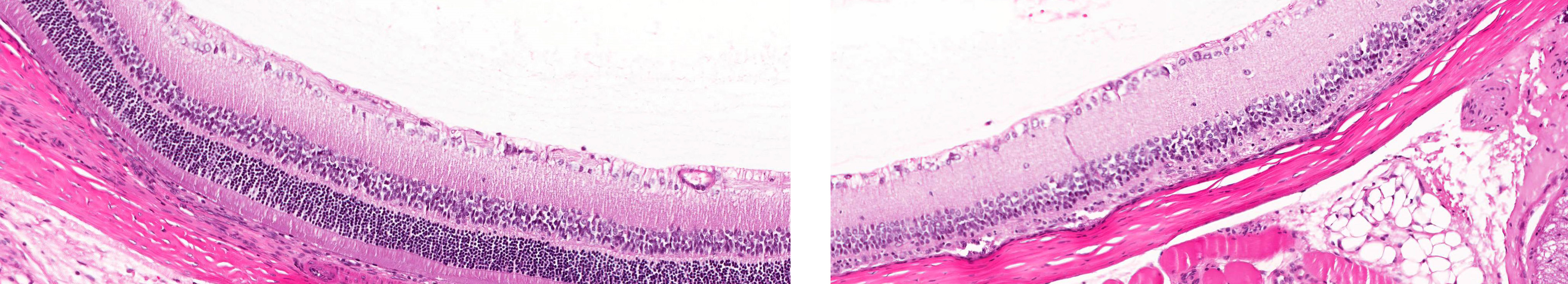
Eye: Unilaterally, a locally extensive area spanning the central to peripheral retina, there is thinning of the retina characterized by vacuolation of the photoreceptor layer, shortening and loss of the photoreceptor layer, decreased cellularity to loss of the outer nuclear layer and occasional infiltrating macrophages.
Contributor’s Morphologic Diagnoses:
Eye: Unilateral, focal, mild retinal degeneration, photoreceptor layer/outer nuclear layer
Contributor’s Comment: Spontaneous or background retinal findings in the Sprague-Dawley (SD) rat have been attributed to heritable and environmental factors, as well as aging. These findings may be present unilaterally or bilaterally. Spontaneous retinal findings may include dysplasia, dystrophy, and degeneration. Dysplasia is defined as disorganization of the sensory retina with or without degeneration and is also known as “linear retinopathy” in the rat. This is the most common background finding in SD rats greater than 11 weeks of age with 3% incidence in one study.1 Dystrophy is defined by photoreceptor and/or retinal pigmented epithelium alterations with variable time to onset and progression.
Degeneration is a non-specific term and can encompass a variety of changes in the retina. In the context of spontaneous changes associated with aging, degeneration is defined by decreased numbers of photoreceptor cells and thinning of the outer nuclear layer especially in the peripheral retina. Degeneration associated with excess light exposure is generally more prominent in the central retina and in albino animals. The development of retinal photooxicity can be influenced by a number of factors including gender, light wavelength and diet.2 The rate of spontaneous degeneration in SD rats over a broad age range (6 to 24 months, males and females) in one study is 3.7% .4 An understanding of the background incidence of retinal degeneration is important since this is the most common manifestation of retinal toxicity. Retinal toxicity can appear microscopically similar to spontaneously occurring retinal degeneration so the incidence and severity must be evaluated to determine the relevance of the finding in given treatment group.2
Contributing Institution:
Wake Forest School of Medicine
Department of Pathology, Section on Comparative Medicine
Medical Center Boulevard
Winston-Salem, NC 27157
www.wakehealth.edu
JPC Diagnosis: Retina: Degeneration of the outer nuclear and photoreceptor layer, focal, severe.
JPC Comment: The lesion described above appears to be consistent with “linear retinopathy” – one of the more frequently observed lesions in the rat retina in toxicologic studies, and reportedly prevalent in the Sprague-Dawley rat. This lesion was first reported in 1975 by Schardein et al, under the name “retinal dystrophy” and has also been recorded in the literature as “retinal or retinochoroidal degeneration and atrophy”, “retinal dysplasia/dystrophy”, or “choroidal defect” in Sprague Dawley rats. While most commonly reported in SD rats, it has also been categorized in Crl:CD BR rats.2
This particular lesion, usually unilateral, is the most frequently observed change in rats from 7-10 weeks of age, achieving maximum frequency (around 3%) in animals from 11-14 weeks of age. This frequency appears to be maintained until 110 weeks of age. Fundoscopic examination of affected rats reveals a sharply demarcated, pale, linear area of pallor in varying locations. Histologically, these lesions result in abutment of the inner nuclear layer directly on the underlying choroid or uvea, much as seen in this case.2 Early reports describe a 38% incidence of diffuse lesions, which appears rare in subsequent studies and may simply reflect evolving diagnostic criteria over time. The earliest study in SD rats theorized the possibility of retinal reattachment as a cause for this particular lesion; although other causes, such as genetic or environmental factors have not been ruled out. 2
Another form of retinal degeneration not discussed by the contributor is associated with aging. age-related degeneration includes retinal thinning with a loss of nuclei in the outer and inner nuclear layers, fusion of the nuclear layers, hypertrophy of the retinal pigmentation and possibly migration of RPE cells or macrophages into the sensory retina. Changes are noticed first in the RPE, and often in the peripheral retina, as it is thinner than the central portion.2
Other retinal diseases of a focal nature which may be seen in the retinas of Sprague-Dawley rats. Retinal folds are unilateral lesions that occur at a prevalence of 0.3% at 7-10 weeks of age. They are difficult to see on funduscopic exam as liner elevations. Histologically, they present as multilayered rosettes.3 Colobomas may be seen at a frequency of approximately 0.5% at 7-10 weeks, and are also generally unilateral. On funduscopic exam, they often appear as a white discoloration of the fundus in a ventral location, with an abnormal optic disc. Histologically, they present with disordered retinal layers, rosette formation, and ectasia of the optic disk.1,3
References:
- Hubert M.F., Gillet J.P., Durand-Cavagna G. (1994). Spontaneous retinal changes in Sprague Dawley rats. Lab Anim Sci. Dec;44(6):561-7.
- Render J.A., Schafer K.A. and Altschuler R.A. (2013). Special senses: Eye and ear. In Toxicologic Pathology: Nonclinical Safety Assessment (P.S. Sahota, J.A. Popp, J.F. Hardisty and C. Gopinath, eds) pp. 942-948. CRC Press, Boca Raton.
- Render, J. The eye in safety studies – ocular conditions and issues. L. Davis and S.W. Thompson Foundation, NE Division Symposium on Toxicologic Pathology of the Eye. https://www.cldavis.org/cgi-bin/download.cgi?pid=331.
- Weisse I. (1994). Aging and Ocular Changes. In Pathobiology of the Aging Rat, vol. 2 (U. Mohr, D.L. Dungworth and C.C. Capen, eds.) p.81