Signalment:
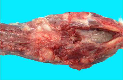
Gross Description:
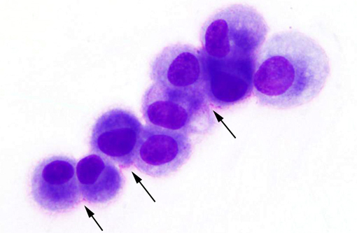
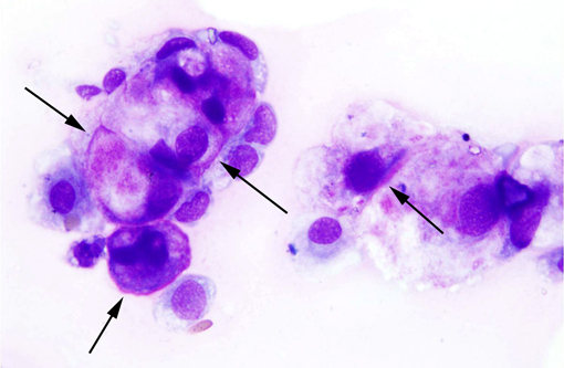
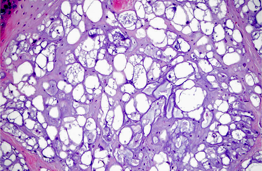
Cytologic Description: Fine needle aspirate, cervical mass: The sample is of good quality with minimal hemodilution. There are moderate numbers of nucleated cells on a moderately thick, grainy, eosinophilic background. Nucleated cells are present individually or in small clusters, showing marked (up to 5-fold) anisocytosis. The smaller cells are approximately 10 microns in diameter with a small amount of basophilic cytoplasm. The larger cells (physaliphorous cells) are markedly distended up to approximately 80 microns by cytoplasmic vacuolation that is clear or contains pink, grainy material. Nuclei are round to oval and often eccentric, with reticular chromatin, typically lacking obvious nucleoli. Mitotic figures are not observed.
Morphologic Diagnosis:
Condition:
Contributor Comment:
Chordomas are the most common neoplasm of the musculoskeletal system of the ferret,(4) but have also been described in other species, such as rats,(11) cats,(2) humans,(10) mink(6) and dogs.(8) The embryonic notochord degenerates early in fetal development and remains as the nucleus pulposus within intervertebral disks.(7) In some cases, residual notochord cells remain outside the intervertebral disks and may become a chordoma.
In ferrets, chordomas arise primarily in or adjacent to the caudal vertebra,(4) but they also have been described elsewhere along the spine.(9,12) Chordomas are locally aggressive, destroy the vertebral body, and invade adjacent tissues. Cutaneous metastases of chordomas have been reported in ferrets.(7,12) Although chordomas of the tail may be treated by amputation, at other locations along the axial skeleton, chordomas may not be amenable to surgical therapy and can cause spinal cord compression, as seen in this case.
The main differential diagnosis for a chordoma is chondrosarcoma. The large, severely vacuolated, physaliphorous cells of chordomas are not a feature of chondrosarcoma;(8) however, differentiation of these tumors is further aided by immunohistochemistry. Chordomas express vimentin, cytokeratin, and S-100 protein, while chondrosarcomas do not express cytokeratin. Combining the midline location of the tumor, cytological criteria such as physaliphorous cells, and immunohistochemical expression of cytokeratin confirms a diagnosis of chordoma.
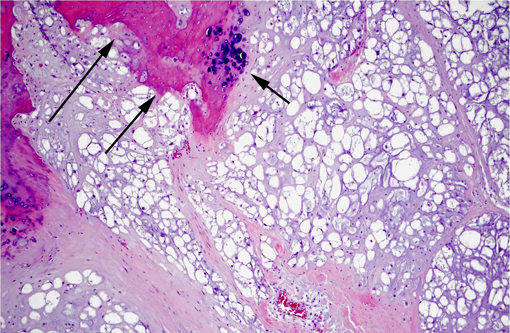
JPC Diagnosis:
Conference Comment:
Conference participants briefly discussed the occurrence of chordomas in various veterinary species. In ferrets, the most common location is on the tip of the tail; however, in most domestic species, chordomas tend to occur in the sacrococcygeal and cervical regions. In dogs, chordomas have also been reported in the brain, spinal cord, and skin.(5) Of the two reported cases of feline chordoma, one was initially diagnosed as chronic granulomatous inflammation due to the interpretation of the characteristic physaliphorous cells as atypical, foamy macrophages, but was later shown to be a classic chordoma, which subsequently metastasized to multiple lymph nodes.(2) The second feline case was classified histologically and immunohistochemically as a chondroid chroma.(1) Chondroid chordomas similar to the human subtype have been reported in ferrets, mink, cats and dogs.(1,5) Similarly to human medicine, the distinction between conventional and chondroid chordomas may also have some degree of prognostic significance in animals. Although all chordomas are potentially locally invasive, chondroid chordomas in ferrets, dogs and cats have not been reported to metastasize, while conventional chordomas in rats(11) and cats(2) (but not dogs)(5) seem to have a higher rate of metastasis. Additionally, chordomas arising from the tail appear to have a good prognosis in all cases.(1)
An alternate spelling of physaliferous may be seen in the literature, and is considered a correct spelling as well.
References:
2. Carpenter JL, Stein BS, King NW Jr, Dayal YD, Moore FM. Chordoma in a cat. J Am Vet Med Assoc. 1990;197:240-242.
3. Chugh R, Tawbi H, Lucas DR, et al. Chordoma: the nonsarcoma primary bone tumor. The Oncologist. 2007;12(11):1344-1350.
4. Dunn DG, Harris RK, Meis JM, Sweet DE. A histomorphologic and immunohistochemical study of chordoma in twenty ferrets (Mustela putorius furo). Vet Pathol. 1991;28(6):467-473.
5. Gruber A, Kneissi S, Vidoni B, Url A. Cervical spinal chordoma with chondromatous component in an dog. Vet Pathol. 2008;45(5):650653.
6. Hadlow WJ. Vertebral chordoma in two ranch mink. Vet Pathol. 1984;21(5):533-536.
7. Munday JS, Brown CA, Richey LJ. Suspected metastatic coccygeal chordoma in a ferret (Mustela putorius furo). J Vet Diagn Invest. 2004;16(5):454-458.
8. Munday JS, Brown CA, Weiss R. Coccygeal chordoma in a dog. J Vet Diagn Invest. 2003;15(3):285-288.
9. Pye GW, Bennett RA, Roberts GD, Terrell SP. Thoracic vertebral chordoma in a domestic ferret (Mustela putorius furo). J Zoo Wildl Med. 2000;31(1):107-111.
10. Rich TA, Schiller A, Suit HD, Mankin HJ. Clinical and pathologic review of 48 cases of chroma. Cancer. 1985;56(1):182-187.
11. Stefanski SA, Elwell MR, Mitsumori K, Yoshitomi K, Dittrich K, Giles HD. Chordomas in Fischer 344 rats. Vet Pathol. 1988;25(1):42-47.
12. Williams BH, Eighmy JJ, Berbert MH, Dunn DG. Cervical chordoma in two ferrets (Mustela putorius furo). Vet Pathol. 1993;30(3):204-206.