Signalment:
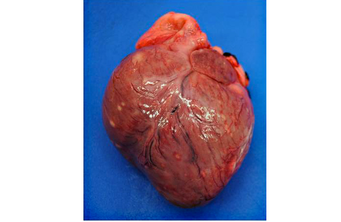
Gross Description:
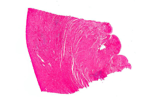
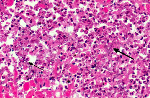
Histopathologic Description:
Morphologic Diagnosis:
Lab Results:
Condition:
Contributor Comment:
Scedosporium prolificans (previously Scedosporium inflatum) is a filamentous fungus within the family Microascaceae.(1) In the environment it has been isolated from soil and potted plants.(1) The organism is increasingly recognized as a cause of disseminated fungal infections in immunocompromised human patients. Treatment of infections is challenging due to the resistance of S. prolificans to most antifungal agents.(1) There are rare reports of S. prolificans infection in animals. In dogs S. prolificans has been isolated from a German Shepherd dog with a disseminated infection involving kidney, heart, bone marrow, skeletal muscle, liver, lung, spleen, lymph nodes and pancreas.(5) In addition one case has been reported of a beagle with osteomyelitis in which S. prolificans was found to have disseminated to the lungs.(7)
One case of S. prolificans infection in a horse associated with osteomyelitis and arthritis has also been described.(9) Musculoskeletal infections are also a common presentation in humans.(1) Further isolates characterized as S. prolificans were obtained from eye scrapings of two horses and from a draining sinus in a cat.(8)
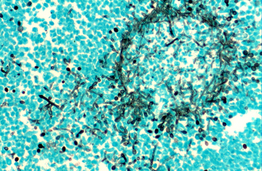
In tissues the morphology of Scedosporium spp. is similar to that of Aspergillus spp. although Scedosporium spp. exhibit haphazard branching with less frequent dichotomous branching.(6) Culture of the fungus allows more definitive identification; the colonies grow rapidly and exhibit a moist, felty appearance with initially a white color that becomes olive-grey to black. Microscopically the conidiophores display distinctly swollen bases (hence the previous name S. inflatum) with ovoid conidia.Â
If material for culture is not available, identification of Scedosporium spp. by PCR is also possible.(4)
Disseminated infections with opportunistic fungi in dogs have been associated with a number of different fungal species. In particular, systemic infections with Aspergillus terreus have commonly been described in German Shepherd dogs. Breed-associated abnormalities in IgA levels or function have been reported but have not been conclusively proven to be the cause of the increased susceptibility to fungal infections.(2,11)
The Border collie presented in the current case had shown poor growth from birth and persistent upper respiratory disease; however, an underlying immunodeficiency was not established.Â
Other fungal species isolated from dogs with disseminated infections include Penicillium sp., Paecilomyces sp., Chrysosporium sp and Pseudoallescheria boydii or Scedosporium apiospermum (the asexual form of P. boydii).(10)
JPC Diagnosis:
Conference Comment:
S. prolificans is a filamentous, non-pigmented, parallel-walled fungus with septate, 3-5 μm, haphazardly branching hyphae (sometimes described as a letter-H pattern) with lemon-shaped conidiophores from which a small cluster of single-cell conidia emerges.(1) Scedosporium will produce conidia in solid non-aerated tissues,(5) such as the myocardium in the present case. Aspergillus has a similar size and tissue morphology, and it also produces conidiophores, or fruiting bodies, but unlike Scedosporium, these are generally more round than lemon-shaped and they only occur in aerated tissues like ectatic bronchi or the surface of skin wounds.(6) Candida sp. appears in tissue in both hyphal and budding yeast forms, which could be confused with Scedosporium, however the presence of pseudohyphae is relatively common in candidiasis and rare in S. prolificans infection.(6) Like S. prolificans, zygomycete hyphae may also appear in a letter-H pattern,(6) though their hyphae are more broad (6-25 μm) and pauciseptate with non-parallel walls and non-dichotomous branching.(3) S. prolificans may also be mistaken for Pythium insidiosum, which, although it is an oomycete rather than a true fungus, produces 2-10 μm, pauciseptate hyphae with non-parallel walls and non-dichotomous branching.(3)
S. prolificans is resistant to many anti-fungal drugs and generally carries a grave prognosis, so differentiating it from other opportunistic fungi or fungal-like organism is imperative.(6) Although Scedosporium has several unique morphologic characteristics, definitive diagnosis with histopathology alone is often difficult to achieve, so culture and/or PCR are critical.
References:
2. Day MJ, Penhale WJ, Eger CE, et al. Disseminated aspergillosis in dogs. Aust Vet J. 1986;63:55-59.
3. Ginn PE, Mansell JEKL, Rakich PM. Skin and appendages. In: Maxie MG, ed. Jubb, Kennedy, and Palmers Pathology of Domestic Animals. Vol 1. 5th ed. Philadelphia, PA: Elsevier Saunders. 2007:695-708.
4. Harun A, Blyth CC, Gilgado F, Middleton P, Chen SC-A, Meyer W. Development and validation of a multiplex PCR for detection of Scedosporium spp. in respiratory tract specimens from patients with cystic fibrosis. J Clin Microbiol. 2011;49:1508-1512.
5. Haynes SM, Hodge PJ, Tyrrell D, Abraham LA. Disseminated Scedosporium prolificans infection in a German Shepherd dog. Aust Vet J. 2012;90:34-38.
6. Kimura M, Maenishi O, Ito H, Ohkusu K. Unique histological characteristics of Scedosporium that could aid in its identification. Pathology International. 2010;60:131-136.
7. Salkin IF, Cooper CR, Bartges JW, Kemna ME, Rinaldi MG. Scedosporium inflatum osteomyelitis in a dog. J Clin Microbiol. 1992;30:2797-2800.
8. Salkin IF, McGinnis MR, Dykstra MJ, Rinaldi MG. Scedosporium inflatum, an emerging pathogen. J Clin Microbiol. 1988;26:498-503.
9. Swerczek TW, Donahue JM, Hunt RJ. Scedosporium prolificans infection associated with arthritis and osteomyelitis in a horse. Journal of the American Veterinary Medical Association. 2001;218:1800-1802,1779.
10. Watt PR, Robins GM, Galloway AM, O'Boyle DA. Disseminated opportunistic fungal disease in dogs: 10 cases (1982-1990). Journal of the American Veterinary Medical Association. 1995;207:67-70.
11. Whitbread TJ, Batt RM, Garthwaite G. Relative deficiency of serum IgA in the German Shepherd dog: a breed abnormality. Res Vet Sci. 1984;37:350-352.