Signalment:
4-year-old spayed female mixed breed dog (
Canis familiaris)3-week history of seizures, nystagmus, strabismus, lateral recumbency at about weekly intervals. On each occasion, supportive care with intravenous fluid, administrated at the referring veterinarian was sufficient to bring about temporary recovery from symptoms. The dog was admitted to the University of Florida Veterinary Medical Center (UF VMC) obtunded with lateral nystagmus, anisocoria, and strabismus. She had patellar hyperreflexia, good palpebral reflexes, miosis of the right pupil, and mydriasis of the left pupil with no pupillary light responses, and good reflexes in all four limbs. Cystic lesions in the brain were diagnosed at the referring veterinarian. The dog began to decline and the owner opted to euthanize.
Gross Description:
The lateral ventricles in the brain were moderately enlarged. There was a 2cm diameter, poorly-demarcated red focus in the meninges over the right lateral occipital area. One gyrus in this region was expanded and contained an 8x5 mm grey focus.
The kidneys contained numerous irregular depressions on the subcapsular surface that extend into the cortex. The capsule did not peel off easily.
Histopathologic Description:
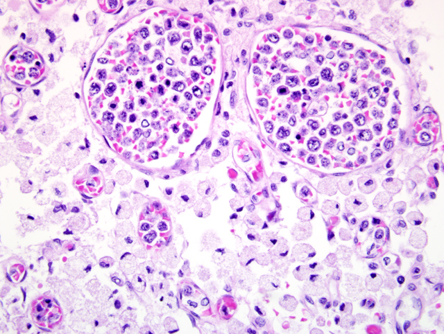
Vessels in the meninges are frequently markedly distended with a neoplastic population of round cells (lymphocytes) (
Fig. 3-1)which are predominantly confined to the lumina of capillaries, arteries, and veins, but also extend into the perivascular tissue. The lymphocytes are large, have large round to oval nuclei, one to several nucleoli, coarsely clumped chromatin, and small amounts of amphophilic cytoplasm. There is marked anisocytosis and anisokaryosis, and an average of 1-2 mitotic figures per 400X field. There is frequent individual cell necrosis. Frequently, there are scant erythrocytes in the vessels along with the neoplastic lymphocytes. There are thrombi present in a low number of affected vessels. Throughout the parenchyma, in both the gray and white matter, there are a low number of vessels which contain neoplastic lymphocytes. There are several areas in the cerebral cortex which contain increased numbers of small vessels with prominent endothelial cells and neoplastic cells often present within the vessels. In these foci, there is rarefaction of the neuropil, necrotic neurons and/or loss of neurons, swollen axons, and accumulation of gitter cells (infarcts). Neoplastic lymphocytes rarely extend into the neuropil in these foci.Â
Morphologic Diagnosis:
1. Intravascular lymphoma, with thrombosis and subacute to chronic infarction, brain
2. Moderate hydrocephalus
Lab Results:
Sodium mEq/L = 137 (145-154), potassium = 5.6 mEq/L (4.1-5.3), BUN = 102 mg/dL (8-31), Creatinine = 4.6 mg/dL (0.8-1.6).
Condition:
Intravascular lymphoma
Contributor Comment:
Canine intravascular lymphoma is a rare large cell lymphoma in which lymphocytes proliferate within blood vessels in the absence of a primary extravascular mass or leukemia.(1) Blood vessels in nearly all tissues examined contained luminal neoplastic lymphocytes, which often largely filled the vessel lumen. Thrombosis was also identified in numerous tissues, and thrombosis resulted in infarction in the brain, spleen and heart. Infarcts were variably prominent in submitted sections. Multifocal cerebral cortical infarctions were the cause of neurologic clinical signs. The hydrocephalus diagnosed on gross examination was most likely congenital and an incidental finding.Â
Canine intravascular lymphoma is also known as malignant angioendotheliomatosis. Although most human cases are of B cell origin, (3) canine cases are typically classified as T cell or non-B, non-T cell.(1) Most human cases present with CNS or cutaneous signs.(3) CNS signs are most common in affected dogs.(1)
In a retrospective study of 17 cases of canine intravascular lymphoma, neoplastic lymphocytes rarely extended outside of vessels into the surrounding parenchyma and were not present in lymph nodes or the bone marrow in the 4 cases where these organs were examined.(1) Bone marrow and lymph node involvement were described in an additional canine case report (2) and bone marrow involvement was reported in 4/5 human patients in which neoplastic lymphocytes were also identified in peripheral blood smears.(4) While neoplastic lymphocytes were observed outside of vessels in multiple locations in this case (brain, kidney, pancreas, lung, lymph node, and bone marrow), the overwhelming distribution of neoplastic lymphocytes was within vessels, with comparatively little involvement of the organ parenchyma, suggesting the diagnosis of intravascular lymphoma. Additionally, leukemia was not identified previously while this animal was having neurologic signs, which presumably were due to intravascular lymphoma, thrombosis and infarction. It was uncertain if this animal was leukemic at the time of death, as no bloodwork was done at the UF VMC. The absence of neoplastic lymphocytes in peripheral blood smears in these dogs makes antemortem diagnosis extremely difficult and the diagnosis was made antemortem in a single dog in this study.(1)
JPC Diagnosis:
Brain, cerebrum: Intravascular lymphoma, with fibrin thrombi and multifocal infarcts
Conference Comment:
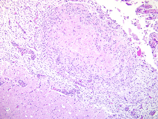
In addition to the diagnosis listed above, some but not all sections contain an interesting well-demarcated focus within an area of rarefaction. This nodular focus is composed of a central core of coagulative necrosis bounded by numerous fibroblasts and glial cells, interpreted as a sequestrum (
Fig. 3-2).Â
The majority of cases of intravascular lymphoma in humans can be placed into four distinct presentations: 1) central nervous system form, 2) cutaneous form, 3) fever of unknown origin, and 4) hemophagocytic form.(4) Most human patients present with neurologic signs, while about 1/3 of human patients present with cutaneous lesions. Cutaneous lesions are not as common in animals with IVL as they are in their human counterparts.(1)
As mentioned by the contributor, most human cases are of B cell origin, while IVL in animals has been shown to be derived primarily from T cells or non-B, non-T lymphocytes with rare cases of B cell tumors.(1) The source of neoplastic cells in IVL is still uncertain, but there is speculation that they may originate from the red pulp of the spleen.(1) The behavior of these neoplasms suggest that initial movement into the bloodstream is possible with subsequent problems related to adhesion and emigration resulting in intravascular accumulations of neoplastic cells as opposed to deposition within organs.(1)
References:
1. McDonough SP, Van Winkle TJ, Valentine BA, van Gessel YA, Summers BA: Clinicopathological and Immunophenotypical features of canine intravascular lymphoma (Malignant Angioendotheliomatosis). J Comp Pathol
126:277-288, 2002
2. Steinberg, H: Multisystem angiotropic lymphoma (malignant angioendotheliomatosis) involving the humerus in a dog. J Vet Diag Invest
8:502-505, 1996
3. Xanthopoulos V, Galanopoulos, AG, Paterakis G, Apessou D, Argyrakos T, Goumakou E, Papadhimitriou SI, Savvidou I, Georgiakaki M, Anagnostopoulos NI: Intravascular B-cell lymphoma with leukemic presentation: case report and review of the literature. Eur Jour Haematol, 2007
4. Zuckerman D, Seliem R, Hochberg E: Intravascular Lymphoma: The Oncologists Great Imitator. The Oncologist,
11:496-502, 2006