Signalment:
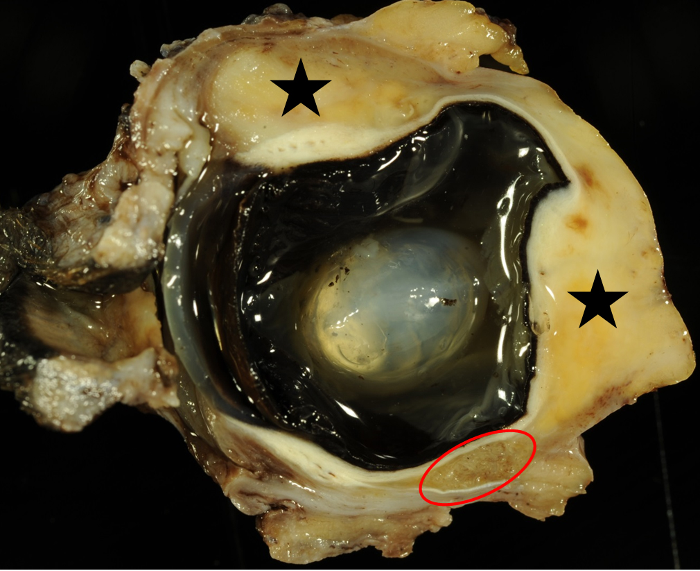
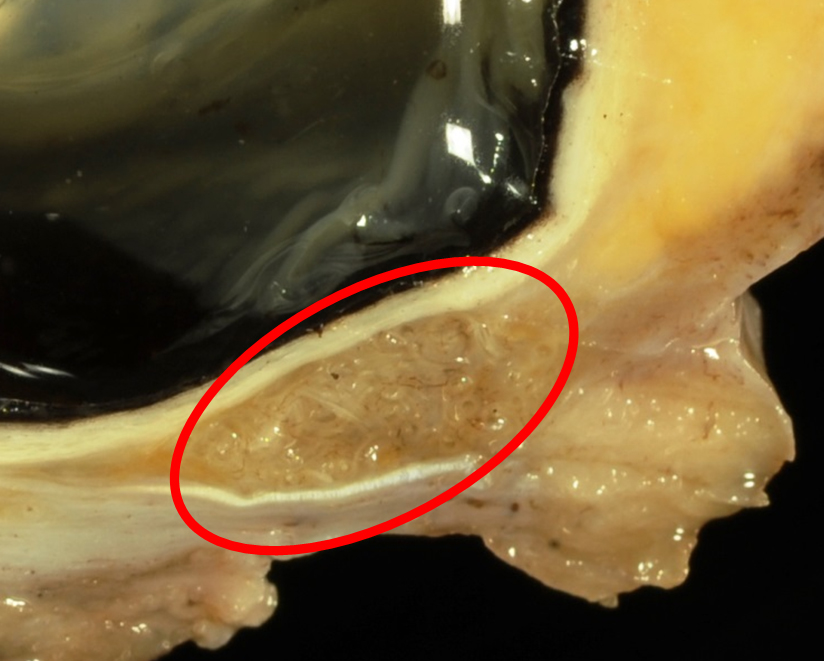
Gross Description:
Histopathologic Description:
Morphologic Diagnosis:
Lab Results:
Condition:
Contributor Comment:
For a long time, ocular onchocercosis in dogs was thought to be an aberrant infection with Onchocerca (O.) lienalis, commonly found in cattle. The assumption that canine ocular onchocerciasis is associated with O. lienalis was only based on the microscopic morphology.6 In contrast, a previously unrecognized species of Onchocerca was suspected to be spilling over from wild ungulates to canines, but it turned out that the nucleotide sequences of Onchocerca species found in canines are unique within the genus.13 An analysis of the molecular genetic data suggested that the causative agent is rather O. lupi than O. lienalis.6
A couple of nematodes are known to affect the eyes or the periorbital tissue. Thelazia, Onchocerca, Ancylostoma, Dirofilaria, Angiostrongylus vasorum, Toxocara canis and Trichinella sp. have been identified within ocular helminthic infections of dogs.11 O. lupi is a vector borne nematode that was first described in 1967 in a Caucasian wolf (Canis lupus) from Russia.5 The life cycle as well as the precise host range of canine Onchocerca is still not fully known, but it might be similar to those of other Onchocerca species. Within other Ochocerca species, blackflies and/or biting mites serve as intermediate hosts, for example Simulium tribulatum, was found to be a putative vector for O. lupi in Southern California.12 All other Onchocerca sp. have a long prepatent period and patency of several months or even years within their hosts.12
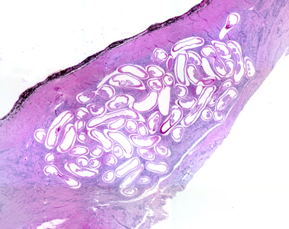
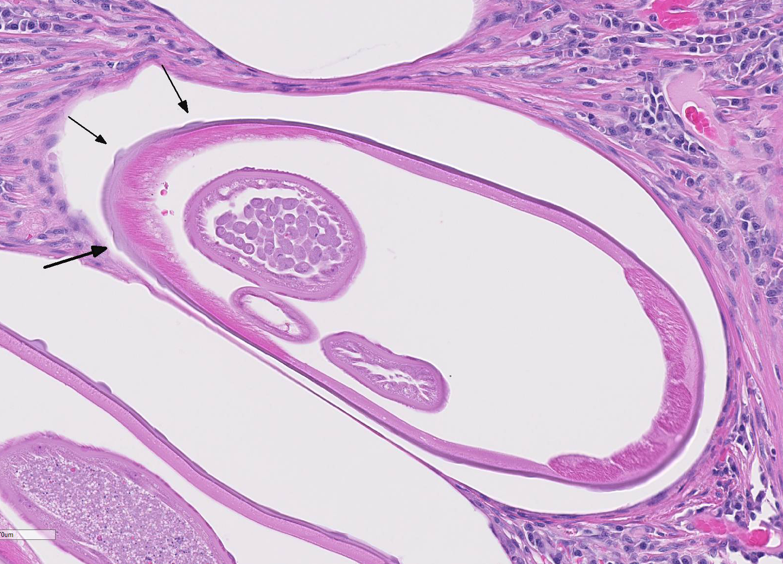
In canine cases, gravid females, mature males, and microfilaria can be found within affected animals. Acute or chronic ocular disease with conjunctivitis, exophthalmos, periorbital swelling, photophobia, discomfort, lacrimation and discharge during the acute phase were reported as clinical signs.8, 12 Chronic cases are characterized by granuloma formation in different parts of the eye and the periorbital tissue. The cuticle of females is a striking feature for the light microscopic identification of Onchocerca sp. It consists of two layers with an outer layer bearing ring-like ridges. In the anterior part of the nematode, these ridges are small and close together, becoming taller and more divergent in posterior direction. At the posterior part, they again get smaller with no visible ridges at the very end of the body.12 The two distinct cuticular layers are hardly identifiable within the presented case most likely due to insufficient preservation. Histopathological examination of affected tissue may reveal coiled female nematodes within a mixture of fibrotic tissue admixed with mononuclear cells. Male nematodes as well as microfilariae may occasionally be present in the periocular tissue.7
Affected dogs are usually older than 1 year, which is most likely owed to the indirect life cycle of Onchocerca spp. as well as the slow development of these nematodes in their final hosts. Microfilariae can be isolated from skin samples, even if the animal has no clinical signs of onchocercosis. Micro-filariae do not occur within the bloodstream, but larval concentration is high within the skin (50 - 3600 per g tissue), which might serve as a suitable screening method. Until today there is no serological test commercially available for the detection of O. lupi.10,13
O. lupi seems to be endemic in the United States. There have been also two proven cases of feline onchocercosis associated with O. lupi. Moreover, several cases of ocular onchocercosis have been reported in humans worldwide.5 Onchocerca sp. infect humans in tropical regions causing severe ocular inflammation known as river blindness.14 Nevertheless, O. lupi has been identified to act as the causative agent in two human ocular cases with similar clinical features compared to those in dogs.8, 9Onchocerca sp. represent a zoonotic pathogen and should be considered as differential diagnosis in canine ocular diseases.
JPC Diagnosis:
Conference Comment:
Conference participants focused largely on the morphologic features distinguishing Onchocerca sp. in tissue section from other metazoan parasites. Filarid nematodes are small parasites that infect a number of different domestic animals. They are easily identified by their coelomyarian musculature, very small digestive tract, and larval microfilaria in the reproductive tract of gravid females.3 In Onchocerca sp., the coelomyarian muscles atrophy and are replaced hypodermal tissue because adults reside within a fibrous capsule and are sedentary. The key diagnostic feature that differentiates Onchocerca sp. from other filarid nematodes is the presence of evenly spaced ring-like circumferential annular cuticular ridges seen on longitudinal section.2,3 This is the only nematode that has this cuticular manifestation. In contrast, Dirofilaria immitis, the canine heartworm, is a relatively common intraocular filarial nematode of dogs and has evenly spaced ridges that run longitudinally and can only be seen on cross section.2,3 These characteristics also help differentiate Onchocerca sp. from Thelazia sp., a spirurid nematode parasite and another common ocular parasite in a wide range of mammalian hosts.3
References:
16. Cullen CL, Webb AA. Ocular manifestations of systemic disease. In: Gelatt KN, Gilger BC, Kern TJ, eds. Veterinary Ophthalmology. Vol 2. 5th ed. Ames, IA: Blackwell Publishing; 2013:1931.
17. Dubielzig RR, Ketring KL, McLellan GJ, Albert DM. Veterinary Ocular Pathology: A comparative review. St. Louis, MO: Saunders Elsevier; 2010:118-120..
18. Gardiner, CH, Poynton, SL. An Atlas of Metazoan Parasites in Animal Tissues. Washington, D.C: Armed Forces Institute of Pathology, American Registry of Pathology, 1999:3538.
19. Hassan KH, Bolcen S, Kubofcik J, et al. Isolation of Onchocerca lupi in dogs and black flies, California, USA. Emerg Infect Dis. 2015; 21(5):789796.
20. Labelle AL, Maddox CW, Daniels JB, et al. Canine ocular onchocercosis in the United States is associated with Onchocerca lupi. Vet Parasitol. 2013; 193(1-3):297301.
21. Mutafchiev Y, Dantas-Torres F, Gianelli A., et al. Redescription of Onchocerca lupi (Spirurida: Onchocercidae) with histopathological observations. Parasit Vectors. 2013; 6:309316.
22. Otranto D, Dantas-Torres F, Giannelli A, et al. Zoonotic Onchocerca lupi infection in dogs, Greece and Portugal, 20112012. Emerg Infect Dis. 2013; 19:20002003.
23. Otranto D, Giannelli A, Scotty Trumble N, et al. Clinical case presentation and a review of the literature of canine onchocercosis by Onchocerca lupi in the United States. Parasit Vectors. 2015; 8:8996.
24. Otranto D, Giannelli A, Latrofa MS, et al. Canine infections with Onchocerca lupi nematodes, United States, 20112014. Emerg Infect Dis. 2015; 21:868871.
25. Sreter T, Szell Z, Egyed Z, et al. Ocular onchocercosis in dogs: A review. Vet Rec. 2002; 151:176180.
26. Sréter T, Széll Z. Onchocercosis: A newly recognized disease in dogs. Vet Parasitol. 2008; 151:113.
27. Széll Z, Sréter T, Erdélyi I., et al. Ocular onchocercosis in dogs: Aberrant infection in an accidental host or lupi onchocercosis? Vet Parasitol. 2001; 101:115125.
28. Zimmermann PA, Dadzie KY, De Sole G, et al. Onchocerca volvulus DNA probe classification correlates with epidemiologic patterns of blindness. J Infect Dis. 1992; 165:946968.