Signalment:
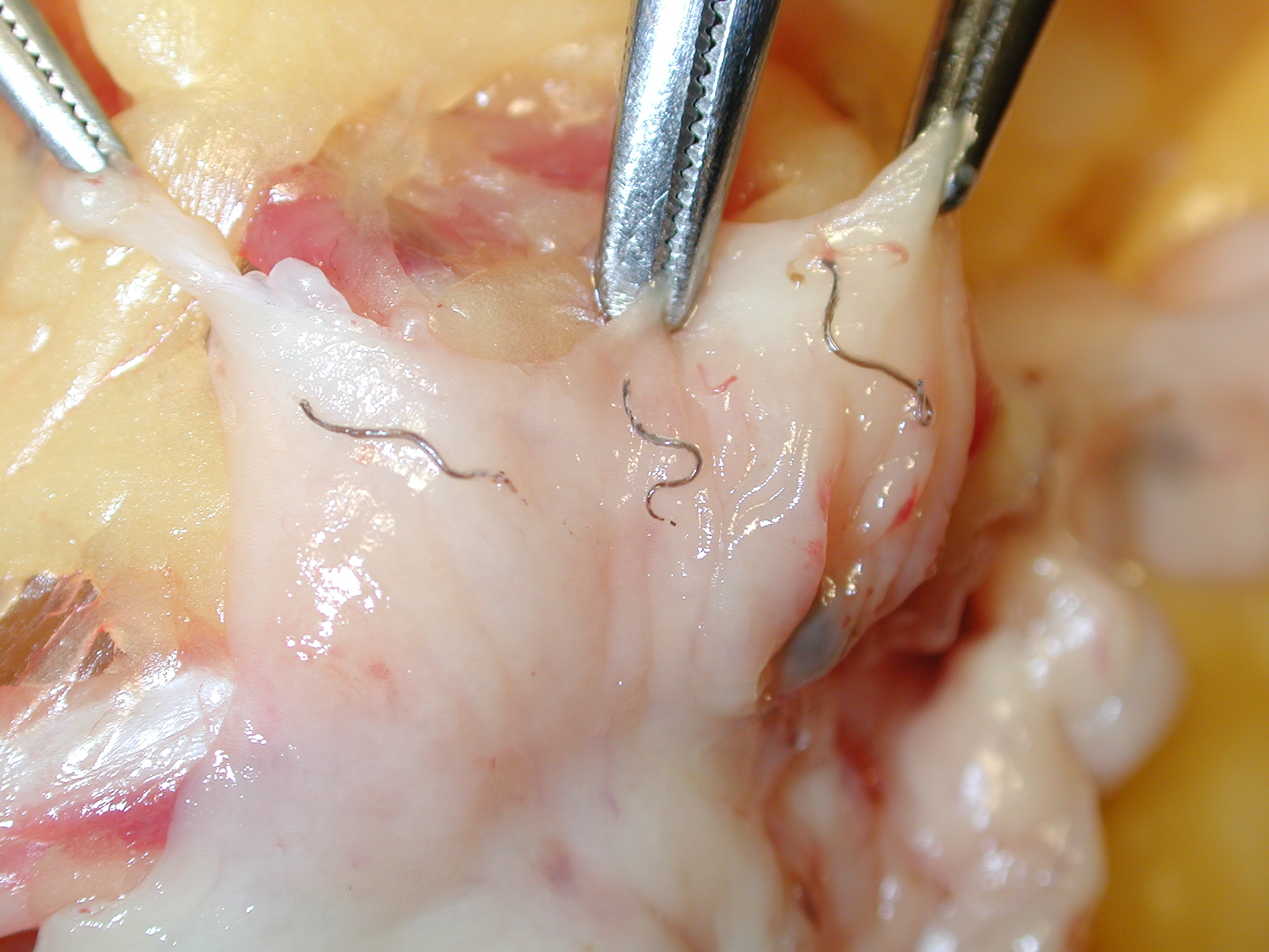
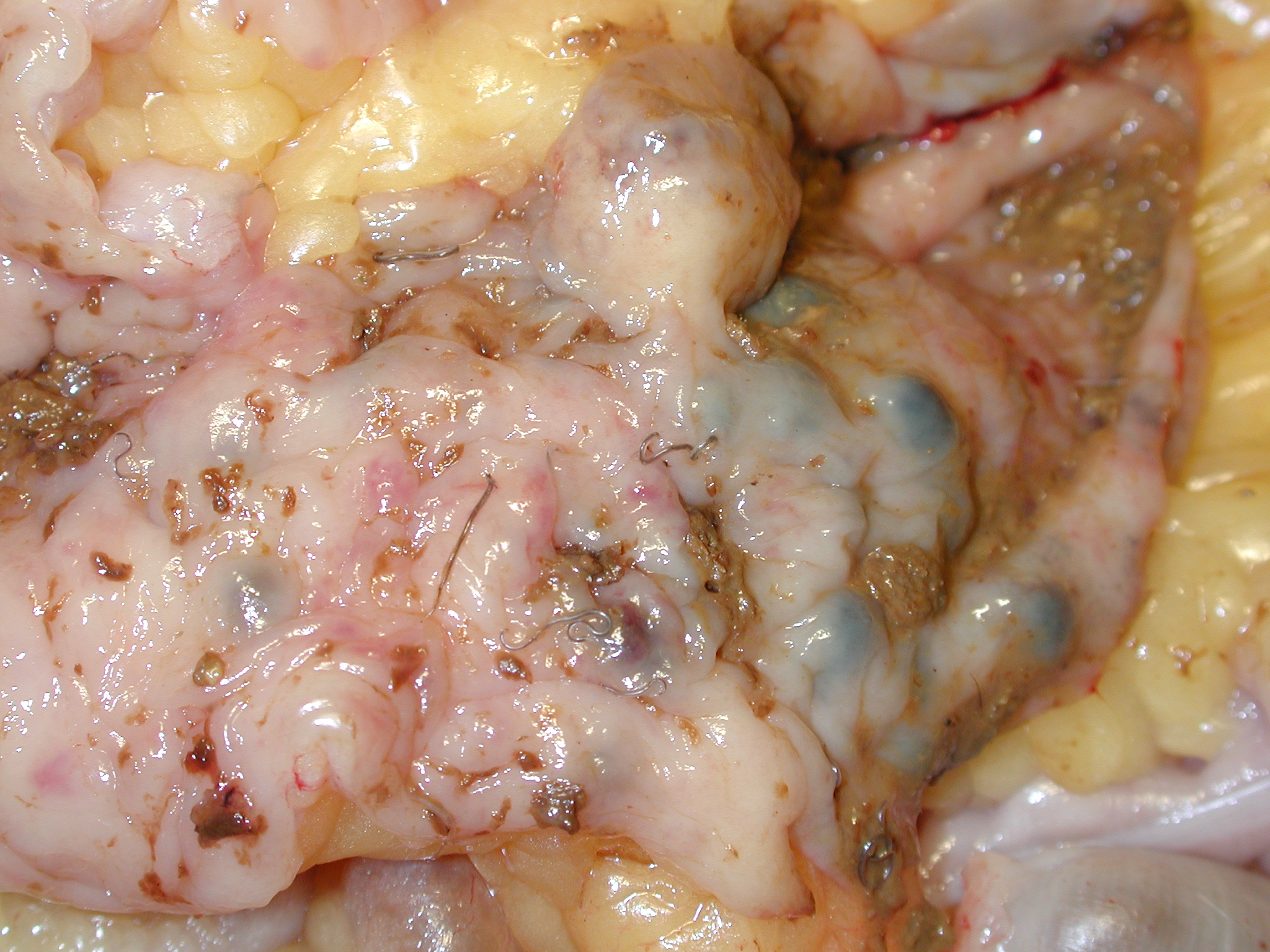
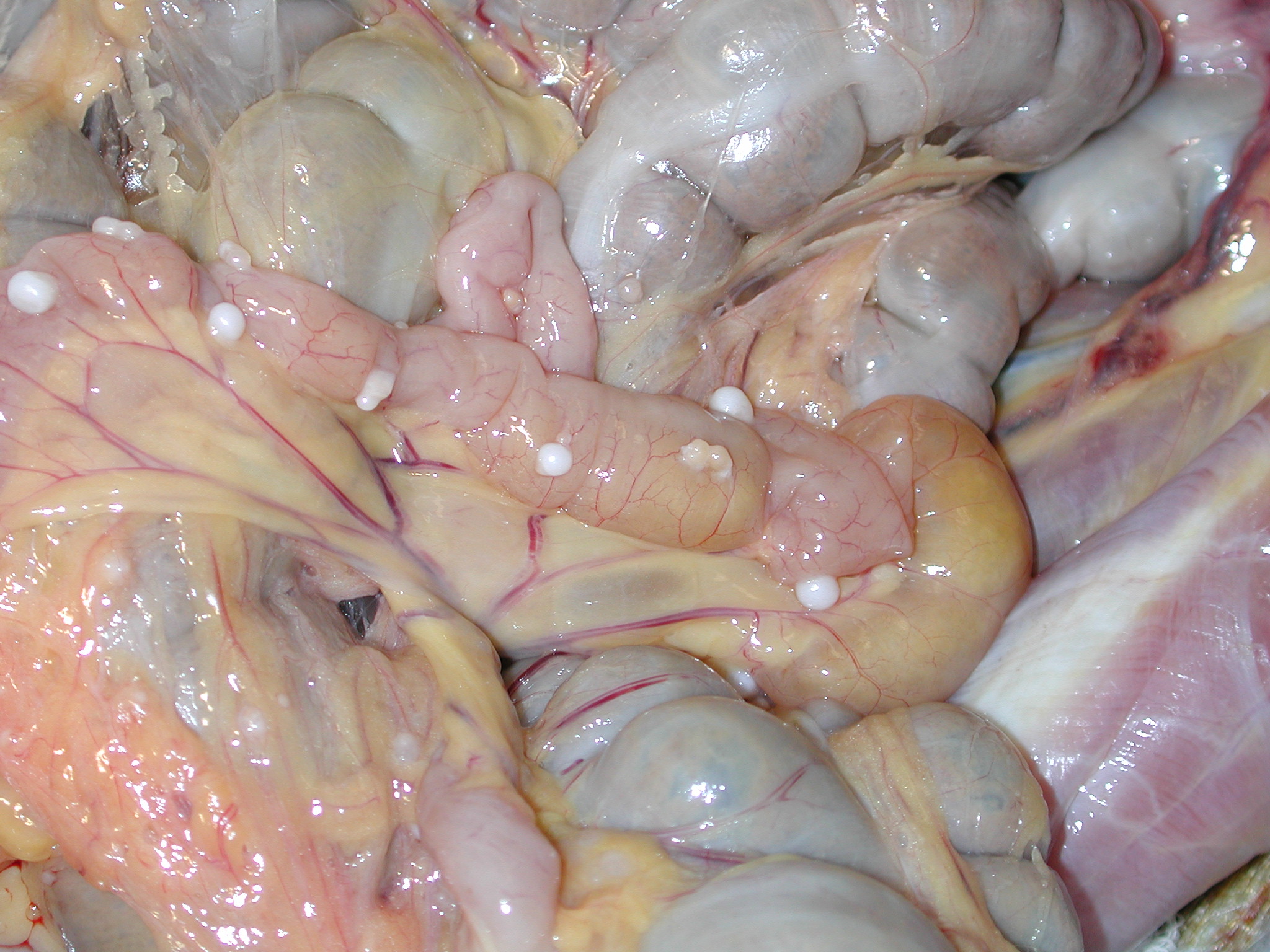
Gross Description:
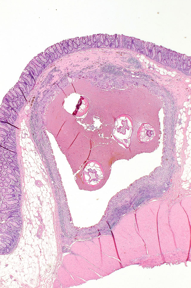
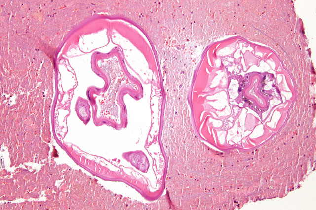
Histopathologic Description:
Further histological characteristics present allows identification as Strongyles, including the presence of platymyarian musculature, prominent vacuolated lateral chords and characteristic intestinal tract with brush borders and iron pigment sometimes visible within intestinal cells.Â
Morphologic Diagnosis:
Condition:
Contributor Comment:
The oesophagostomes, sometimes referred to as nodular worms, are among the most common and injurious parasites of monkeys and apes.(6) Worms characteristically produce nodules or cysts in the submucosa or muscularis of the large intestine and less frequently in ectopic sites. Although confusion exists about species identification, apiostomum, bifurcum, aculeatum and stephanostomum are recognized in the genus Oesophagostomum.(1)
Adult worms live in the lumen of the bowel in their definitive host. Eggs are passed in the feces, hatch and release larvae that mot twice to become infective. Third stage larvae when swallowed by a new host, burrow into the submucosa of the small or large intestine, molt again to fourth stage larvae and return to the lumen of the large intestine, where they molt again to become mature worms.(2)
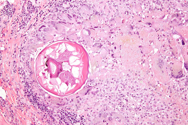
Seen not uncommonly in baboons, mangabeys, macaques and great apes, infestation in New World monkeys is rare. Prior to the influx of feral, recently imported Chinese macaques in recent years, the chronic, healed lesions from these parasites were occasionally recognized as discrete and circumscribed, highly mineralized nodules visible on the serosal margin of the bowel (See submitted gross image 3). Such lesions generally did not demonstrate histologic evidence of residual recognizable parasite structures. The submitted case demonstrates an active nonhuman primate infection.
Oesophagostomum infestation from a variety of species is of course well recognized in numerous other animal species including pigs (O. dentatum), cattle (O. radiatum), sheep (O. columbianus), and a several wild ruminants in which such nodular worm disease may be associated with significant morbidity and mortality. (8,5)
Human Oesophagostomiasis is an infrequently described and recognized parasite infection in humans, generally caused by Oesophagostomum bifurcum.(3) It is a regional and very localized public health problem in Africa, but is considered common in northern Togo and Ghana.(7) Human infestation may cause localized abdominal pain and discomfort, commonly in the right lower quadrant and this is often accompanied by epigastric or periumbilical masses.(2)
JPC Diagnosis:
Conference Comment:
The contributor did a magnificent job describing not only the identification features and life cycle of this nematode parasite, but also gave an excellent summary of comparative pathology.
For the pathologist, it is important to systematically describe nematode parasites in tissue section. One satisfactory method it to start at the outer layers and work ones way in. A brief review of the major histologic identifiable features is presented below and is based on Dr. Chris Gardiners guidelines in An Atlas of Metazoan Parasites in Animals Tissues. (4)
Cuticle: The cuticle is the outermost covering of a nematode, which can range in thickness from being very prominent to almost imperceivable. Alae, which are winglike extensions of the cuticle, can also be used to identify certain nematodes. (4)
Hypodermis: The hypodermis is immediately internal to the cuticle and extends into the body cavity, or pseudocoelom. Projections of the hypodermis into the pseudocoelom are called lateral chords. These chords can have many different shapes and are helpful in parasite identification.(4)
Musculature: Muscle cells extend from the hypodermis into the pseudocoelom and are composed of a contractile element and a cytoplasmic element. On a normal H&E slide, the cytoplasmic portion is usually clear, and the contractile portion is bright pink to red. Muscles are categorized as being either coelomyarian or platymyarian. Coelomyarian muscles extend into the body cavity in a circular manner, whereas platymyarian muscles are often flattened against the hypodermis and do not extend into the body cavity. Coelomyarian muscles are often numerous and with many being present in a single section of a nematode, and this explains the second portion of the muscle naming nomenclature, polymyarian (e.g., coelmyarian polymyarian musculature). Platymyarian cells usually extend along the length of the worm and are few in number, and their arrangement is described as meromyarian.(4)
Digestive Tract: Nematodes have a digestive tract composed of the following structures: a mouth, buccal cavity, esophagus, intestine, and anus. The digestive tract size is described relative to the diameter of the nematode, and thus the descriptors large, medium, and small are used. The number of cells lining the intestine are commonly described as either -�-�few multinucleate cells or -�-�many uninucleate cells. Often the intestinal cells contain pigment from digested blood or bile, and this can also be helpful when present to identify them as intestinal cells.(4)
Reproductive tract: Excluding the group of nematodes known as Rhabditoids, both males and females are represented in an active parasitic infection with females being the larger of the two sexes. Females have two genital tracts and the male nematode has one. Depending on the nematode species, some produce eggs and other produce larvae. (4)
References:
2. Binford CH, Connor DH. Pathology of Tropical and Extraordinary Diseases Vol. II, pp 440-445. Armed Forces Institute of Pathology, Washington D.C., 1976
3. Bogers JJ, Storey PA, Faile G, Hewitt E, Yelifari L, Polderman A, Van Marck EA. Human oesophagostomiasis: a histomorphometric study of 13 new cases in northern Ghana. Virchows Arch 439:21-26, 2001
4. Gardiner CH, Poynton SL: An Atlas of Metazoan Parasites in Animal Tissues, pp. 1-43. Armed Forces Institute of Pathology, Washington, DC, 1999
5. Jones TC, Hunt RD, King NW. Veterinary Pathology Sixth Ed. pp 612-613. William & Wilkins, Baltimore MD, 1997
6. Orihel TC, Sebold. Nematodes of the Bowel and Tissues. In: Pathology of Simian Primates Part II: Infectious and Parasitic Diseases, ed. Fiennes R, pp. 76-99. S. Karger, New York, 1972
7. Polderman AM, Anemana SD, Asigri V. Parasitology Today 15(4): 129-30 Apr 1999
8. Soulsby EJL. Helminths, Arthropods and Protozoa of Domesticated Animals 6th Ed. pp 195-200. Williams and Wilkins Company, Baltimore MD, 1971
9. Yamaguti, S. Systema Helminthum, The Nematodes of Vertebrates Vol. III, pp. 393-397. Interscience Publishers, New York, NY, 1961