Signalment:
Gross Description:
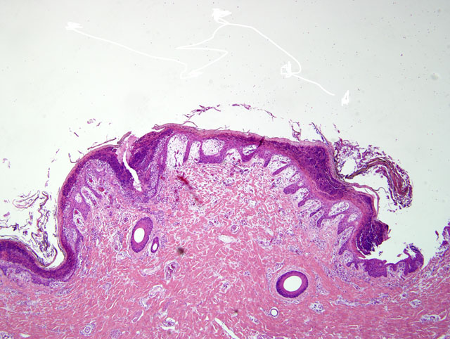
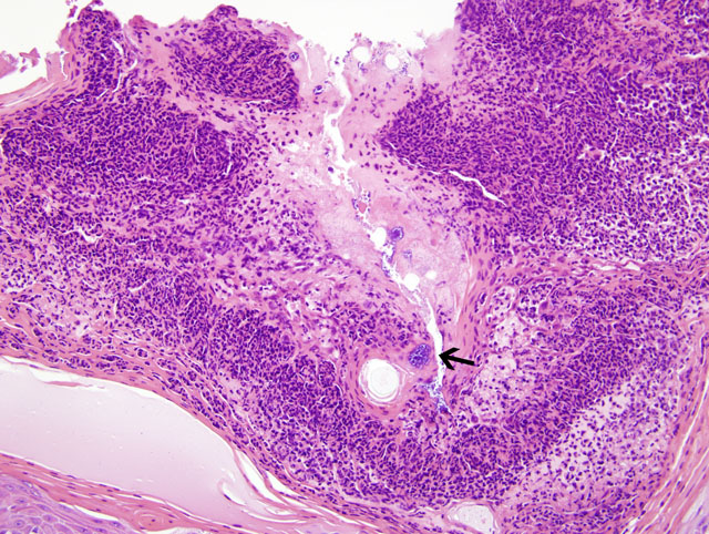
Histopathologic Description:
Morphologic Diagnosis:
Condition:
Contributor Comment:
The presence of cocci bacteria in the epidermal pustules of pigs affected by exudative epidermitis makes this condition similar to human bullous impetigo. Human bullous impetigo is an infection caused by staphylococcal exotoxins and characterized by epidermal pustules which contain many cocci bacteria.1
JPC Diagnosis:
Conference Comment:
Exudative epidermitis of pigs is similar to two human conditions: staphylococcal scalded skin disease and bullous impetigo. Both of these conditions have exfoliotoxins that cause separation between the stratum spinosum and stratum granulosum. However, in bullous impetigo the cocci are present within the intact pustules like in exudative epidermitis, whereas in scalded skin syndrome the cocci are located at a distant often extracutaneous site.2 Staphylococcus intermedius exfoliotoxins cause impetigo, bullous impetigo and superficial spreading pyoderma in dogs.3
Review of this slide led to a discussion of various cutaneous lesions in swine. Sarcoptes scabiei causes erythematous allergic dermatitis with secondary self trauma and crusting. Lesions are primarily located on the rump, flank and abdomen.2 Zinc-responsive dermatosis in swine occurs in 2-4 month old growing pigs, and causes thick, dry scales and crusts that can produce deep fissures. Roughly symmetrical lesions are often found on the lower limbs, around the eyes, ears, snout, scrotum and tail. The microscopic lesion is marked hyperplastic dermatitis with parakeratotic hyperkeratosis.2 Dermatosis vegetans is an inherited disorder in Landrace pigs that results in vegetating skin lesions, hoof malformations and giant cell pneumonia. Skin lesions range from brown-black plaques with a raised border and depressed center to dry-horny papillomatous lesions. The head is typically spared.2 Erysipelothrix rhusiopathiae causes septicemia with cutaneous vasculitis and rhomboidal dermal infarcts, and is also known as diamond back skin disease. Additional lesions in acute cases include fibrinoid glomerular necrosis, renal intracapsular hemorrhage and fibrinous polyarthritis. The most common lesions in chronic cases include vegetative valvular endocarditis, chronic proliferative arthritis and diskospondylitis.6 Blue discoloration of the ears, tail and snout are due to venous thrombosis most commonly caused by Salmonella cholerasuis, but also other septicemic and viral infections such as Erysipelothrix rhusiopathiae, porcine pestivirus (Classical swine fever) and porcine asfarvirus (African swine fever).1 Porcine dermatitis and nephropathy syndrome causes a systemic necrotizing vasculitis with hemorrhagic dermal infarcts, exudative glomerulonephritis and interstitial nephritis in feeder pigs. Skin lesions typically occur on the perineal area of the hindquarters, limbs, dependent areas of the abdomen and thorax, and ear margins. The condition has been associated with Porcine circovirus-2 and Porcine reproductive and respiratory syndrome virus (PRRS).5 Porcine juvenile pustular psoriasiform dermatitis is most common in weaned Landrace pigs, and causes erythematous serpigenous plaques on the ventral abdomen and inner thighs. Histological lesions include eosinophilic perivascular inflammation, spongiform pustules and psoriasiform hyperplasia.2 Swinepox is caused by suipoxvirus. Typical poxviral lesions include proliferative and necrotizing skin lesions with large eosinophilic intracytoplasmic inclusions. Lesions primarily occur on the ventral and lateral abdomen, lateral thorax and medial legs of young, growing pigs. Mucosal surfaces are rarely affected. Hematopinus suis, the sucking louse, acts as a mechanical vector.2 Melanomas are often congenital in the Duroc, Sinclair minipig and in Hormel crosses.2
References:
2. Ginn PE, Mansell JEKL and Rakich PM: Skin and appendages. In: Jubb, Kennedy, and Palmers Pathology of Domestic Animals, ed. Maxie MG, 5th ed. Vol. 1, pp. 591-744. Saunders, Edinburgh, Scotland, 2007
3. Gross TL, Ihrke PJ, Walder EJ, Affolter VK: Skin Diseases of the Dog and Cat, 2nd ed., pp. 4-9. Blackwell Publishing, Ames, Iowa, 2005
4. Taylor DF: Exudative epidermitis. In: Diseases of Swine, eds. Leman AD, Straw BE, Mengeling WL, et al., 7th ed., pp. 522 525. Iowa State Press, Ames, Iowa, 1992
5. Thibault S, Drolet R, Germain M-C, DAllaire S, Larochelle, and Magar R: Cutaneous and systemic necrotizing vasculitis in swine. Vet Pathol 35:108-116, 1998
6. Thompson K: Bones and Joints. In: Jubb, Kennedy, and Palmers Pathology of Domestic Animals, ed. Maxie MG, 5th ed., vol. 1, pp.163-164. Saunders, Edinburgh, Scotland, 2007