Signalment:
Twelve-week-old female Chihuahua dog (
Canis familiaris)This puppy presented for evaluation of non-responsive hypoglycemia of several weeks duration. She subsequently developed diarrhea. On presentation blood glucose was 38 mg/dL (73-116), total protein was 2.6 g/dL (5.5-7.2), albumin was <1.0 g/dL (2.8-4) and PCV was 18%. Coccidial oocysts were found on fecal flotation and therapy was initiated. The puppy deteriorated over the next two days and eventually died.
Gross Description:
The examined puppy was in decreased nutritional condition (BCS 1.5/5) with moderately to markedly reduced subcutaneous and intra-abdominal adipose tissue. There was scant adipose tissue present in the peri-renal mesentery and within the coronary groove of the heart. The stomach contained a small amount of tan mucoid ingesta, the small intestines segmentally contained scant beige pasty ingesta and there was no fecal material present in the colon. No other remarkable gross lesions were present.
Histopathologic Description:
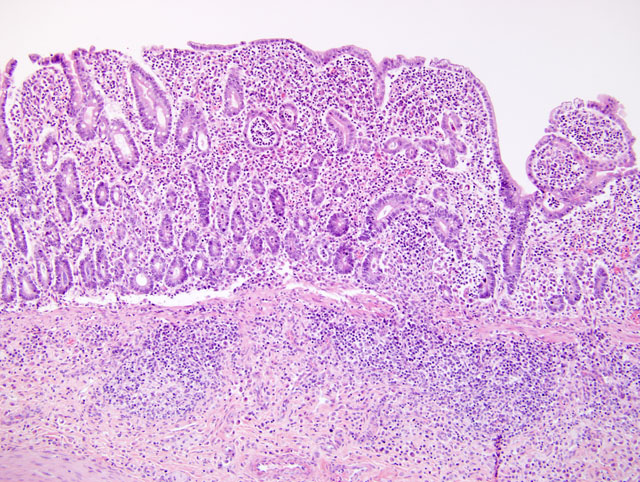
Jejunum: Diffusely there is marked blunting and fusion of villi (
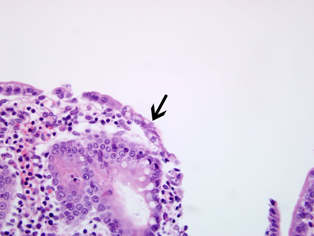
Fig. 1-1). The lamina propria is expanded by a cellular infiltrate composed of numerous neutrophils, lymphocytes, plasma cells, and macrophages that multifocally extend into the submucosa. Multifocally crypts are distended and filled with similar inflammatory cells admixed with pyknotic and karyorrhectic debris (crypt abscesses). Segmentally along villi there are small aggregates of 1x2 um bacilli that are intimately attached to the apical tips of enterocytes (
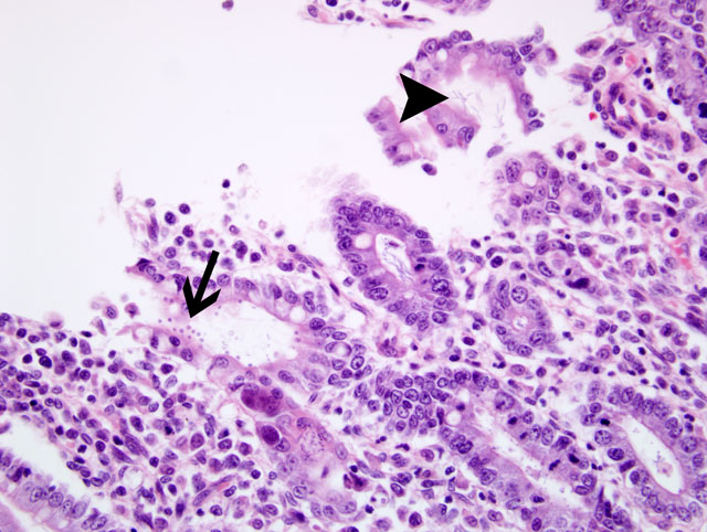
Fig. 1-2). Multifocally crypts are filled with aggregates of spirochete bacteria. Free in the lumen of the jejunum and attached to the apical surface of remaining villous epithelium are scattered 3-6um diameter round, amphophilic protozoal organisms (cryptosporidium) (
Fig. 1-3).
Morphologic Diagnosis:
Diffuse, severe neutrophilic and lymphoplasmacytic enteritis with bacilli, cryptosporidia and spirochetes
Condition:
Attaching and effacing E. coli
Contributor Comment:
This puppy suffered from a severe, malabsorptive protein-losing enteropathy secondary to multiple pathogens. The most pathogenic organism in this case is likely enteroadherent (attaching-effacing)
Escherichia coli. These bacteria are categorized as enteropathogenic
E. coli (EPEC) and colonize the mucosa by a nonpilus adhesion termed EPEC adhesive factor(1). Concurrent infection with cryptosporidium and coccidia has been reported in an immunosuppressed puppy and they were considered opportunistic pathogens(6). Cryptosporidium in dogs is seldom reported in the United States and typically occurs in immunosuppressed puppies.Â
Cryptosporidium parvum and
C. canis have been isolated from naturally infected dogs(4). There was prominent lymphoid depletion present in the spleen and mesenteric lymph nodes, indicating this puppy was likely immunosuppressed secondary to viral infection or a primary underlying immunosuppression. There were no lesions of parvovirus or distemper.Â
No coccidial organisms were seen in multiple sections of intestine; however, there may have been low numbers of organisms due to the previous treatment. The numbers of spirochete bacteria are impressive in this case, but they are not a primary pathogen and this likely represents secondary opportunistic overgrowth. Weakly beta-haemolytic intestinal spirochaetes identified as
Brachyspira pilosicoli have been isolated from puppies and dogs with diarrhea(3,5).Â
Brachyspira canis has been isolated from clinically healthy dogs, suggesting it is a commensal organism(5).
JPC Diagnosis:
Small intestine (jejunum): Enteritis, subacute, diffuse, severe, with marked villus atrophy, fusion, and blunting, crypt necrosis and loss, and attaching bacilli, apicomplexans and intracrypt helical bacteria
Conference Comment:
There are several different types of
E. coli that affect domestic species and each type has virulence characteristics that manifest as varying disease entities. Enteropathogenic
E. coli (EPEC) attaches to the mucosa and causes a malabsorptive diarrhea. Some strains of EPEC do not produce toxins, but they do cause blunting and fusion of villi with subsequent diarrhea. The nomenclature for
E. coli can be extremely confusing, and this type of
E. coli is also known as enteroadherent
E. coli (EAEC), and attaching and effacing
E. coli (2)
EPEC attaches to a host enterocyte via long fimbria and subsequently releases proteins known as adhesins to form a secure attachment to the surface epithelium. Translocated intimin receptor, another protein produced by EPEC, is transported from the bacteria into the host cell. This causes a conformational change in the host cells cytoskeleton. The affected enterocyte forms a pedestal-like structure beneath the bacteria and this unfortunate cell also loses its surface microvilli. The pathogenicity of EPEC is largely determined by the density of organisms on the surface of enterocytes. As the contributor mentioned, co-infections are common and lead to a much worse clinical picture. Bacterial attachment is most prolific in the distal small intestine and large intestine. Profuse diarrhea with EPEC results from a combination of maldigestion and malabsorption.(2)
References:
1. Barker IK, Van Dreumel AA, Palmer N: The alimentary system.Â
In: Pathology of Domestic Animals, eds. Jubb KVF, Kennedy PC, Palmer N, vol. 2, 4th ed., pp. 200-213. Academic Press, Philadelphia, PA, 1993
2. Brown CC, Baker DC, Barker IK: Alimentary system.Â
In: Jubb, Kennedy, and Palmer's Pathology of Domestic Animals, ed. Maxie MG, vol 2 ed., pp. 183-193. Elsevier Limited, Philadelphia, PA, 2007
3. Manabe M, Suenaga I, Ogawa Y, Adachi Y: Brachyspira pilosicoli isolated from two beagles and one mongrel in Japan. J Vet Med Sci.Â
66(5):589-92, 2004
4. Miller DL, Liggett A, Radi ZA, Branch LO: Gastrointestinal cryptosporidiosis in a puppy. Vet Parasitol.Â
115(3):199-204, 2003
5. Oxberry SL, Hampson DJ: Colonisation of pet shop puppies with Brachyspira pilosicoli. Vet Microbiol.Â
93(2):167-74, 2003
6. Willard MD, Bouley D: Cryptosporidiosis, coccidiosis, and total colonic mucosal collapse in an immunosuppressed puppy. J Am Anim Hosp Assoc.Â
35(5):405-9, 1999